Downloaded 158 times
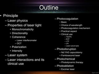
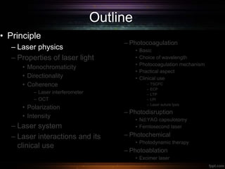
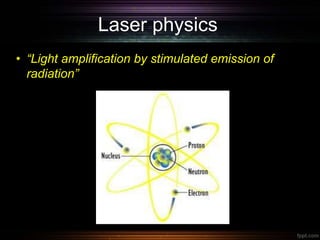
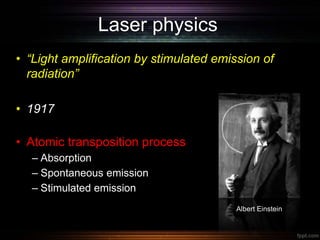
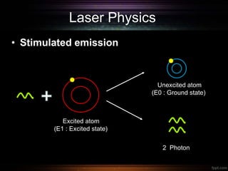
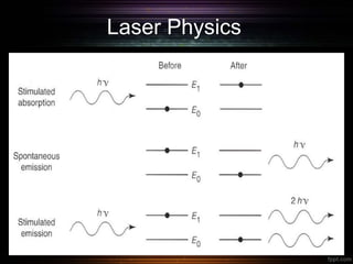
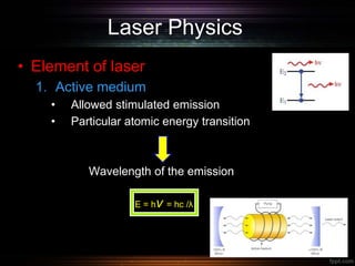
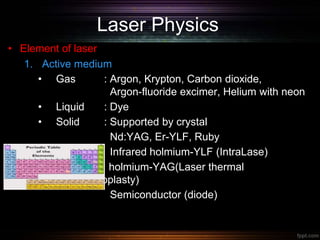
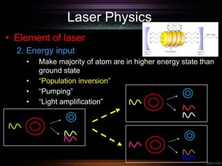
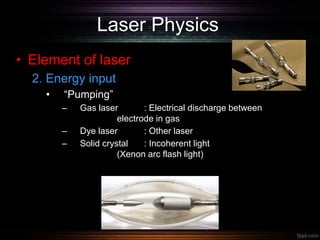

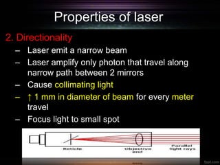
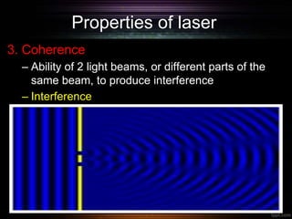
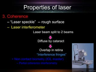
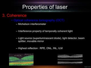
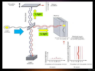
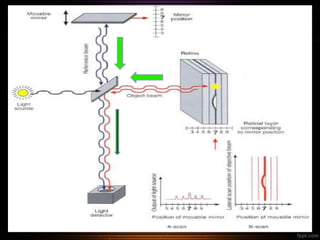
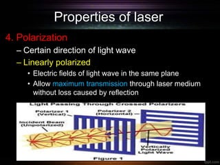
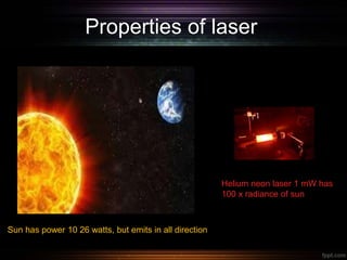
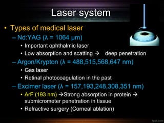
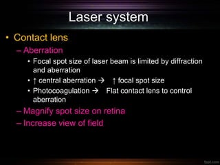
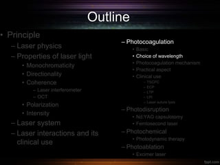
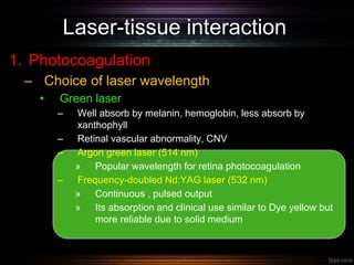
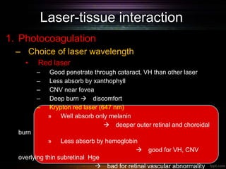
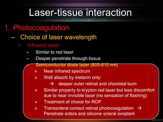
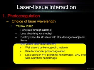
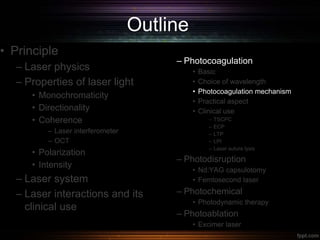
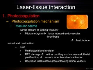
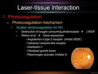
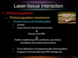
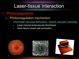
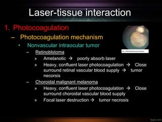
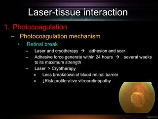
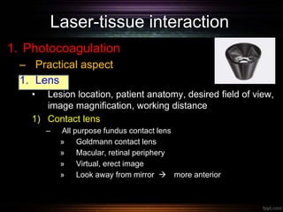
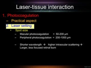
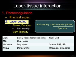
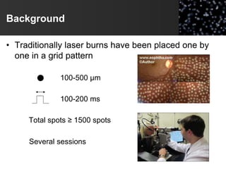
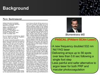
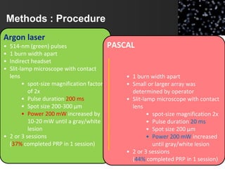
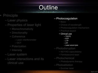
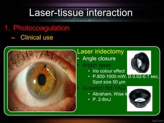
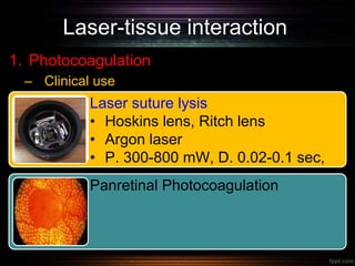
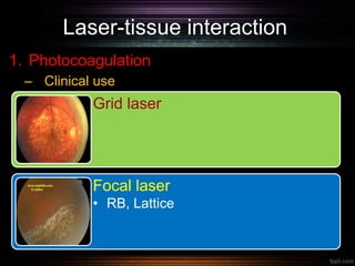
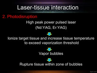
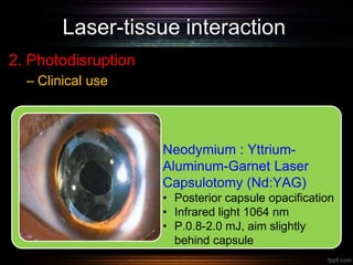
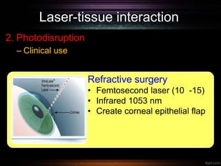
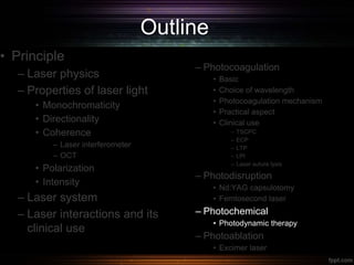
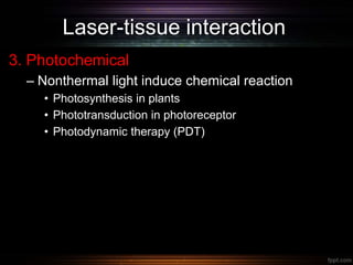
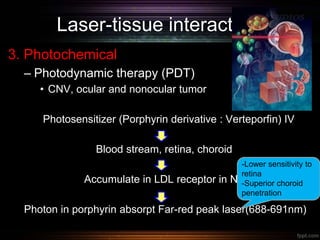
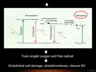
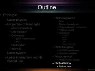
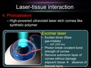


This document provides an overview of fundamental laser ophthalmology. It begins with laser physics, describing the principles of stimulated emission and population inversion required for laser action. It then discusses the properties of laser light including monochromaticity, directionality, coherence, polarization and intensity. The document outlines different laser systems used in ophthalmology and laser-tissue interactions, focusing on photocoagulation. It explains the basic mechanism of photocoagulation, factors in choosing the appropriate wavelength, and discusses photocoagulation in the clinical context of conditions like diabetic macular edema and retinal neovascularization.


![Karimi understanding lasers[1]](https://cdn.slidesharecdn.com/ss_thumbnails/karimiunderstandinglasers1-160907050417-thumbnail.jpg?width=640&height=640&fit=bounds)


![CTEV [ clubfoot] DR ARUN LAL ,DR MOHAMED ASHRAF travancore medical college k...](https://cdn.slidesharecdn.com/ss_thumbnails/ctevclubfootdrarunlaldrmohamedashraftravancoremedicalcollegekollamkeralaindia-260208063247-18fc466c-thumbnail.jpg?width=640&height=640&fit=bounds)


![PERI-PROSTHETIC FRACTURE NAIL-PLATE CONSTRUCT [NPC].pptx](https://cdn.slidesharecdn.com/ss_thumbnails/drarunkumardrmohamedashrafperiprostheticfrasturenail-plateconstructnpc-260209164459-7e9d15a1-thumbnail.jpg?width=640&height=640&fit=bounds)

