Downloaded 304 times


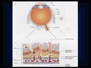

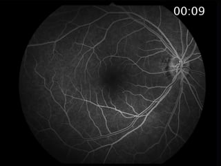
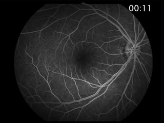
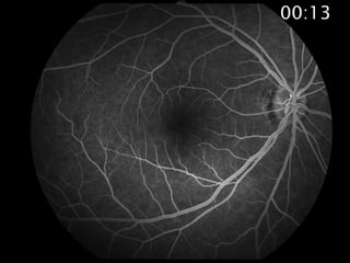
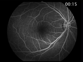
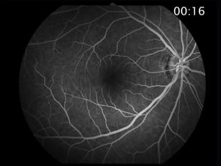
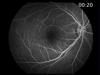
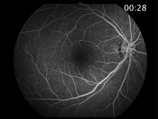
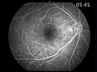
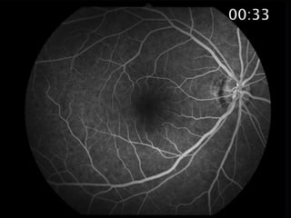
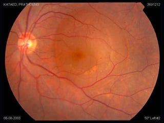
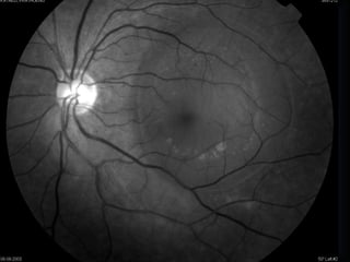
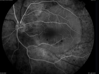
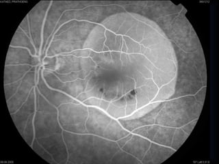
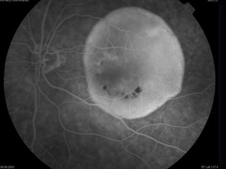
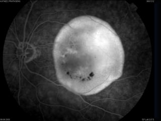
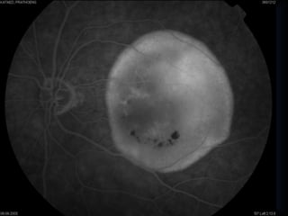
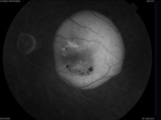
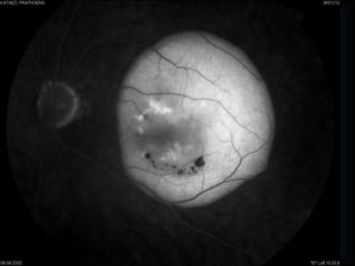
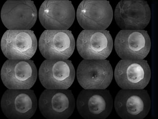
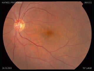
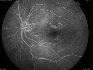
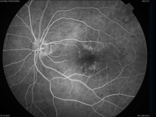
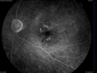
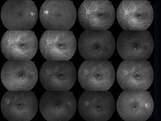
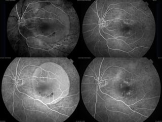
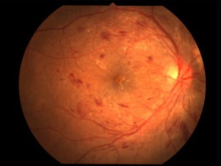
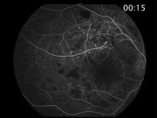
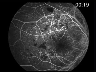
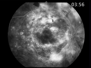
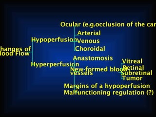
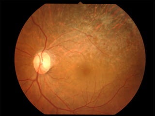
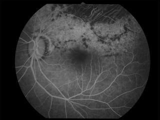
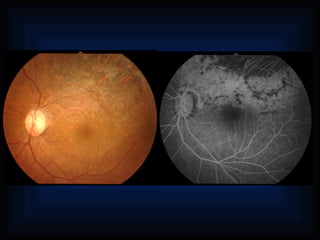
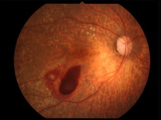
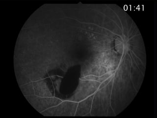
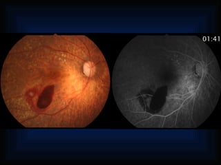
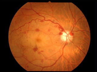
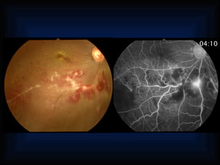
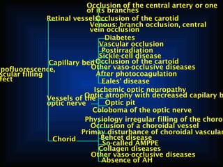
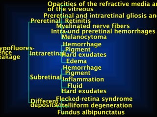
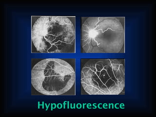


This document describes various ocular diseases and abnormalities and how they may appear on angiography. It discusses changes that can occur in blood flow and vessels in the choroid, retina, vitreous, optic nerve, and associated with various conditions like diabetes, vascular occlusion, inflammation and tumors. Specific abnormalities mentioned include neovascularization, tortuosity, dilation, cystoid edema, detachments, drusen, scarring and abnormalities in fluorescence patterns.


