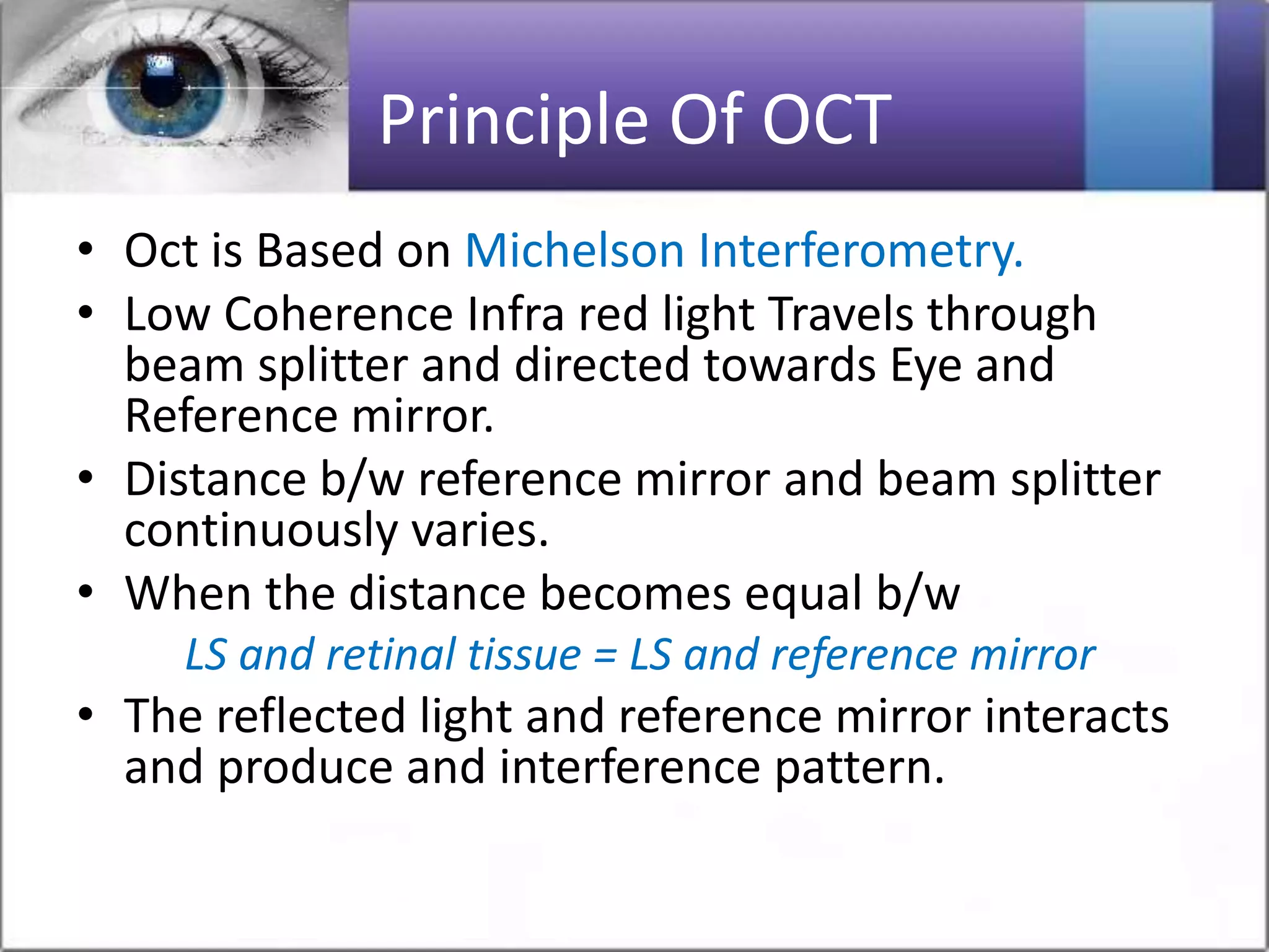
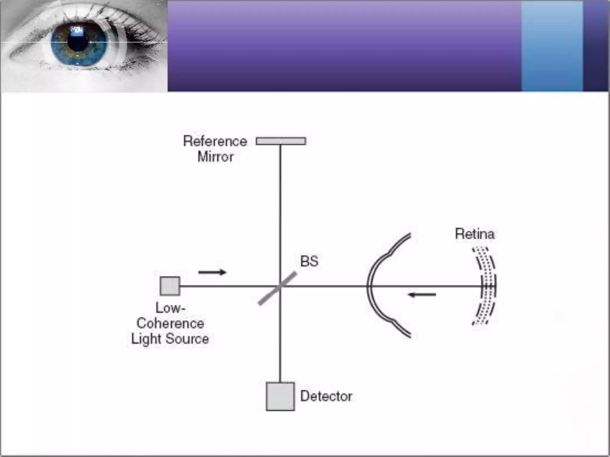
Optical coherence tomography (OCT) is a non-invasive imaging technique that uses light waves to capture high-resolution cross-sectional images of the retina, proving vital for diagnosing and guiding treatment for various eye conditions, particularly glaucoma and retinal diseases like age-related macular degeneration. The technology involves two main types: time-domain OCT and spectral-domain OCT, with the latter offering superior sensitivity and speed. OCT provides critical insights into retinal anatomy and pathology, allowing for effective monitoring and management of eye diseases.