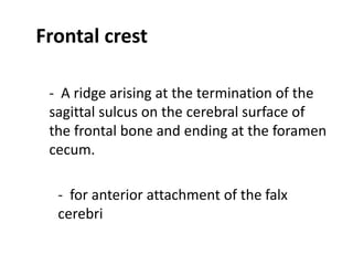
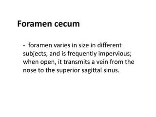
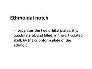
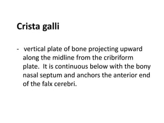
Downloaded 338 times


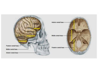
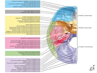
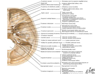
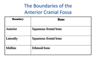
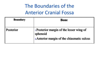
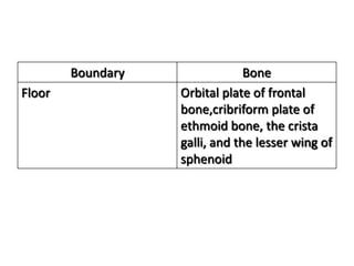
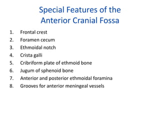
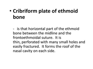
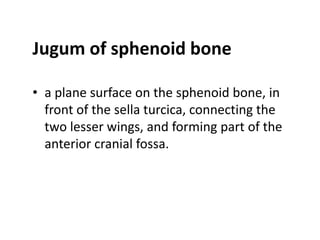
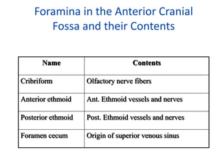
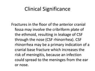
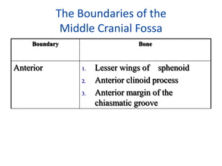
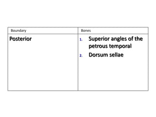
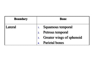
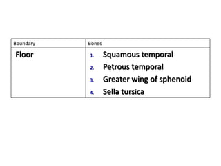
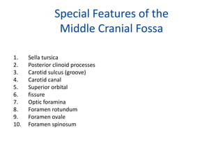
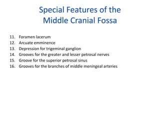
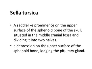
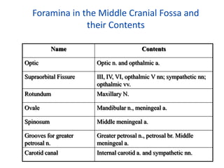
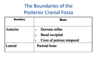
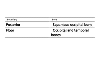
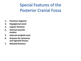
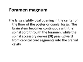
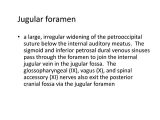
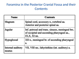


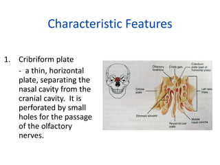
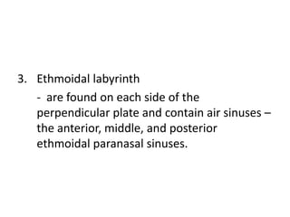
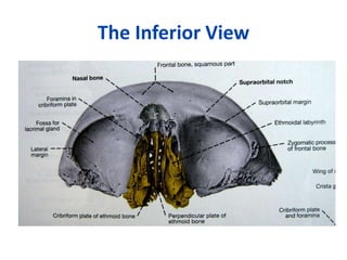
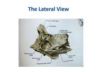
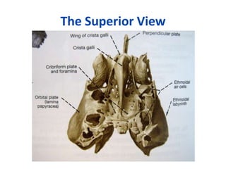


The document describes the anatomy of the anterior, middle, and posterior cranial fossae. It details the boundaries, special features, and foramina of each fossa. Key structures mentioned include the cribriform plate, sella turcica, foramen magnum, optic foramina, and jugular foramen. Clinical significance is noted for fractures involving the cribriform plate which can lead to CSF rhinorrhea and risk of meningitis.


![CTEV [ clubfoot] DR ARUN LAL ,DR MOHAMED ASHRAF travancore medical college k...](https://cdn.slidesharecdn.com/ss_thumbnails/ctevclubfootdrarunlaldrmohamedashraftravancoremedicalcollegekollamkeralaindia-260208063247-18fc466c-thumbnail.jpg?width=640&height=640&fit=bounds)


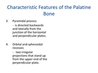
![ONFH[AVN HIP] -TRIPLE REGIME -A NOVAL SURGICAL CONCEPT .pptx](https://cdn.slidesharecdn.com/ss_thumbnails/onfhavnhip2026koaconcalicutdrgokuldevdrmashraf-260210064517-213ec005-thumbnail.jpg?width=640&height=640&fit=bounds)
