Downloaded 307 times

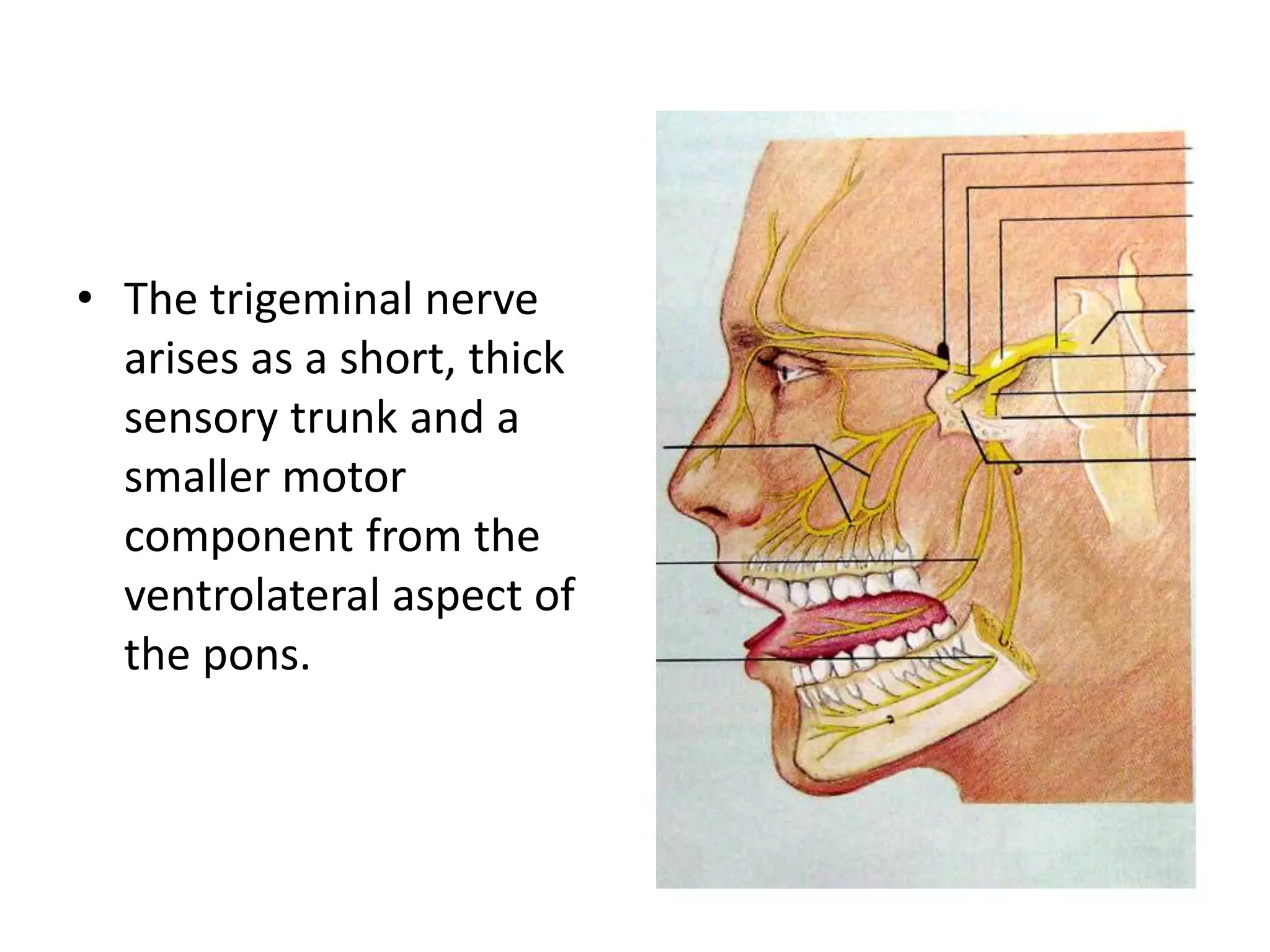
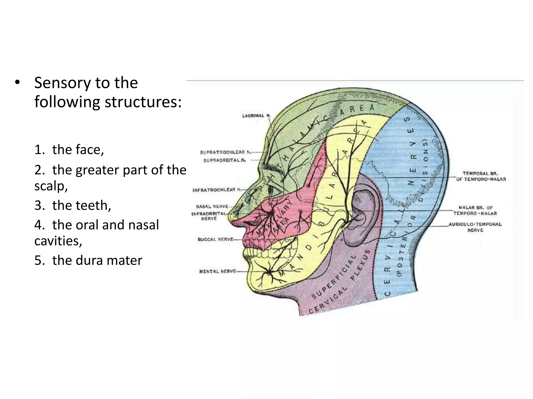

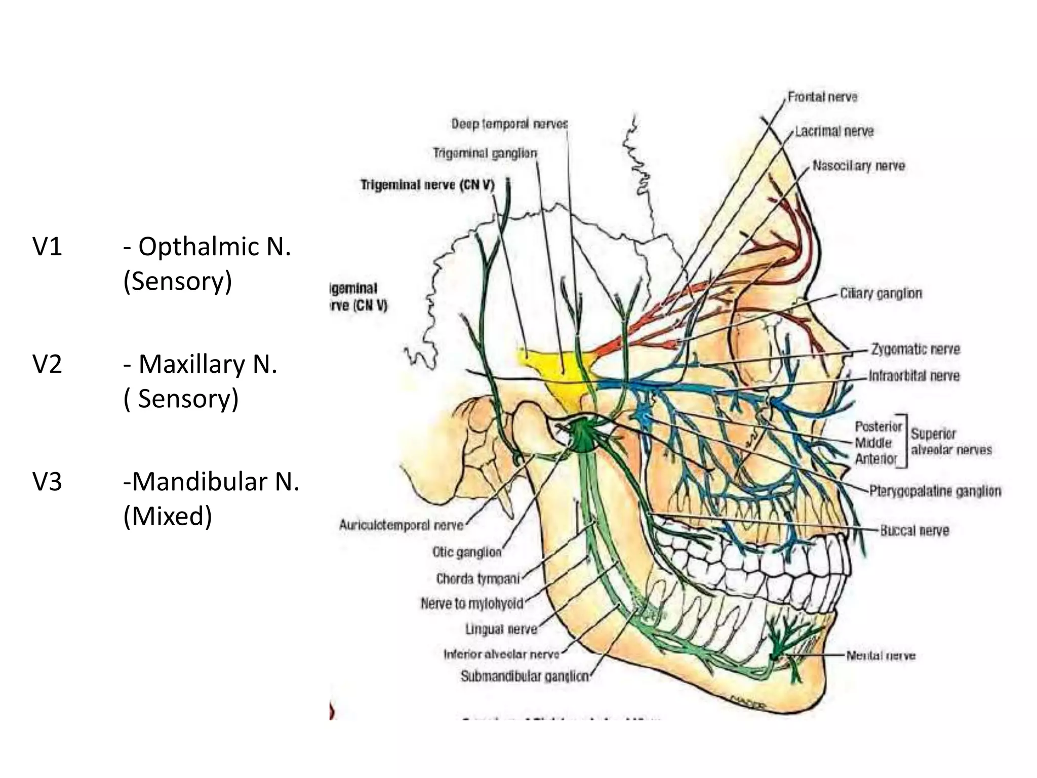

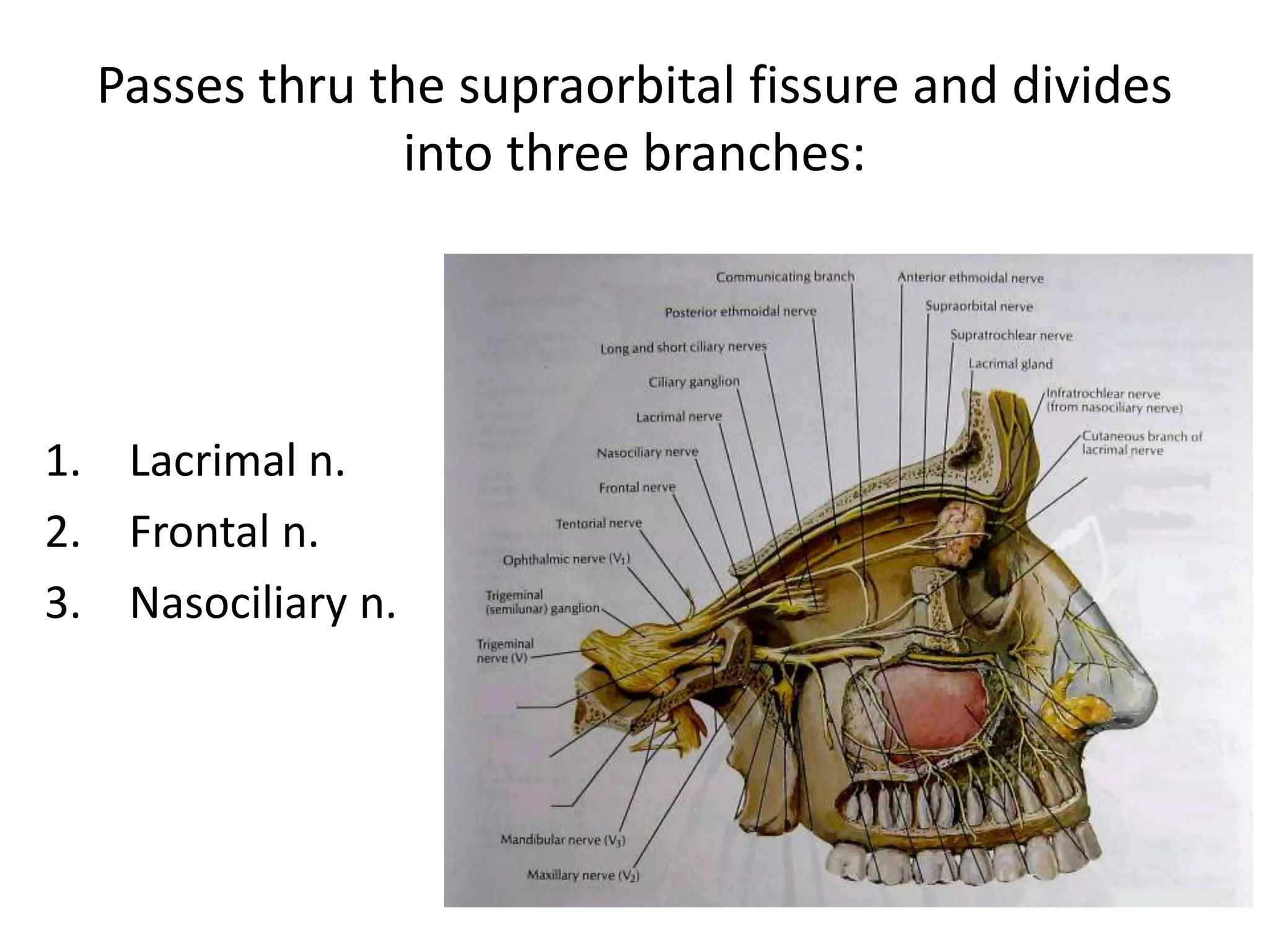
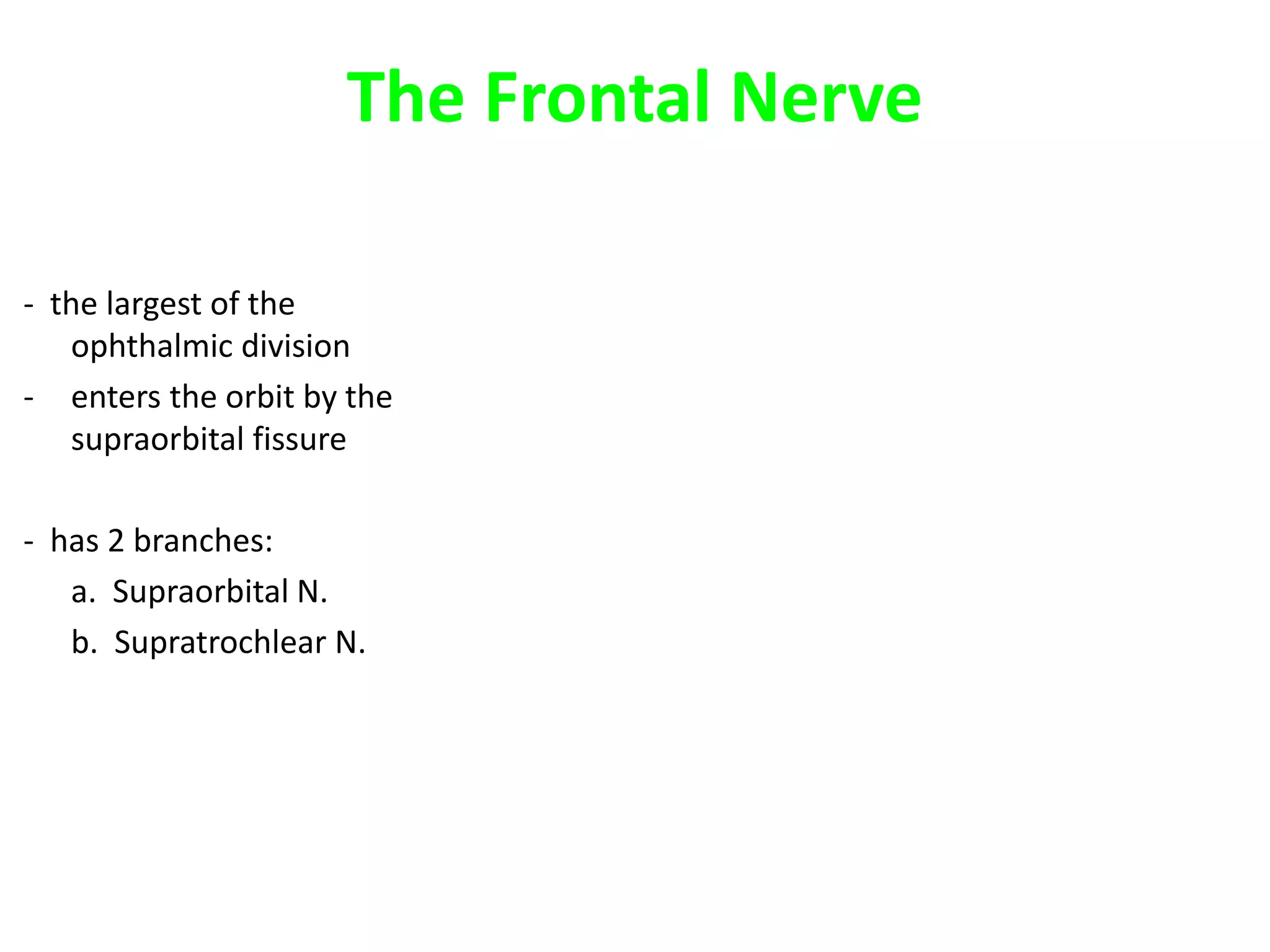
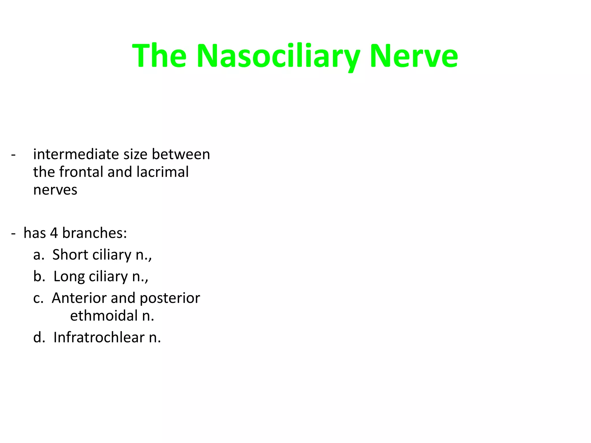
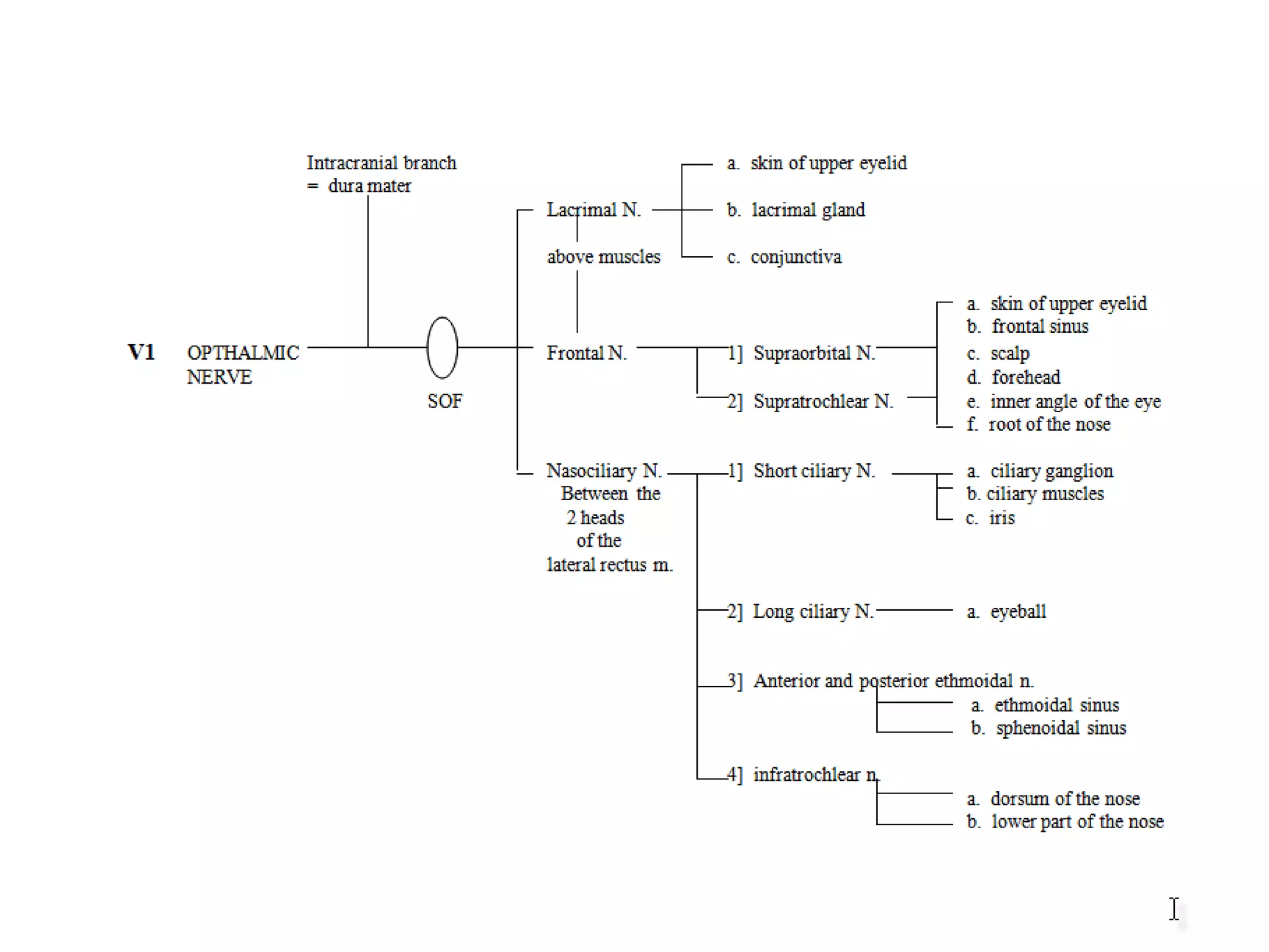

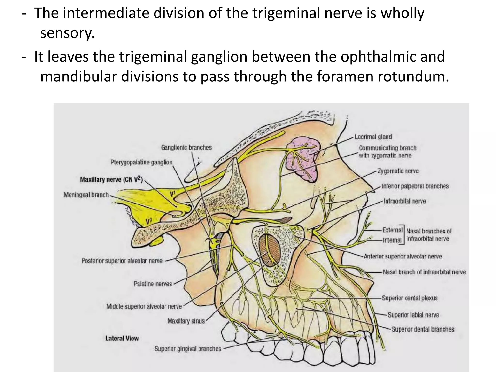

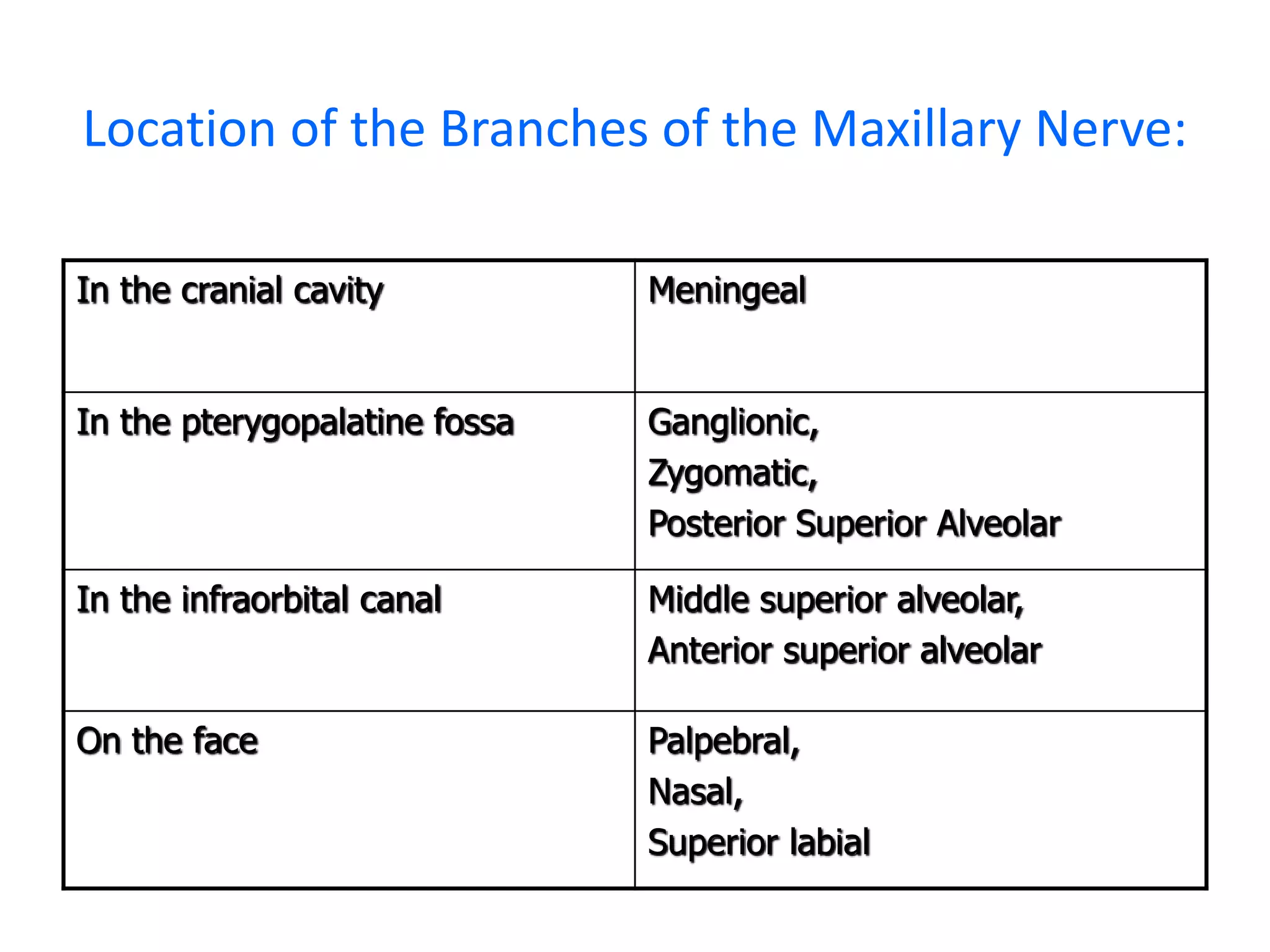
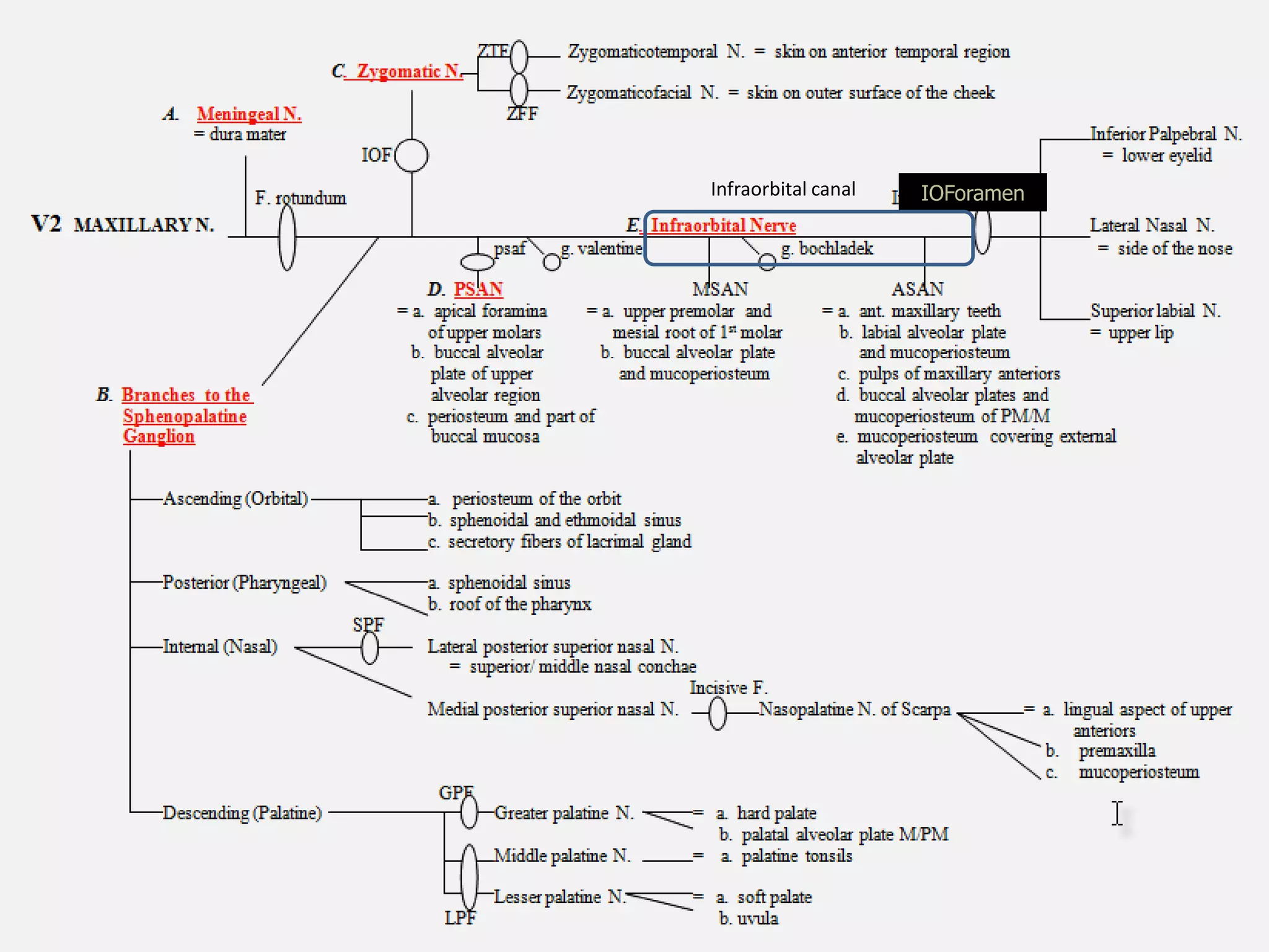

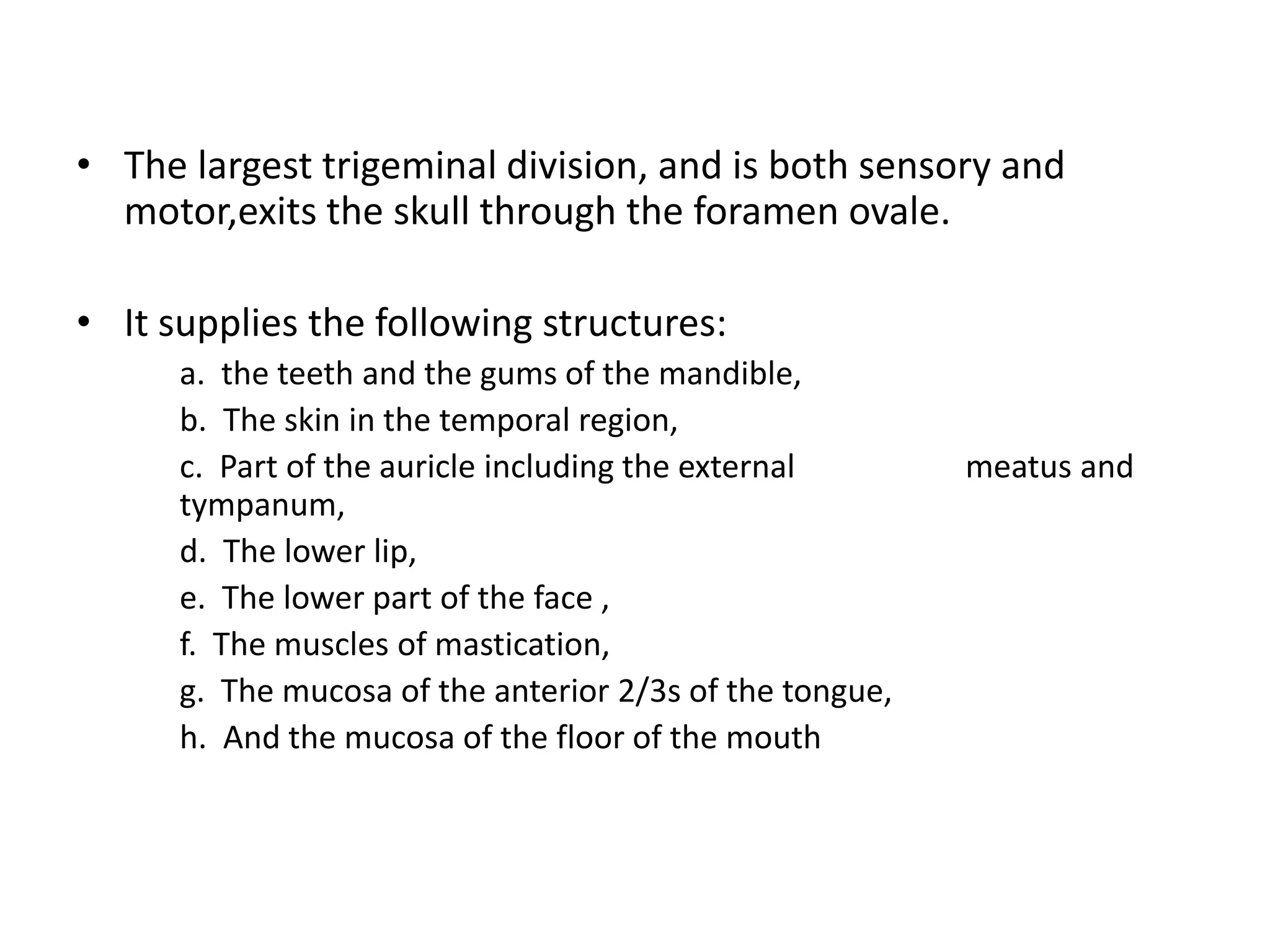

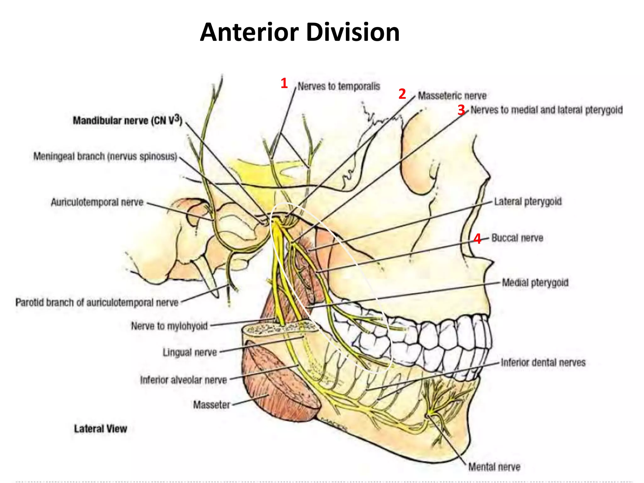
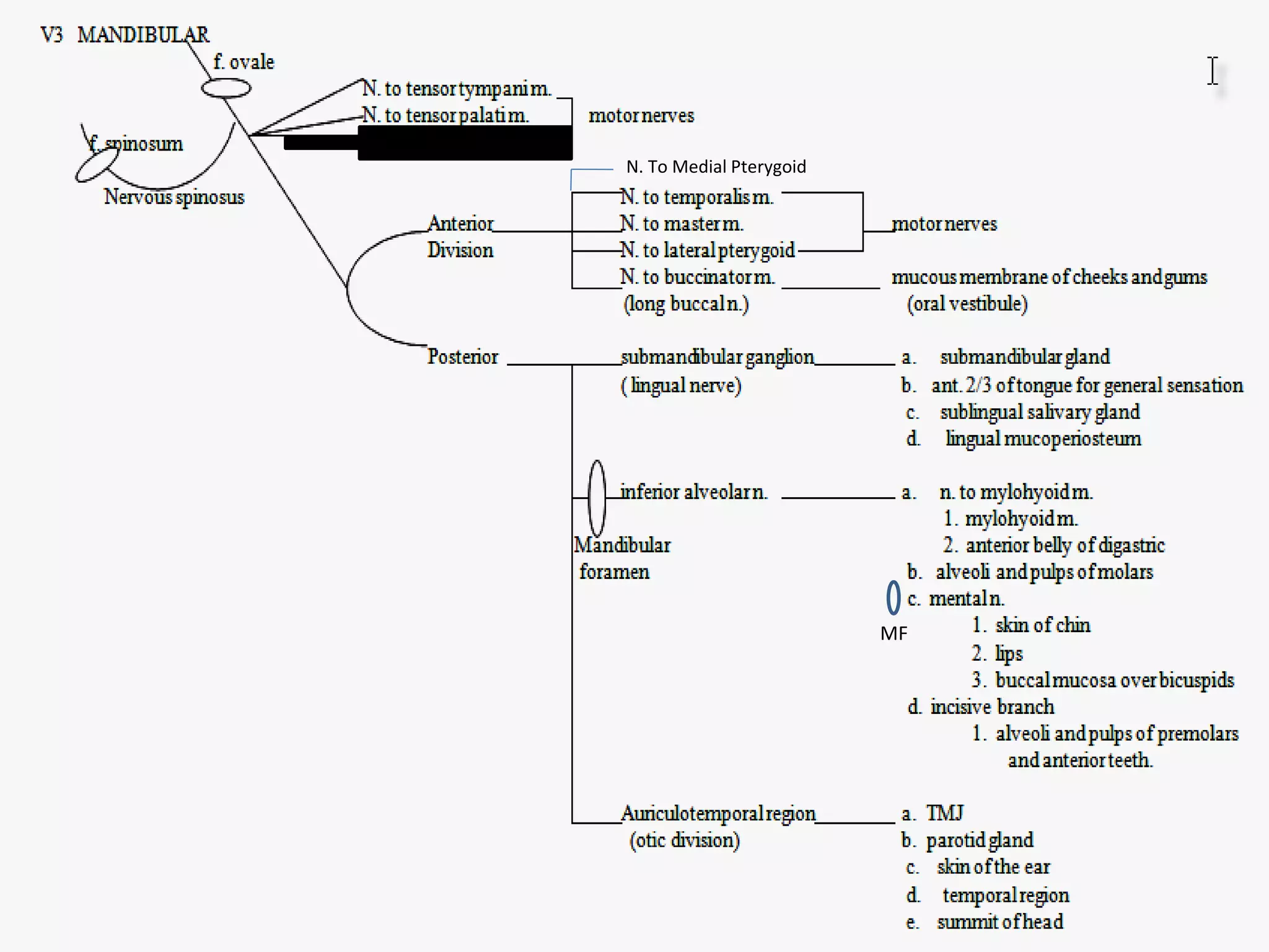
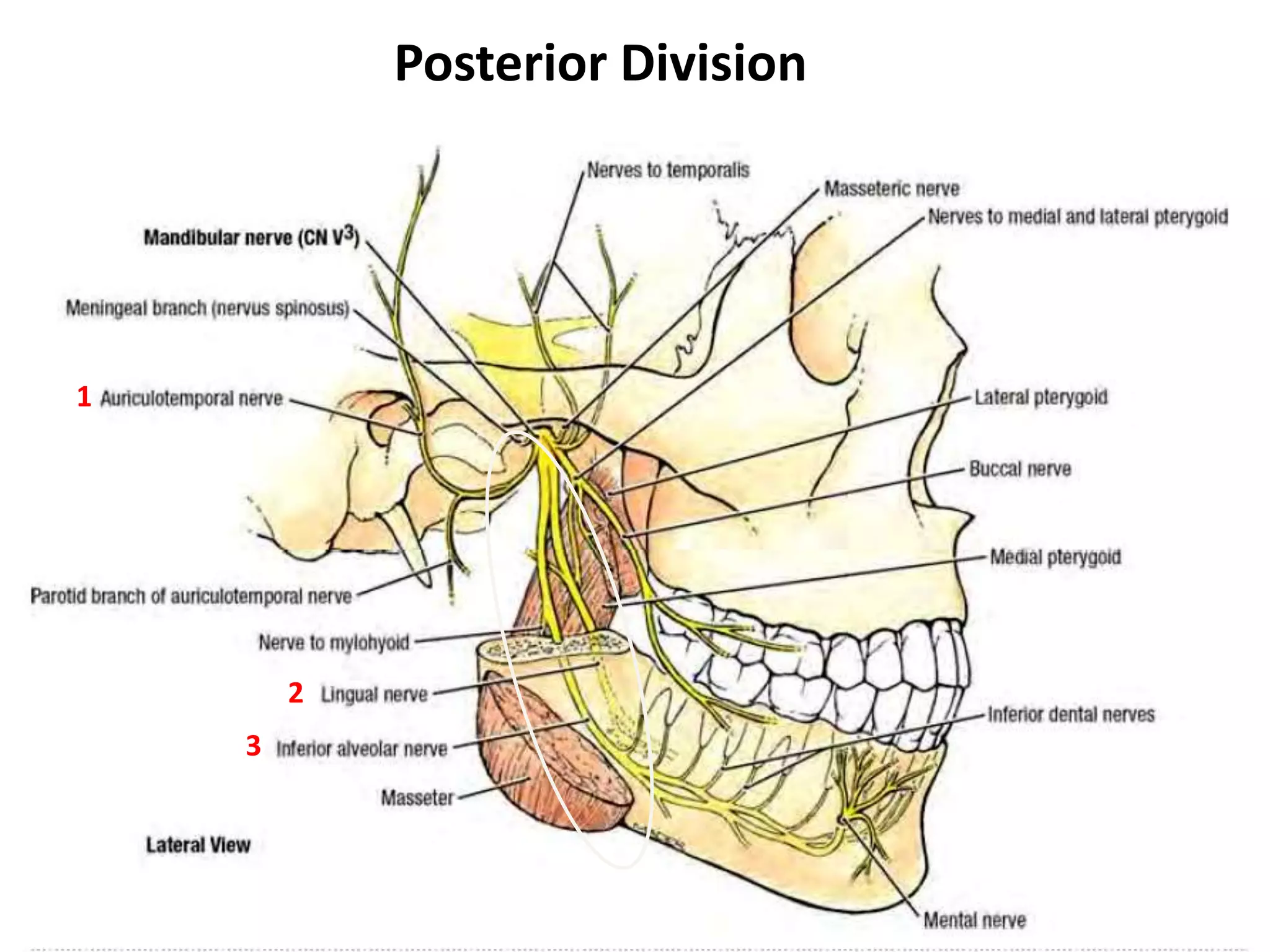
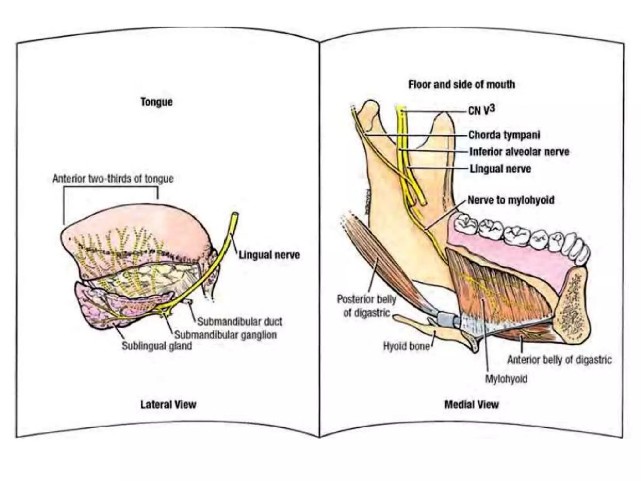


The trigeminal nerve arises from the pons and has three main divisions - the ophthalmic, maxillary, and mandibular nerves. The ophthalmic nerve is sensory to the eye and parts of the nose and forehead. The maxillary nerve is sensory to parts of the face and upper teeth. The mandibular nerve is mixed sensory and motor, innervating the lower face, teeth, and muscles of mastication. It exits through the foramen ovale. The document then describes the branches and distributions of each trigeminal nerve division.


