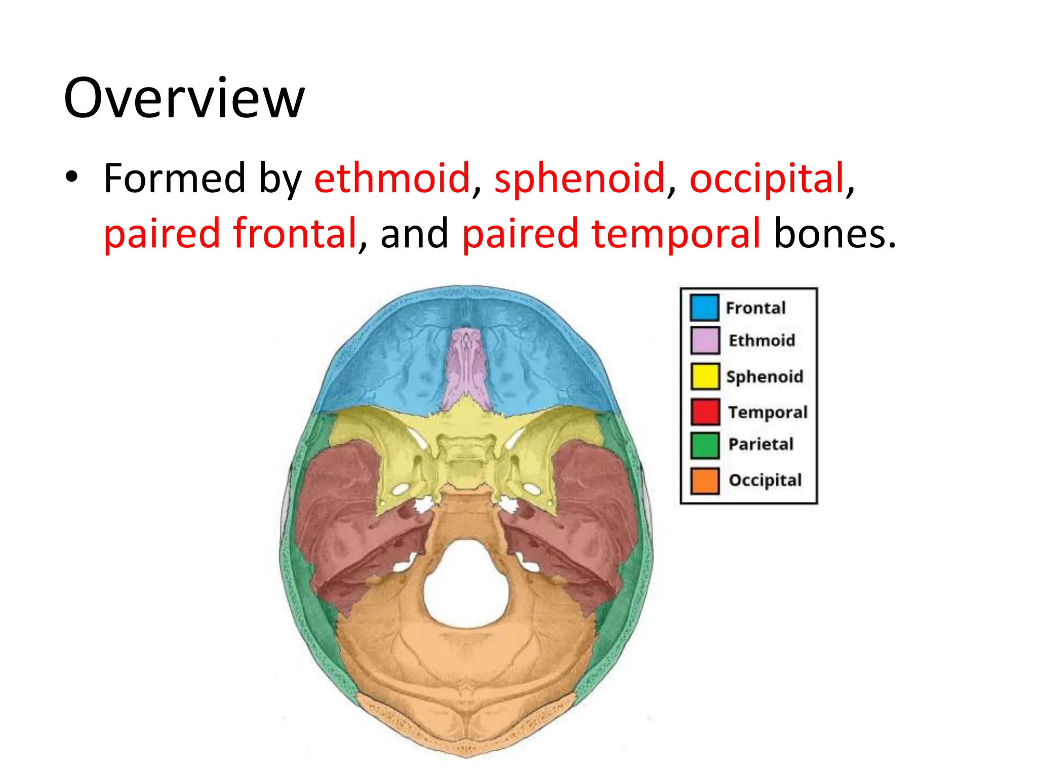
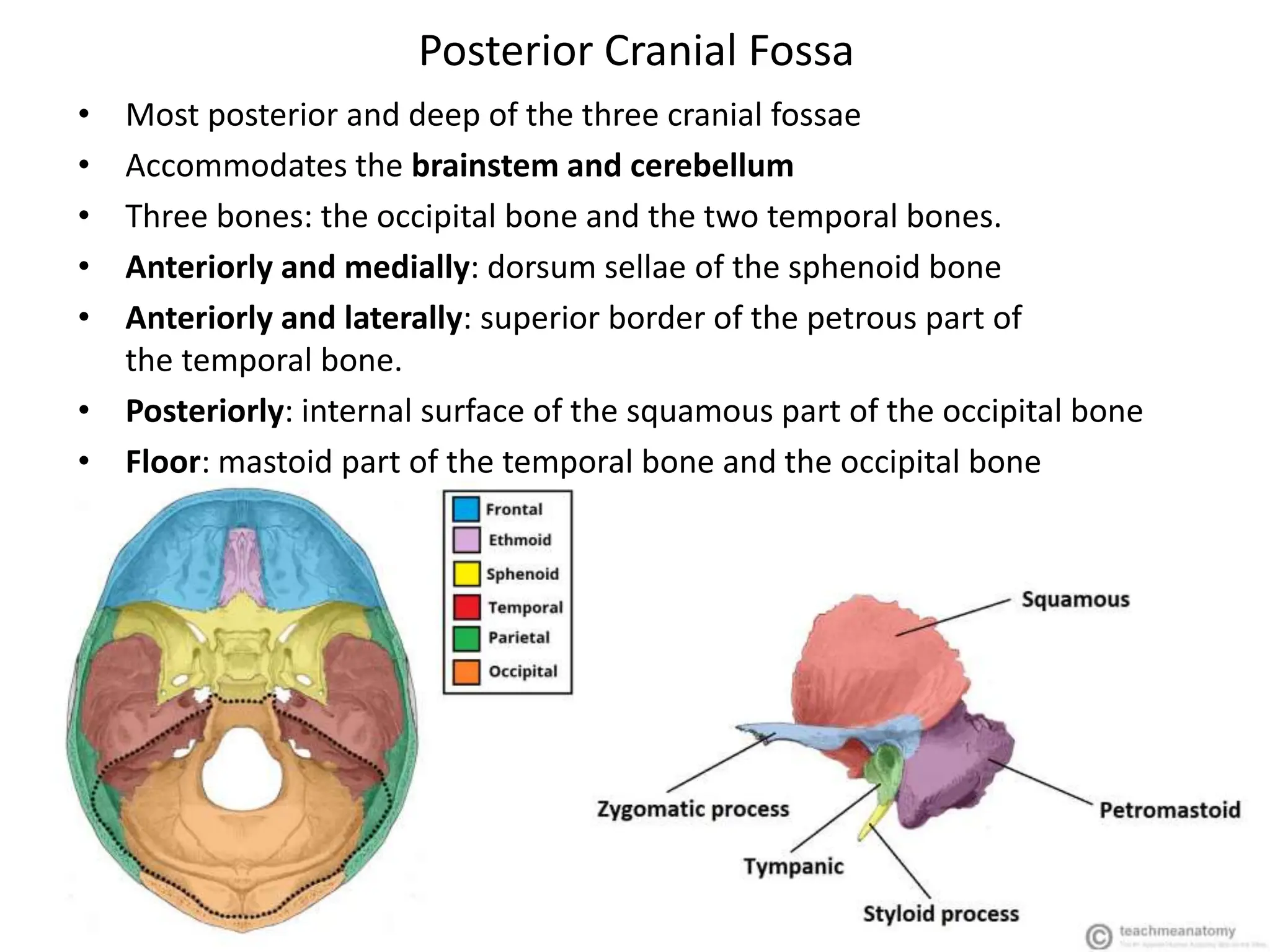
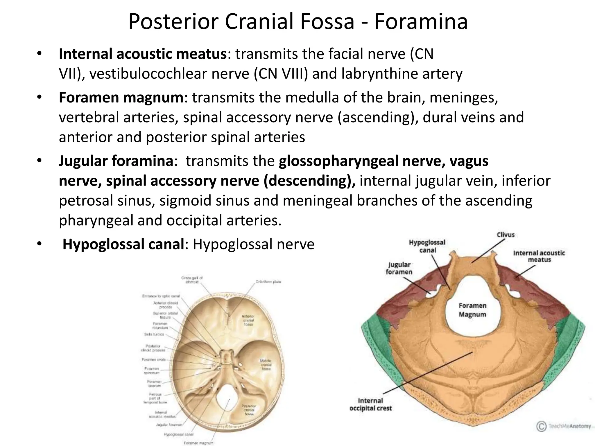
SlideShare a Scribd company logo
1 of 19
Skull Base Anatomy
Overview
• Forms the floor of the cranial cavity
• Separates the brain from other facial
structures
Overview
• Formed by ethmoid, sphenoid, occipital,
paired frontal, and paired temporal bones.
Overview
• Subdivided into 3 regions: the anterior,
middle, and posterior cranial fossae.
Anterior Cranial Fossa
• Most shallow and superior of the three cranial fossae
• Lies superiorly over the nasal and orbital cavities
• Accommodates the anteroinferior portions of the frontal lobes of the
brain
Anterior Cranial Fossa: Borders
• Anteriorly and laterally bounded by the inner surface of the frontal bone
• Posteriorly and medially bounded by the limbus of the sphenoid bone
• Posteriorly and laterally it is bounded by the lesser wings of
the sphenoid bone
• The floor consists of the frontal bone, ethmoid bone and the anterior
aspects of the body and lesser wings of the sphenoid bone
Anterior Cranial Fossa : Contents
• Frontal crest: acts as a site of attachment for the falx cerebri (a sheet of
dura mater that divides the two cerebral hemispheres)
• Crista galli: midline of ethmoid bone, acts as another point of attachment
for the falx cerebri
• Cribriform plate: on either site of the crista galli, supports the olfactory
bulb and has numerous foramina that transmit vessels and nerves.
• Anterior clinoid processes: rounded ends of the lesser wings, serve as a
place of attachment for the tentorium cerebelli (a sheet of dura mater
that divides the cerebrum from the cerebellum)
Anterior Cranial Fossa : Foramina
• Cribriform plate: numerous small foramina, transmiting olfactory nerve
fibers. 2 larger foramen;
• Anterior ethmoidal foramen transmits the anterior ethmoidal artery,
nerve and vein
• Posterior ethmoidal foramen transmits the posterior ethmoidal artery,
nerve and vein
Anterior Cranial Fossa : Clinical Relevance
• Cribriform plate: thinnest part, most likely to fracture
• Anosmia
• CSF rhinorrhoea: leakage of CSF into the nasal cavity
Middle Cranial Fossa
• As its name suggests, centrally in the cranial floor
• Butterfly shaped
• Anteriorly and laterally: lesser wings of the sphenoid bone
• Anteriorly and medially: limbus of the sphenoid bone
• Posteriorly and laterally: superior border of the petrous part of the temporal bone
• Posteriorly and medially: dorsum sellae of the sphenoid bone
• Floor: the body and greater wing of the sphenoid, and the squamous and petrous
parts of the temporal bone.
Middle Cranial Fossa: Central Part
• Formed by the body of the sphenoid bone
• Contains the sella turcica(turkish saddle), acts to hold and support the
pituitary gland
• Tuberculum sellae (horn of the saddle): anterior wall of the sella turcica,
and the posterior aspect of the chiasmatic sulcus
• Hypophysial fossa or pituitary fossa (seat of the saddle): a depression in
the body of the sphenoid, which holds the pituitary gland
• Dorsum sellae (back of the saddle): posterior wall of the sella turcica,
separates the middle cranial fossa from the posterior cranial fossa.
Middle Cranial Fossa: Lateral Parts
• Formed by the greater wings of the sphenoid bone,
and the squamous and petrous parts of the temporal bones
• Support the temporal lobes of the brain
• The site of many foramina
Foramina of the Sphenoid Bone
• Optic canals: optic nerves (CN II) and ophthalmic arteries, connected by
the chiasmatic sulcus
• Superior orbital fissure: opens anteriorly into the orbit;
contains the oculomotor nerve (CN III), trochlear nerve (CN IV),
ophthalmic branch of the trigeminal nerve (CN V1), abducens nerve (CN
VI), opthalmic veins and sympathetic fibers
• Foramen rotundum: transmits the maxillary branch of
the trigeminal nerve (CN V2).
• Foramen ovale: the mandibular branch of the trigeminal nerve (CN V3)
and accessory meningeal artery
• Foramen spinosum: transmits the middle meningeal artery, middle
meningeal vein and a meningeal branch of CN V3
Foramina of the Temporal Bone
• Hiatus of the greater petrosal nerve: transmits the greater petrosal nerve
(a branch of the facial nerve), and the petrosal branch of the middle
meningeal artery
• Hiatus of the lesser petrosal nerve – transmits the lesser petrosal nerve (a
branch of the glossopharyngeal nerve).
• Carotid canal – located posteriorly and medially to the foramen ovale. This
is traversed by the internal carotid artery, the deep petrosal nerve also
passes through this canal
• Foramen lacerum: At the junction of the sphenoid, temporal and occipital
bones, filled with cartilage
Clinical Relevance: Pituitary Surgery
• The pituitary gland lies in the sella turcica of the sphenoid bone
• In cases of a pituitary tumour, it may need to be removed surgically.
• usually by a endoscopic transsphenoidal approach
• The sphenoid sinus is opened and the endoscope passes through to the
pituitary gland
• Complications of pituitary surgery include CSF rhinorrhoea, meningitis,
diabetes insipidis, haemorrhage and visual disturbances
Posterior Cranial Fossa
• Most posterior and deep of the three cranial fossae
• Accommodates the brainstem and cerebellum
• Three bones: the occipital bone and the two temporal bones.
• Anteriorly and medially: dorsum sellae of the sphenoid bone
• Anteriorly and laterally: superior border of the petrous part of
the temporal bone.
• Posteriorly: internal surface of the squamous part of the occipital bone
• Floor: mastoid part of the temporal bone and the occipital bone
Posterior Cranial Fossa - Foramina
• Internal acoustic meatus: transmits the facial nerve (CN
VII), vestibulocochlear nerve (CN VIII) and labrynthine artery
• Foramen magnum: transmits the medulla of the brain, meninges,
vertebral arteries, spinal accessory nerve (ascending), dural veins and
anterior and posterior spinal arteries
• Jugular foramina: transmits the glossopharyngeal nerve, vagus
nerve, spinal accessory nerve (descending), internal jugular vein, inferior
petrosal sinus, sigmoid sinus and meningeal branches of the ascending
pharyngeal and occipital arteries.
• Hypoglossal canal: Hypoglossal nerve
Clinical Relevance: Cerebellar Tonsillar Herniation
• Downward displacement of the cerebellar tonsils through the foramen
magnum
• Produced by a raised intracranial pressure; causes include hydrocephalus,
space occupying lesions, and a malformed posterior cranial fossa
• Cerebellar tonsillar herniation results in the compression of
the pons and medulla, which contain the cardiac and respiratory centers
References
• TeachMeAnatomy. (2017). The Anterior
Cranial Fossa. [online] Available at:
http://teachmeanatomy.info/head/areas/crani
al-fossa/anterior/ [Accessed 30 Mar. 2017].
• Skull Base Anatomy. (2016, June 28). Retrieved
March 30, 2017, from
http://emedicine.medscape.com/article/8826
27-overview

More Related Content

Similar to Skull base anatomy.pptx

Review Of Skull And Cranial Cavity
Review Of Skull And Cranial CavityReview Of Skull And Cranial Cavity
Review Of Skull And Cranial CavityDr. Noura El Tahawy
 
anatomy of The orbit (1)
anatomy of The orbit (1)anatomy of The orbit (1)
anatomy of The orbit (1)ddert
 
Module-1-Bones-of-the-Skull.pptx
Module-1-Bones-of-the-Skull.pptxModule-1-Bones-of-the-Skull.pptx
Module-1-Bones-of-the-Skull.pptxhehehehe62
 
Anatomy of Skullbase
Anatomy of SkullbaseAnatomy of Skullbase
Anatomy of SkullbaseJinu Iype
 
anatomy of nose&paranasal sinuses.pdf
anatomy of nose&paranasal sinuses.pdfanatomy of nose&paranasal sinuses.pdf
anatomy of nose&paranasal sinuses.pdfHusseinWali1
 
Pons anatomy and syndromes
Pons anatomy and syndromesPons anatomy and syndromes
Pons anatomy and syndromesAmruta Rajamanya
 
Base of skull by dr kifayat
Base of skull by dr kifayatBase of skull by dr kifayat
Base of skull by dr kifayatKifayat Khan
 
“BASE OF SKULL: ANATOMY and APPLIED ASPECT”
“BASE OF SKULL: ANATOMY and APPLIED ASPECT”“BASE OF SKULL: ANATOMY and APPLIED ASPECT”
“BASE OF SKULL: ANATOMY and APPLIED ASPECT”RAKESHTALUKDAR2
 
Basic Skull Anatomy
Basic Skull AnatomyBasic Skull Anatomy
Basic Skull AnatomyHadi Munib
 
Trigeminal Nerve Dr.AD.pptx
Trigeminal Nerve Dr.AD.pptxTrigeminal Nerve Dr.AD.pptx
Trigeminal Nerve Dr.AD.pptxArunCreations
 
brainstem cerebellum cranial nerves forr
brainstem cerebellum cranial nerves forrbrainstem cerebellum cranial nerves forr
brainstem cerebellum cranial nerves forromarkhaled833960
 
Nose and Paranasal Sinus.pptx
Nose and Paranasal Sinus.pptxNose and Paranasal Sinus.pptx
Nose and Paranasal Sinus.pptxMathew Joseph
 

Similar to Skull base anatomy.pptx (20)

Review Of Skull And Cranial Cavity
Review Of Skull And Cranial CavityReview Of Skull And Cranial Cavity
Review Of Skull And Cranial Cavity
 
Anatomy of skull base
Anatomy of skull baseAnatomy of skull base
Anatomy of skull base
 
anatomy of The orbit (1)
anatomy of The orbit (1)anatomy of The orbit (1)
anatomy of The orbit (1)
 
Anatomy of the orbit
Anatomy of the orbit Anatomy of the orbit
Anatomy of the orbit
 
Module-1-Bones-of-the-Skull.pptx
Module-1-Bones-of-the-Skull.pptxModule-1-Bones-of-the-Skull.pptx
Module-1-Bones-of-the-Skull.pptx
 
Nervous system anatomy
Nervous system anatomyNervous system anatomy
Nervous system anatomy
 
Anatomy of Skullbase
Anatomy of SkullbaseAnatomy of Skullbase
Anatomy of Skullbase
 
Overview of Neck.pdf
Overview of Neck.pdfOverview of Neck.pdf
Overview of Neck.pdf
 
anatomy of nose&paranasal sinuses.pdf
anatomy of nose&paranasal sinuses.pdfanatomy of nose&paranasal sinuses.pdf
anatomy of nose&paranasal sinuses.pdf
 
Pons anatomy and syndromes
Pons anatomy and syndromesPons anatomy and syndromes
Pons anatomy and syndromes
 
Base of skull by dr kifayat
Base of skull by dr kifayatBase of skull by dr kifayat
Base of skull by dr kifayat
 
“BASE OF SKULL: ANATOMY and APPLIED ASPECT”
“BASE OF SKULL: ANATOMY and APPLIED ASPECT”“BASE OF SKULL: ANATOMY and APPLIED ASPECT”
“BASE OF SKULL: ANATOMY and APPLIED ASPECT”
 
Basic Skull Anatomy
Basic Skull AnatomyBasic Skull Anatomy
Basic Skull Anatomy
 
Cranial fossa-SCALP
Cranial fossa-SCALPCranial fossa-SCALP
Cranial fossa-SCALP
 
lecture ear.pptx
lecture ear.pptxlecture ear.pptx
lecture ear.pptx
 
human anatomy -Back of thigh
human anatomy -Back of thighhuman anatomy -Back of thigh
human anatomy -Back of thigh
 
Anatomy of Orbit
Anatomy of Orbit Anatomy of Orbit
Anatomy of Orbit
 
Trigeminal Nerve Dr.AD.pptx
Trigeminal Nerve Dr.AD.pptxTrigeminal Nerve Dr.AD.pptx
Trigeminal Nerve Dr.AD.pptx
 
brainstem cerebellum cranial nerves forr
brainstem cerebellum cranial nerves forrbrainstem cerebellum cranial nerves forr
brainstem cerebellum cranial nerves forr
 
Nose and Paranasal Sinus.pptx
Nose and Paranasal Sinus.pptxNose and Paranasal Sinus.pptx
Nose and Paranasal Sinus.pptx
 

Recently uploaded

Luciferase in rDNA technology (biotechnology).pptx
Luciferase in rDNA technology (biotechnology).pptxLuciferase in rDNA technology (biotechnology).pptx
Luciferase in rDNA technology (biotechnology).pptxAleenaTreesaSaji
 
Cultivation of KODO MILLET . made by Ghanshyam pptx
Cultivation of KODO MILLET . made by Ghanshyam pptxCultivation of KODO MILLET . made by Ghanshyam pptx
Cultivation of KODO MILLET . made by Ghanshyam pptxpradhanghanshyam7136
 
Traditional Agroforestry System in India- Shifting Cultivation, Taungya, Home...
Traditional Agroforestry System in India- Shifting Cultivation, Taungya, Home...Traditional Agroforestry System in India- Shifting Cultivation, Taungya, Home...
Traditional Agroforestry System in India- Shifting Cultivation, Taungya, Home...jana861314
 
Scheme-of-Work-Science-Stage-4 cambridge science.docx
Scheme-of-Work-Science-Stage-4 cambridge science.docxScheme-of-Work-Science-Stage-4 cambridge science.docx
Scheme-of-Work-Science-Stage-4 cambridge science.docxyaramohamed343013
 
SOLUBLE PATTERN RECOGNITION RECEPTORS.pptx
SOLUBLE PATTERN RECOGNITION RECEPTORS.pptxSOLUBLE PATTERN RECOGNITION RECEPTORS.pptx
SOLUBLE PATTERN RECOGNITION RECEPTORS.pptxkessiyaTpeter
 
Recombination DNA Technology (Nucleic Acid Hybridization )
Recombination DNA Technology (Nucleic Acid Hybridization )Recombination DNA Technology (Nucleic Acid Hybridization )
Recombination DNA Technology (Nucleic Acid Hybridization )aarthirajkumar25
 
Isotopic evidence of long-lived volcanism on Io
Isotopic evidence of long-lived volcanism on IoIsotopic evidence of long-lived volcanism on Io
Isotopic evidence of long-lived volcanism on IoSérgio Sacani
 
Labelling Requirements and Label Claims for Dietary Supplements and Recommend...
Labelling Requirements and Label Claims for Dietary Supplements and Recommend...Labelling Requirements and Label Claims for Dietary Supplements and Recommend...
Labelling Requirements and Label Claims for Dietary Supplements and Recommend...Lokesh Kothari
 
Unlocking the Potential: Deep dive into ocean of Ceramic Magnets.pptx
Unlocking  the Potential: Deep dive into ocean of Ceramic Magnets.pptxUnlocking  the Potential: Deep dive into ocean of Ceramic Magnets.pptx
Unlocking the Potential: Deep dive into ocean of Ceramic Magnets.pptxanandsmhk
 
Stunning ➥8448380779▻ Call Girls In Panchshil Enclave Delhi NCR
Stunning ➥8448380779▻ Call Girls In Panchshil Enclave Delhi NCRStunning ➥8448380779▻ Call Girls In Panchshil Enclave Delhi NCR
Stunning ➥8448380779▻ Call Girls In Panchshil Enclave Delhi NCRDelhi Call girls
 
All-domain Anomaly Resolution Office U.S. Department of Defense (U) Case: “Eg...
All-domain Anomaly Resolution Office U.S. Department of Defense (U) Case: “Eg...All-domain Anomaly Resolution Office U.S. Department of Defense (U) Case: “Eg...
All-domain Anomaly Resolution Office U.S. Department of Defense (U) Case: “Eg...Sérgio Sacani
 
Call Girls in Mayapuri Delhi 💯Call Us 🔝9953322196🔝 💯Escort.
Call Girls in Mayapuri Delhi 💯Call Us 🔝9953322196🔝 💯Escort.Call Girls in Mayapuri Delhi 💯Call Us 🔝9953322196🔝 💯Escort.
Call Girls in Mayapuri Delhi 💯Call Us 🔝9953322196🔝 💯Escort.aasikanpl
 
Animal Communication- Auditory and Visual.pptx
Animal Communication- Auditory and Visual.pptxAnimal Communication- Auditory and Visual.pptx
Animal Communication- Auditory and Visual.pptxUmerFayaz5
 
Biological Classification BioHack (3).pdf
Biological Classification BioHack (3).pdfBiological Classification BioHack (3).pdf
Biological Classification BioHack (3).pdfmuntazimhurra
 
Artificial Intelligence In Microbiology by Dr. Prince C P
Artificial Intelligence In Microbiology by Dr. Prince C PArtificial Intelligence In Microbiology by Dr. Prince C P
Artificial Intelligence In Microbiology by Dr. Prince C PPRINCE C P
 
Analytical Profile of Coleus Forskohlii | Forskolin .pptx
Analytical Profile of Coleus Forskohlii | Forskolin .pptxAnalytical Profile of Coleus Forskohlii | Forskolin .pptx
Analytical Profile of Coleus Forskohlii | Forskolin .pptxSwapnil Therkar
 
G9 Science Q4- Week 1-2 Projectile Motion.ppt
G9 Science Q4- Week 1-2 Projectile Motion.pptG9 Science Q4- Week 1-2 Projectile Motion.ppt
G9 Science Q4- Week 1-2 Projectile Motion.pptMAESTRELLAMesa2
 
Behavioral Disorder: Schizophrenia & it's Case Study.pdf
Behavioral Disorder: Schizophrenia & it's Case Study.pdfBehavioral Disorder: Schizophrenia & it's Case Study.pdf
Behavioral Disorder: Schizophrenia & it's Case Study.pdfSELF-EXPLANATORY
 

Recently uploaded (20)

Luciferase in rDNA technology (biotechnology).pptx
Luciferase in rDNA technology (biotechnology).pptxLuciferase in rDNA technology (biotechnology).pptx
Luciferase in rDNA technology (biotechnology).pptx
 
Cultivation of KODO MILLET . made by Ghanshyam pptx
Cultivation of KODO MILLET . made by Ghanshyam pptxCultivation of KODO MILLET . made by Ghanshyam pptx
Cultivation of KODO MILLET . made by Ghanshyam pptx
 
Traditional Agroforestry System in India- Shifting Cultivation, Taungya, Home...
Traditional Agroforestry System in India- Shifting Cultivation, Taungya, Home...Traditional Agroforestry System in India- Shifting Cultivation, Taungya, Home...
Traditional Agroforestry System in India- Shifting Cultivation, Taungya, Home...
 
Scheme-of-Work-Science-Stage-4 cambridge science.docx
Scheme-of-Work-Science-Stage-4 cambridge science.docxScheme-of-Work-Science-Stage-4 cambridge science.docx
Scheme-of-Work-Science-Stage-4 cambridge science.docx
 
SOLUBLE PATTERN RECOGNITION RECEPTORS.pptx
SOLUBLE PATTERN RECOGNITION RECEPTORS.pptxSOLUBLE PATTERN RECOGNITION RECEPTORS.pptx
SOLUBLE PATTERN RECOGNITION RECEPTORS.pptx
 
Recombination DNA Technology (Nucleic Acid Hybridization )
Recombination DNA Technology (Nucleic Acid Hybridization )Recombination DNA Technology (Nucleic Acid Hybridization )
Recombination DNA Technology (Nucleic Acid Hybridization )
 
Isotopic evidence of long-lived volcanism on Io
Isotopic evidence of long-lived volcanism on IoIsotopic evidence of long-lived volcanism on Io
Isotopic evidence of long-lived volcanism on Io
 
Labelling Requirements and Label Claims for Dietary Supplements and Recommend...
Labelling Requirements and Label Claims for Dietary Supplements and Recommend...Labelling Requirements and Label Claims for Dietary Supplements and Recommend...
Labelling Requirements and Label Claims for Dietary Supplements and Recommend...
 
Unlocking the Potential: Deep dive into ocean of Ceramic Magnets.pptx
Unlocking  the Potential: Deep dive into ocean of Ceramic Magnets.pptxUnlocking  the Potential: Deep dive into ocean of Ceramic Magnets.pptx
Unlocking the Potential: Deep dive into ocean of Ceramic Magnets.pptx
 
Stunning ➥8448380779▻ Call Girls In Panchshil Enclave Delhi NCR
Stunning ➥8448380779▻ Call Girls In Panchshil Enclave Delhi NCRStunning ➥8448380779▻ Call Girls In Panchshil Enclave Delhi NCR
Stunning ➥8448380779▻ Call Girls In Panchshil Enclave Delhi NCR
 
9953056974 Young Call Girls In Mahavir enclave Indian Quality Escort service
9953056974 Young Call Girls In Mahavir enclave Indian Quality Escort service9953056974 Young Call Girls In Mahavir enclave Indian Quality Escort service
9953056974 Young Call Girls In Mahavir enclave Indian Quality Escort service
 
All-domain Anomaly Resolution Office U.S. Department of Defense (U) Case: “Eg...
All-domain Anomaly Resolution Office U.S. Department of Defense (U) Case: “Eg...All-domain Anomaly Resolution Office U.S. Department of Defense (U) Case: “Eg...
All-domain Anomaly Resolution Office U.S. Department of Defense (U) Case: “Eg...
 
Call Girls in Mayapuri Delhi 💯Call Us 🔝9953322196🔝 💯Escort.
Call Girls in Mayapuri Delhi 💯Call Us 🔝9953322196🔝 💯Escort.Call Girls in Mayapuri Delhi 💯Call Us 🔝9953322196🔝 💯Escort.
Call Girls in Mayapuri Delhi 💯Call Us 🔝9953322196🔝 💯Escort.
 
Animal Communication- Auditory and Visual.pptx
Animal Communication- Auditory and Visual.pptxAnimal Communication- Auditory and Visual.pptx
Animal Communication- Auditory and Visual.pptx
 
Biological Classification BioHack (3).pdf
Biological Classification BioHack (3).pdfBiological Classification BioHack (3).pdf
Biological Classification BioHack (3).pdf
 
Artificial Intelligence In Microbiology by Dr. Prince C P
Artificial Intelligence In Microbiology by Dr. Prince C PArtificial Intelligence In Microbiology by Dr. Prince C P
Artificial Intelligence In Microbiology by Dr. Prince C P
 
Analytical Profile of Coleus Forskohlii | Forskolin .pptx
Analytical Profile of Coleus Forskohlii | Forskolin .pptxAnalytical Profile of Coleus Forskohlii | Forskolin .pptx
Analytical Profile of Coleus Forskohlii | Forskolin .pptx
 
G9 Science Q4- Week 1-2 Projectile Motion.ppt
G9 Science Q4- Week 1-2 Projectile Motion.pptG9 Science Q4- Week 1-2 Projectile Motion.ppt
G9 Science Q4- Week 1-2 Projectile Motion.ppt
 
Engler and Prantl system of classification in plant taxonomy
Engler and Prantl system of classification in plant taxonomyEngler and Prantl system of classification in plant taxonomy
Engler and Prantl system of classification in plant taxonomy
 
Behavioral Disorder: Schizophrenia & it's Case Study.pdf
Behavioral Disorder: Schizophrenia & it's Case Study.pdfBehavioral Disorder: Schizophrenia & it's Case Study.pdf
Behavioral Disorder: Schizophrenia & it's Case Study.pdf
 

Skull base anatomy.pptx

  • 2. Overview • Forms the floor of the cranial cavity • Separates the brain from other facial structures
  • 3. Overview • Formed by ethmoid, sphenoid, occipital, paired frontal, and paired temporal bones.
  • 4. Overview • Subdivided into 3 regions: the anterior, middle, and posterior cranial fossae.
  • 5. Anterior Cranial Fossa • Most shallow and superior of the three cranial fossae • Lies superiorly over the nasal and orbital cavities • Accommodates the anteroinferior portions of the frontal lobes of the brain
  • 6. Anterior Cranial Fossa: Borders • Anteriorly and laterally bounded by the inner surface of the frontal bone • Posteriorly and medially bounded by the limbus of the sphenoid bone • Posteriorly and laterally it is bounded by the lesser wings of the sphenoid bone • The floor consists of the frontal bone, ethmoid bone and the anterior aspects of the body and lesser wings of the sphenoid bone
  • 7. Anterior Cranial Fossa : Contents • Frontal crest: acts as a site of attachment for the falx cerebri (a sheet of dura mater that divides the two cerebral hemispheres) • Crista galli: midline of ethmoid bone, acts as another point of attachment for the falx cerebri • Cribriform plate: on either site of the crista galli, supports the olfactory bulb and has numerous foramina that transmit vessels and nerves. • Anterior clinoid processes: rounded ends of the lesser wings, serve as a place of attachment for the tentorium cerebelli (a sheet of dura mater that divides the cerebrum from the cerebellum)
  • 8. Anterior Cranial Fossa : Foramina • Cribriform plate: numerous small foramina, transmiting olfactory nerve fibers. 2 larger foramen; • Anterior ethmoidal foramen transmits the anterior ethmoidal artery, nerve and vein • Posterior ethmoidal foramen transmits the posterior ethmoidal artery, nerve and vein
  • 9. Anterior Cranial Fossa : Clinical Relevance • Cribriform plate: thinnest part, most likely to fracture • Anosmia • CSF rhinorrhoea: leakage of CSF into the nasal cavity
  • 10. Middle Cranial Fossa • As its name suggests, centrally in the cranial floor • Butterfly shaped • Anteriorly and laterally: lesser wings of the sphenoid bone • Anteriorly and medially: limbus of the sphenoid bone • Posteriorly and laterally: superior border of the petrous part of the temporal bone • Posteriorly and medially: dorsum sellae of the sphenoid bone • Floor: the body and greater wing of the sphenoid, and the squamous and petrous parts of the temporal bone.
  • 11. Middle Cranial Fossa: Central Part • Formed by the body of the sphenoid bone • Contains the sella turcica(turkish saddle), acts to hold and support the pituitary gland • Tuberculum sellae (horn of the saddle): anterior wall of the sella turcica, and the posterior aspect of the chiasmatic sulcus • Hypophysial fossa or pituitary fossa (seat of the saddle): a depression in the body of the sphenoid, which holds the pituitary gland • Dorsum sellae (back of the saddle): posterior wall of the sella turcica, separates the middle cranial fossa from the posterior cranial fossa.
  • 12. Middle Cranial Fossa: Lateral Parts • Formed by the greater wings of the sphenoid bone, and the squamous and petrous parts of the temporal bones • Support the temporal lobes of the brain • The site of many foramina
  • 13. Foramina of the Sphenoid Bone • Optic canals: optic nerves (CN II) and ophthalmic arteries, connected by the chiasmatic sulcus • Superior orbital fissure: opens anteriorly into the orbit; contains the oculomotor nerve (CN III), trochlear nerve (CN IV), ophthalmic branch of the trigeminal nerve (CN V1), abducens nerve (CN VI), opthalmic veins and sympathetic fibers • Foramen rotundum: transmits the maxillary branch of the trigeminal nerve (CN V2). • Foramen ovale: the mandibular branch of the trigeminal nerve (CN V3) and accessory meningeal artery • Foramen spinosum: transmits the middle meningeal artery, middle meningeal vein and a meningeal branch of CN V3
  • 14. Foramina of the Temporal Bone • Hiatus of the greater petrosal nerve: transmits the greater petrosal nerve (a branch of the facial nerve), and the petrosal branch of the middle meningeal artery • Hiatus of the lesser petrosal nerve – transmits the lesser petrosal nerve (a branch of the glossopharyngeal nerve). • Carotid canal – located posteriorly and medially to the foramen ovale. This is traversed by the internal carotid artery, the deep petrosal nerve also passes through this canal • Foramen lacerum: At the junction of the sphenoid, temporal and occipital bones, filled with cartilage
  • 15. Clinical Relevance: Pituitary Surgery • The pituitary gland lies in the sella turcica of the sphenoid bone • In cases of a pituitary tumour, it may need to be removed surgically. • usually by a endoscopic transsphenoidal approach • The sphenoid sinus is opened and the endoscope passes through to the pituitary gland • Complications of pituitary surgery include CSF rhinorrhoea, meningitis, diabetes insipidis, haemorrhage and visual disturbances
  • 16. Posterior Cranial Fossa • Most posterior and deep of the three cranial fossae • Accommodates the brainstem and cerebellum • Three bones: the occipital bone and the two temporal bones. • Anteriorly and medially: dorsum sellae of the sphenoid bone • Anteriorly and laterally: superior border of the petrous part of the temporal bone. • Posteriorly: internal surface of the squamous part of the occipital bone • Floor: mastoid part of the temporal bone and the occipital bone
  • 17. Posterior Cranial Fossa - Foramina • Internal acoustic meatus: transmits the facial nerve (CN VII), vestibulocochlear nerve (CN VIII) and labrynthine artery • Foramen magnum: transmits the medulla of the brain, meninges, vertebral arteries, spinal accessory nerve (ascending), dural veins and anterior and posterior spinal arteries • Jugular foramina: transmits the glossopharyngeal nerve, vagus nerve, spinal accessory nerve (descending), internal jugular vein, inferior petrosal sinus, sigmoid sinus and meningeal branches of the ascending pharyngeal and occipital arteries. • Hypoglossal canal: Hypoglossal nerve
  • 18. Clinical Relevance: Cerebellar Tonsillar Herniation • Downward displacement of the cerebellar tonsils through the foramen magnum • Produced by a raised intracranial pressure; causes include hydrocephalus, space occupying lesions, and a malformed posterior cranial fossa • Cerebellar tonsillar herniation results in the compression of the pons and medulla, which contain the cardiac and respiratory centers
  • 19. References • TeachMeAnatomy. (2017). The Anterior Cranial Fossa. [online] Available at: http://teachmeanatomy.info/head/areas/crani al-fossa/anterior/ [Accessed 30 Mar. 2017]. • Skull Base Anatomy. (2016, June 28). Retrieved March 30, 2017, from http://emedicine.medscape.com/article/8826 27-overview