Downloaded 254 times

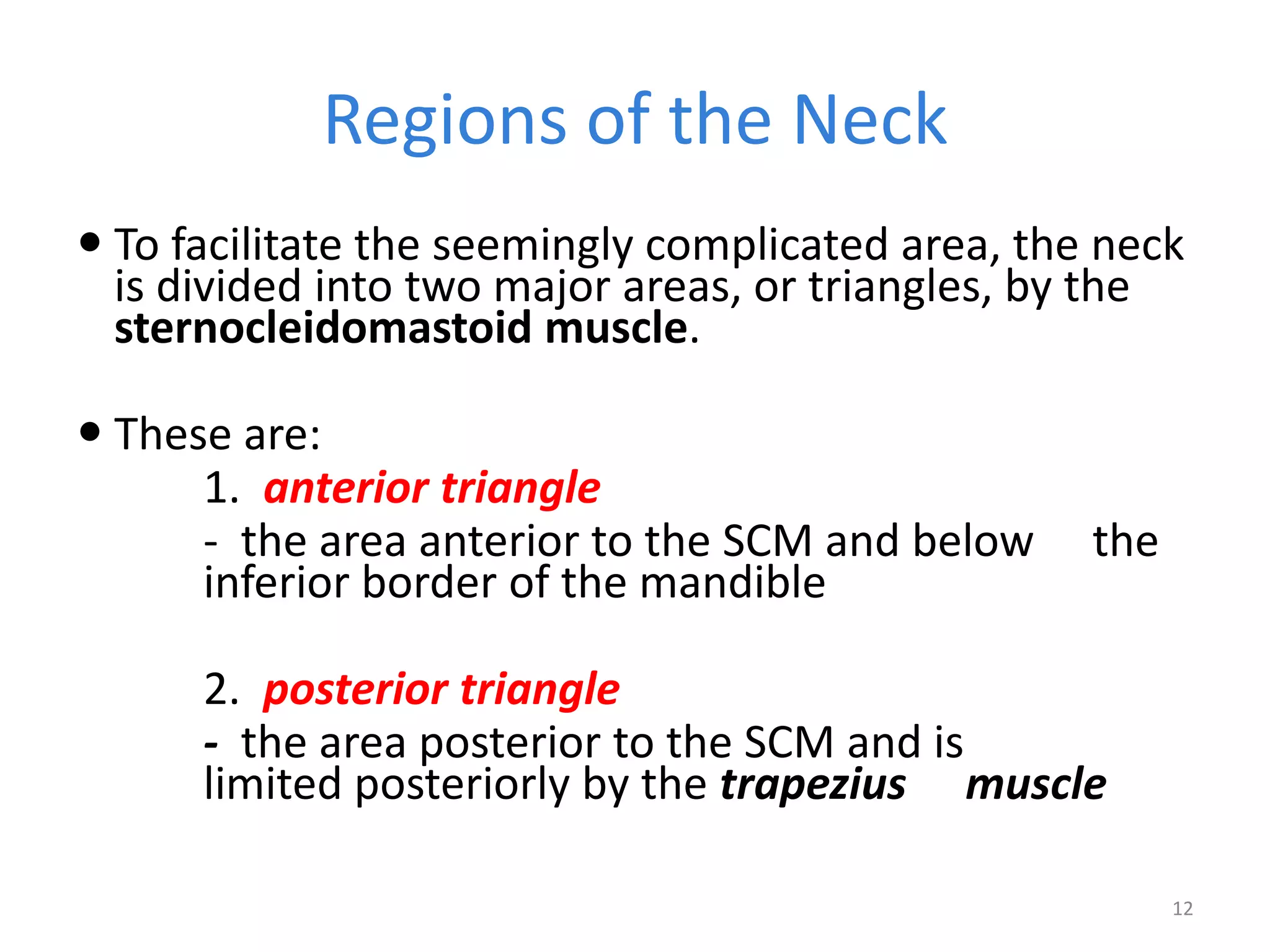
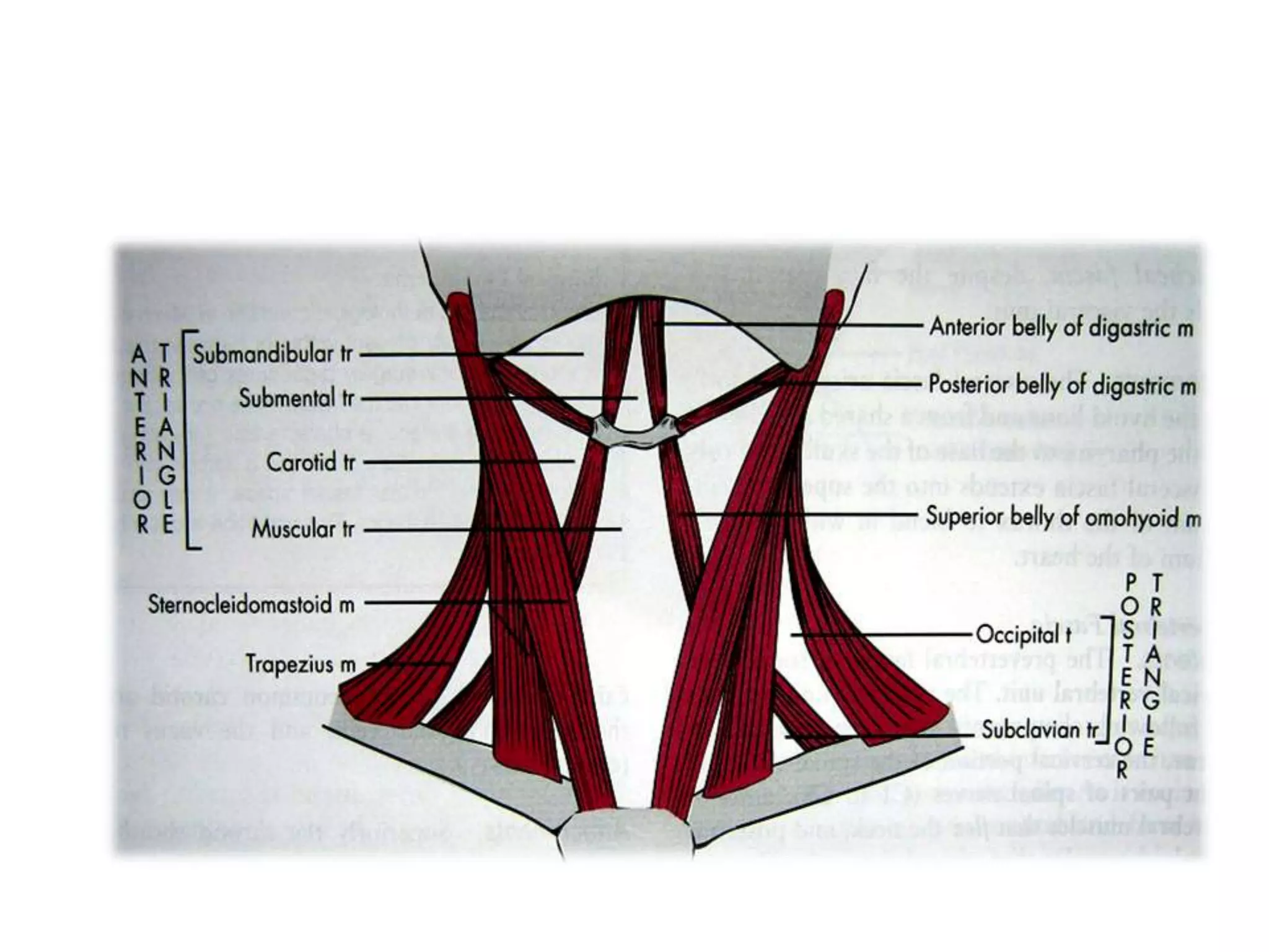

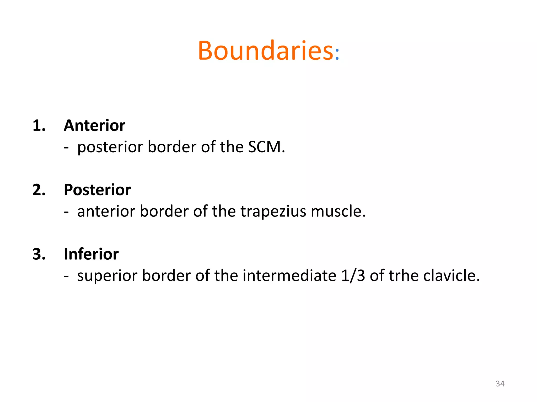

The neck is the region between the head and trunk that contains important structures. It functions to transport the esophagus, trachea, blood vessels, and nerves between the head and chest. The boundaries of the neck include the mandible superiorly, the clavicles inferiorly, and vertebrae posteriorly. The surface anatomy includes palpable structures like the thyroid cartilage. The neck is divided into the anterior and posterior triangles by the sternocleidomastoid muscle. The anterior triangle contains structures like the carotid artery and jugular vein.


