Downloaded 134 times


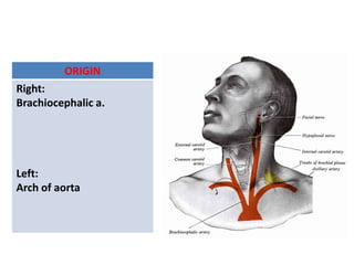
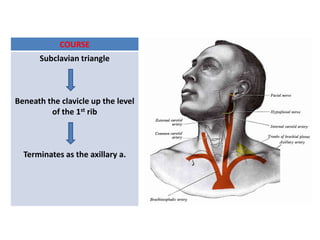
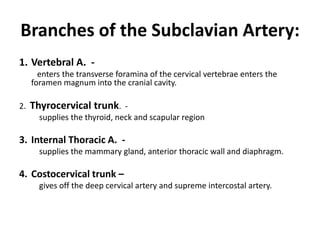
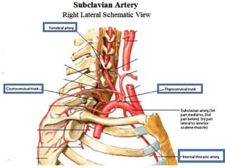

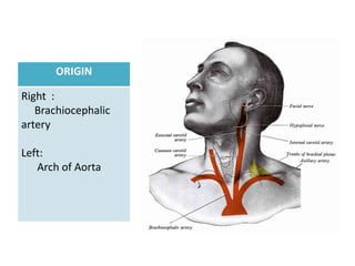
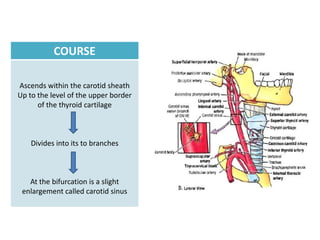
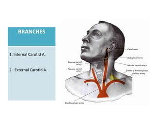
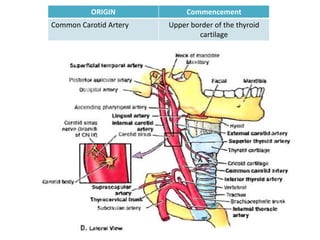
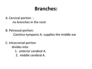
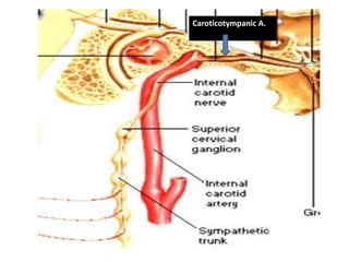
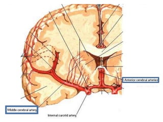
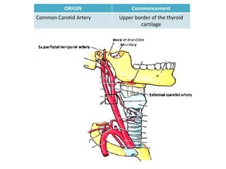
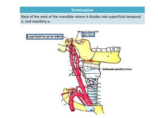
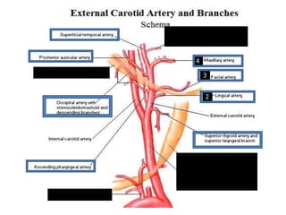

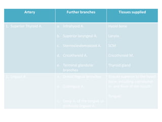
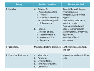
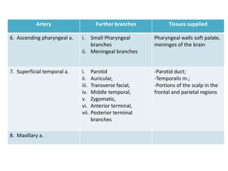
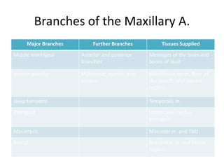
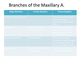
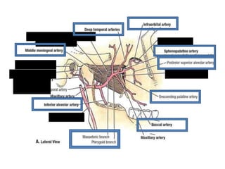
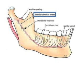

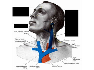

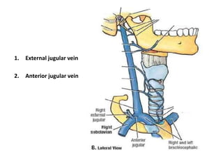
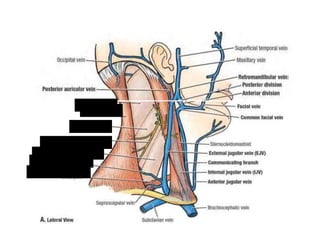

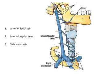
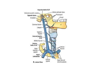
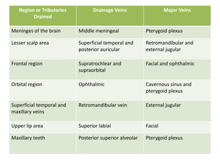
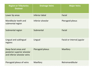
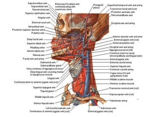

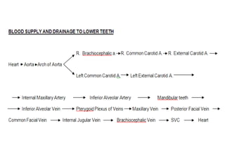
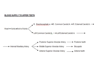
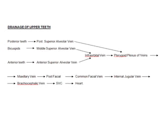

The document describes the arteries and veins of the head and neck region, including the branches of the external carotid artery like the lingual artery and maxillary artery, as well as the drainage veins like the external jugular vein and internal jugular vein. It provides details on the origin, course, and branches of arteries supplying different structures of the head and neck, such as the common carotid artery, internal carotid artery, and subclavian artery. Key veins that drain blood from the head and neck include the pterygoid plexus, retromandibular vein, and external jugular vein.


