Downloaded 125 times


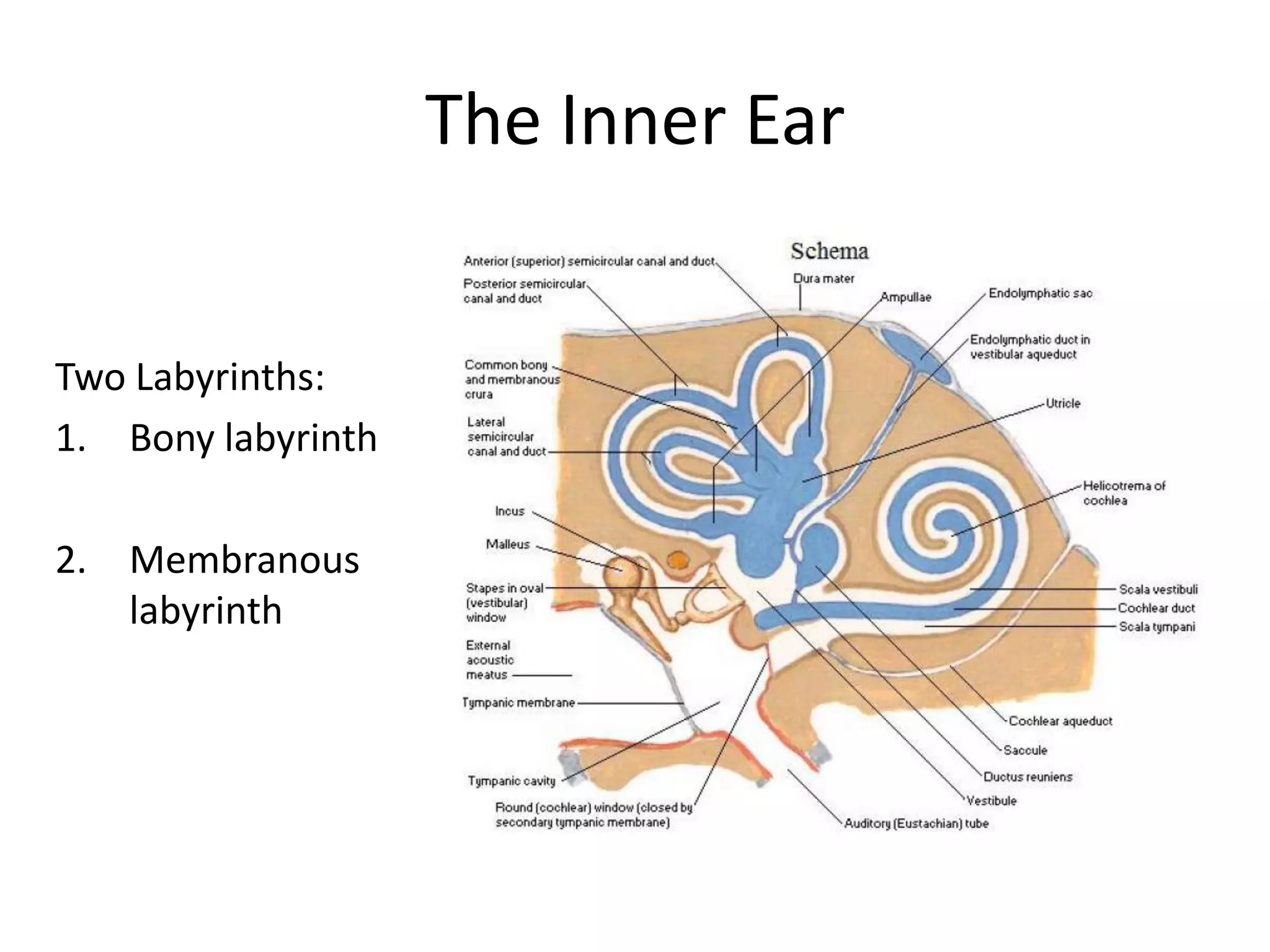


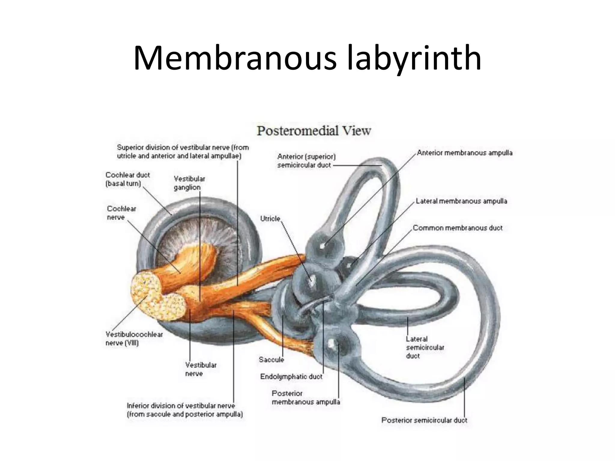
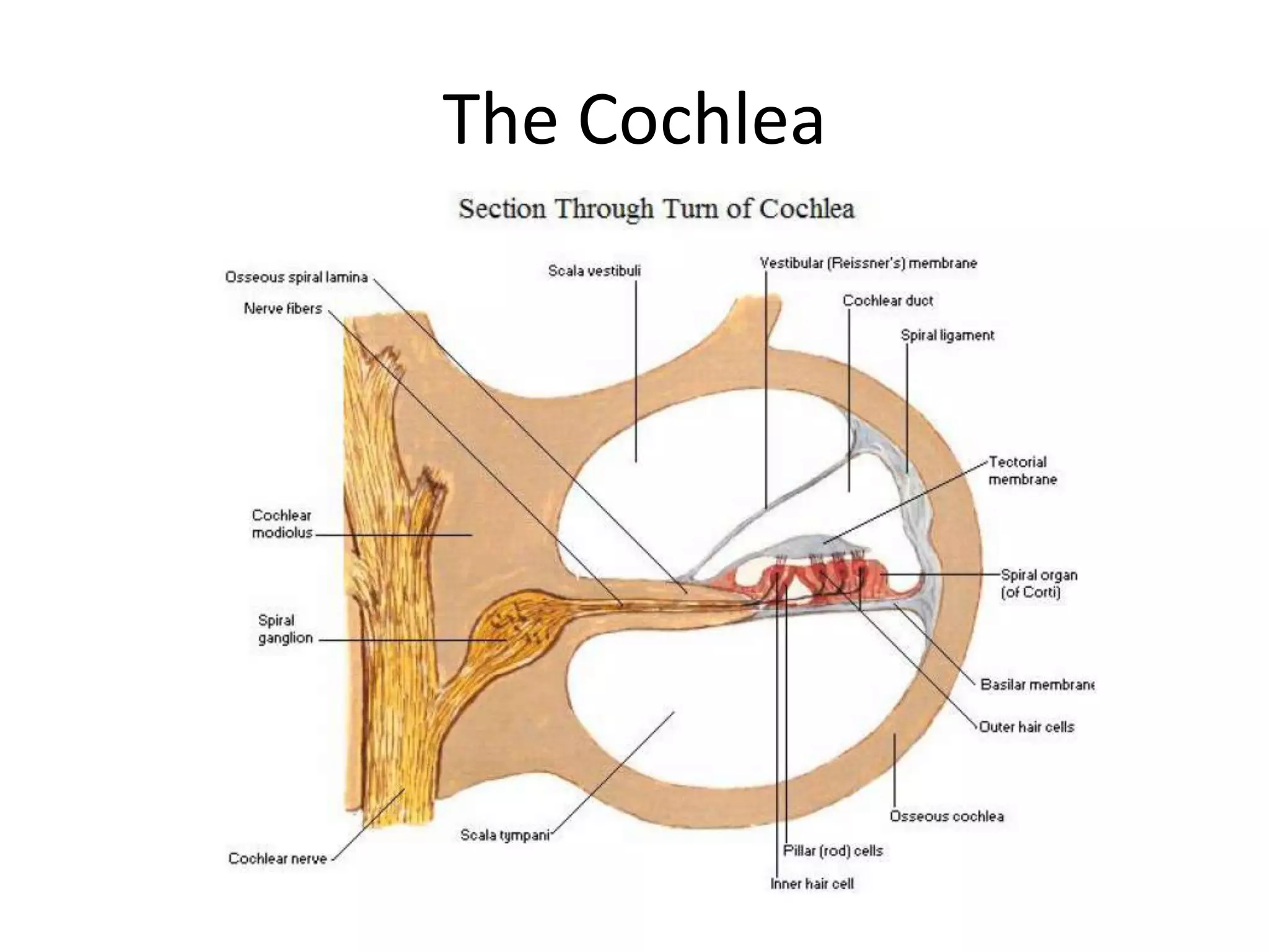

The ear has three main sections - the outer, middle, and inner ear. The outer ear collects and directs sound waves through the external auditory meatus to the eardrum. The middle ear contains the auditory ossicles that transmit vibrations through to the inner ear. The inner ear senses both hearing and equilibrium, containing the cochlea which detects sounds and the vestibular system that monitors movement and orientation. Receptors in the cochlea and vestibular system of the inner ear enable the senses of hearing and balance.


