Downloaded 951 times

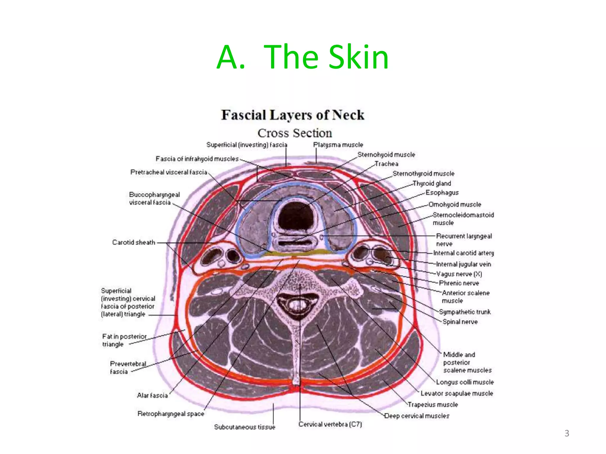

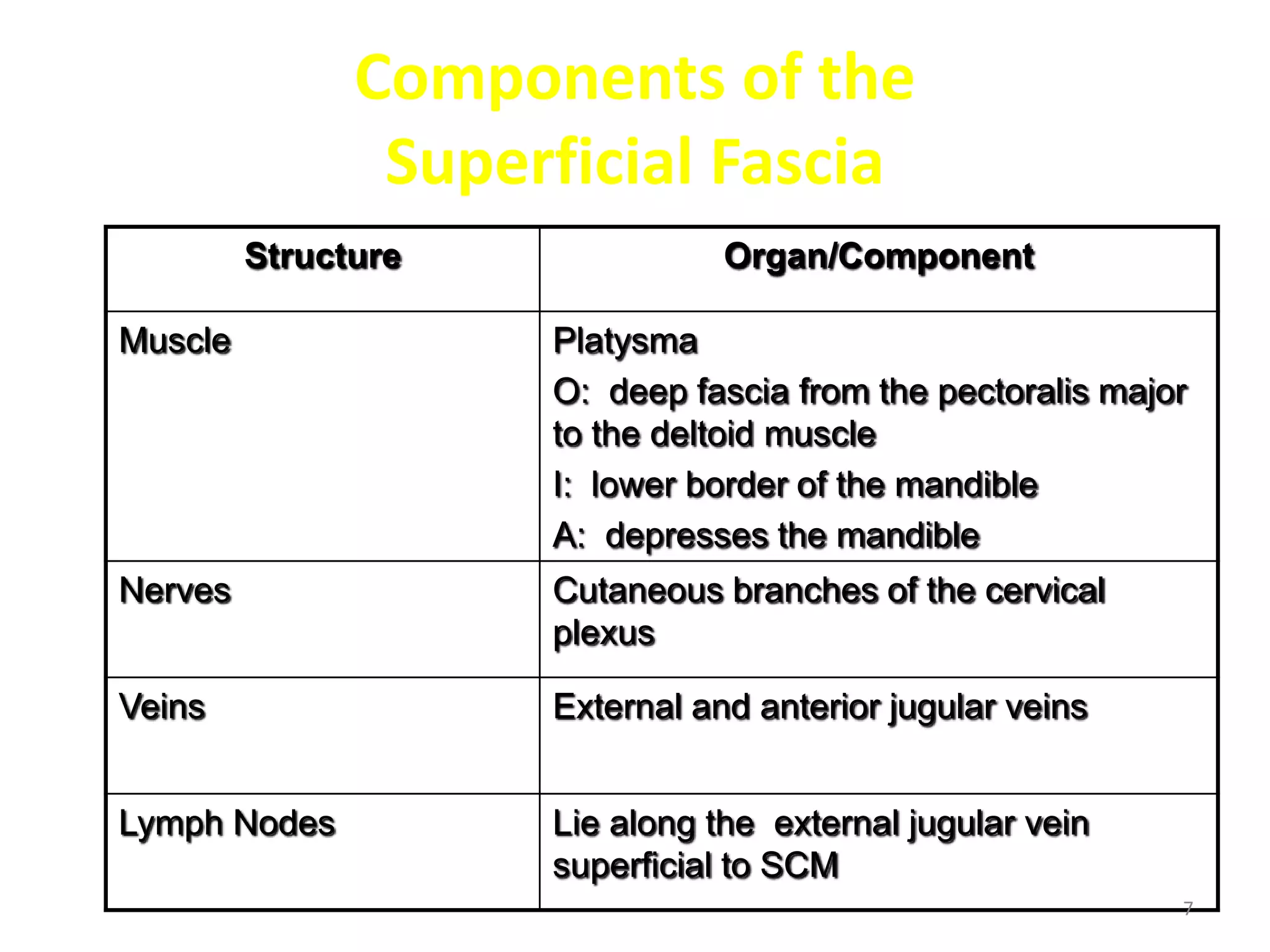

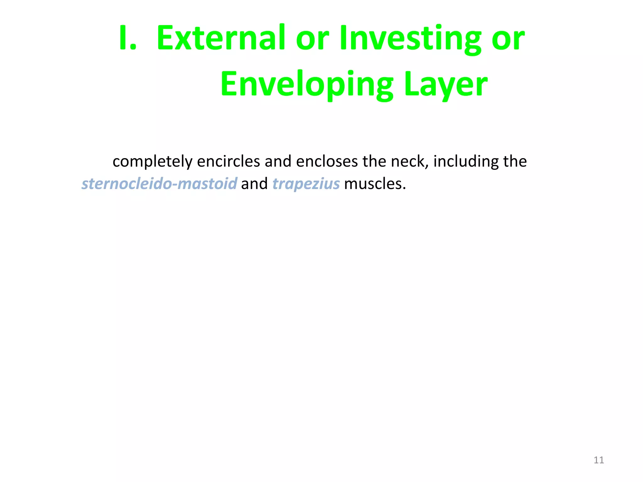


The neck contains three layers of fascia - superficial, deep cervical, and prevertebral. The superficial fascia lies beneath the skin and contains nerves, veins and lymph nodes. The deep cervical fascia has three layers - investing, pretracheal, and prevertebral. It surrounds muscles and structures of the neck. Potential spaces between the fascial layers can allow spread of infection or tumors if invaded.


