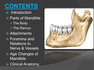
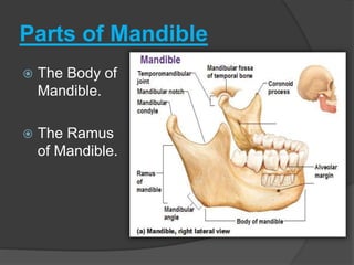
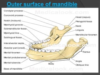
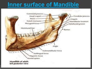
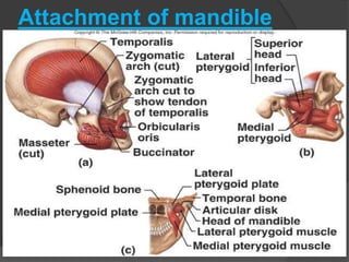
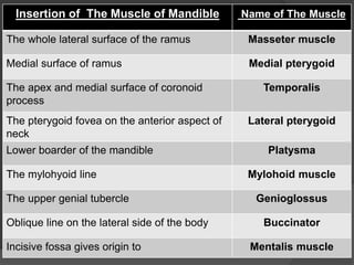
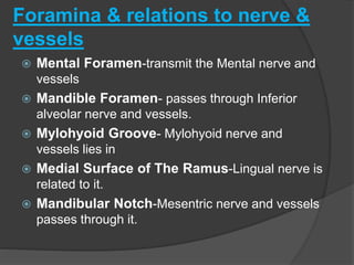
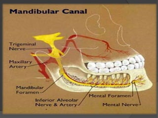
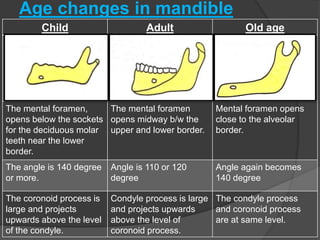
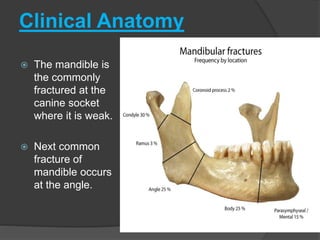
SUJOY KULEY
ROLL NO: 1489
1ST YEAR BDS
CONTENTS
 Introduction
 Parts of Mandible
 The Body
 The Ramus
 Attachments
 Foramina and
Relations to
Nerve & Vessels
 Age Changes of
Mandible
 Clinical Anatomy.
Introduction
 The Mandible, or lower jaw, is the
largest and strongest bone of the face.
 It develops from the First Pharyngeal
Arch.
 It has a Horseshoe-shaped body which
lodges the teeth.
 A pair of Rami which project upwards
from the posterior ends of the body and
provide attachment to muscle.
Parts of Mandible
 The Body of
Mandible.
 The Ramus
of Mandible.
The Body of Mandible
 Each half of the body of Mandible
consist of –
 Two surfaces i.e. Outer and Inner surface.
 Two borders i.e. Upper and Lower border.
Outer surface of Body of
mandible.
 Symphysis Menti-
 the line at which right and left halves of bone
meet each other.
 Mental Protubrence-
 A median triangular projecting area in the lower
part of Symphysis Menti.
 Mental Foramen -
 Lies below the interval between the premolar.
 Oblique Line-
 It runs downwards and forwards towards the
mental tubercle.
Inner surface of the body of
mandible
 Mylohyoid Line-
 A prominent ridge that runs obliquely downwards
and forwards from below 3rd molar.
 Submandibular Fossa-
 Below mylohyoid line forms slightly depressed
area.
 Genial Tubercles-
 The posterior surface of symphysis mentis marked
by four small elevations.
 Mylohyoid Groove-
 Below the posterior end of the mylohyoid line.
Borders of the body of the
mandible
 Upper or alveolar border bears sockets
for the teeth.
 The lower border of the mandible is
called the base of the mandible. Near
the midline the base shows an oval
depression called the Digastric Fossa.
The ramus of mandible
 The ramus has-
• Two surfaces- Lateral And Medial.
• Four border- Upper, Lower, Anterior and Posterior.
• Coronoid and Condyloid Process.
Surface of The Ramus
 Lateral surface, is flat and
bears a numbers of oblique
ridge.
 Medial surface presents-
 Mandibular foramen- lies
little above the center of
the mandible at the level
of the occlusal surface of
the teeth.
 Lingula- anterior margin
of the mandibular foramen
is marked by a tongue
shape projection towards
the head of the mandible.
Borders of the ramus
 The upper border of the ramus is thin and is
curved downwards forming the mandibular
notch.
 The lower border is backward continuous with
the base of the mandible.
 Anterior border is thin .
 Posterior border is thick
Superior part of the
ramus
 Condyloid process- a strong projection
from postero-superior part of ramus. The
head is covered with fibro-cartilage and
articulate with the temporal bone to form
Temporomandibular Joint.
 Coronoid process- is flattened
triangular upward projection from the
antero-superior part of the ramus.
Outer surface of mandible
Inner surface of Mandible
Attachment of mandible
Insertion of The Muscle of Mandible Name of The Muscle
The whole lateral surface of the ramus Masseter muscle
Medial surface of ramus Medial pterygoid
The apex and medial surface of coronoid
process
Temporalis
The pterygoid fovea on the anterior aspect of
neck
Lateral pterygoid
Lower boarder of the mandible Platysma
The mylohyoid line Mylohoid muscle
The upper genial tubercle Genioglossus
Oblique line on the lateral side of the body Buccinator
Incisive fossa gives origin to Mentalis muscle
Foramina & relations to nerve &
vessels
 Mental Foramen-transmit the Mental nerve and
vessels
 Mandible Foramen- passes through Inferior
alveolar nerve and vessels.
 Mylohyoid Groove- Mylohyoid nerve and
vessels lies in
 Medial Surface of The Ramus-Lingual nerve is
related to it.
 Mandibular Notch-Mesentric nerve and vessels
passes through it.
Age changes in mandible
Child Adult Old age
The mental foramen,
opens below the sockets
for the deciduous molar
teeth near the lower
border.
The mental foramen
opens midway b/w the
upper and lower border.
Mental foramen opens
close to the alveolar
border.
The angle is 140 degree
or more.
Angle is 110 or 120
degree
Angle again becomes
140 degree
The coronoid process is
large and projects
upwards above the level
of the condyle.
Condyle process is large
and projects upwards
above the level of
coronoid process.
The condyle process
and coronoid process
are at same level.
Clinical Anatomy
 The mandible is
the commonly
fractured at the
canine socket
where it is weak.
 Next common
fracture of
mandible occurs
at the angle.
The  Mandible

The Mandible

  • 1.
    SUJOY KULEY ROLL NO:1489 1ST YEAR BDS
  • 2.
    CONTENTS  Introduction  Partsof Mandible  The Body  The Ramus  Attachments  Foramina and Relations to Nerve & Vessels  Age Changes of Mandible  Clinical Anatomy.
  • 3.
    Introduction  The Mandible,or lower jaw, is the largest and strongest bone of the face.  It develops from the First Pharyngeal Arch.  It has a Horseshoe-shaped body which lodges the teeth.  A pair of Rami which project upwards from the posterior ends of the body and provide attachment to muscle.
  • 4.
    Parts of Mandible The Body of Mandible.  The Ramus of Mandible.
  • 5.
    The Body ofMandible  Each half of the body of Mandible consist of –  Two surfaces i.e. Outer and Inner surface.  Two borders i.e. Upper and Lower border.
  • 6.
    Outer surface ofBody of mandible.  Symphysis Menti-  the line at which right and left halves of bone meet each other.  Mental Protubrence-  A median triangular projecting area in the lower part of Symphysis Menti.  Mental Foramen -  Lies below the interval between the premolar.  Oblique Line-  It runs downwards and forwards towards the mental tubercle.
  • 7.
    Inner surface ofthe body of mandible  Mylohyoid Line-  A prominent ridge that runs obliquely downwards and forwards from below 3rd molar.  Submandibular Fossa-  Below mylohyoid line forms slightly depressed area.  Genial Tubercles-  The posterior surface of symphysis mentis marked by four small elevations.  Mylohyoid Groove-  Below the posterior end of the mylohyoid line.
  • 8.
    Borders of thebody of the mandible  Upper or alveolar border bears sockets for the teeth.  The lower border of the mandible is called the base of the mandible. Near the midline the base shows an oval depression called the Digastric Fossa.
  • 9.
    The ramus ofmandible  The ramus has- • Two surfaces- Lateral And Medial. • Four border- Upper, Lower, Anterior and Posterior. • Coronoid and Condyloid Process.
  • 10.
    Surface of TheRamus  Lateral surface, is flat and bears a numbers of oblique ridge.  Medial surface presents-  Mandibular foramen- lies little above the center of the mandible at the level of the occlusal surface of the teeth.  Lingula- anterior margin of the mandibular foramen is marked by a tongue shape projection towards the head of the mandible.
  • 11.
    Borders of theramus  The upper border of the ramus is thin and is curved downwards forming the mandibular notch.  The lower border is backward continuous with the base of the mandible.  Anterior border is thin .  Posterior border is thick
  • 12.
    Superior part ofthe ramus  Condyloid process- a strong projection from postero-superior part of ramus. The head is covered with fibro-cartilage and articulate with the temporal bone to form Temporomandibular Joint.  Coronoid process- is flattened triangular upward projection from the antero-superior part of the ramus.
  • 13.
  • 14.
  • 15.
  • 16.
    Insertion of TheMuscle of Mandible Name of The Muscle The whole lateral surface of the ramus Masseter muscle Medial surface of ramus Medial pterygoid The apex and medial surface of coronoid process Temporalis The pterygoid fovea on the anterior aspect of neck Lateral pterygoid Lower boarder of the mandible Platysma The mylohyoid line Mylohoid muscle The upper genial tubercle Genioglossus Oblique line on the lateral side of the body Buccinator Incisive fossa gives origin to Mentalis muscle
  • 17.
    Foramina & relationsto nerve & vessels  Mental Foramen-transmit the Mental nerve and vessels  Mandible Foramen- passes through Inferior alveolar nerve and vessels.  Mylohyoid Groove- Mylohyoid nerve and vessels lies in  Medial Surface of The Ramus-Lingual nerve is related to it.  Mandibular Notch-Mesentric nerve and vessels passes through it.
  • 19.
    Age changes inmandible Child Adult Old age The mental foramen, opens below the sockets for the deciduous molar teeth near the lower border. The mental foramen opens midway b/w the upper and lower border. Mental foramen opens close to the alveolar border. The angle is 140 degree or more. Angle is 110 or 120 degree Angle again becomes 140 degree The coronoid process is large and projects upwards above the level of the condyle. Condyle process is large and projects upwards above the level of coronoid process. The condyle process and coronoid process are at same level.
  • 20.
    Clinical Anatomy  Themandible is the commonly fractured at the canine socket where it is weak.  Next common fracture of mandible occurs at the angle.