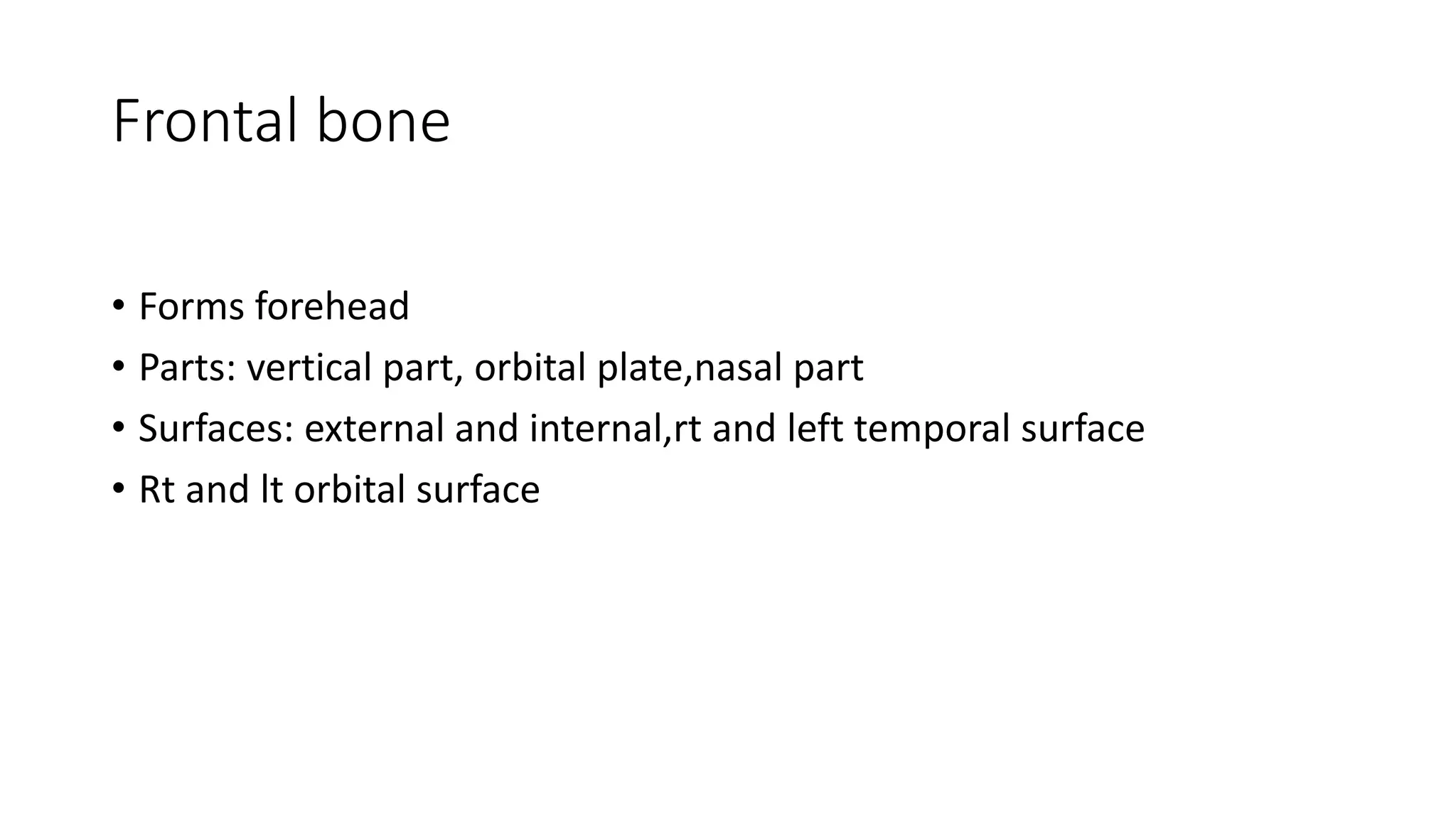
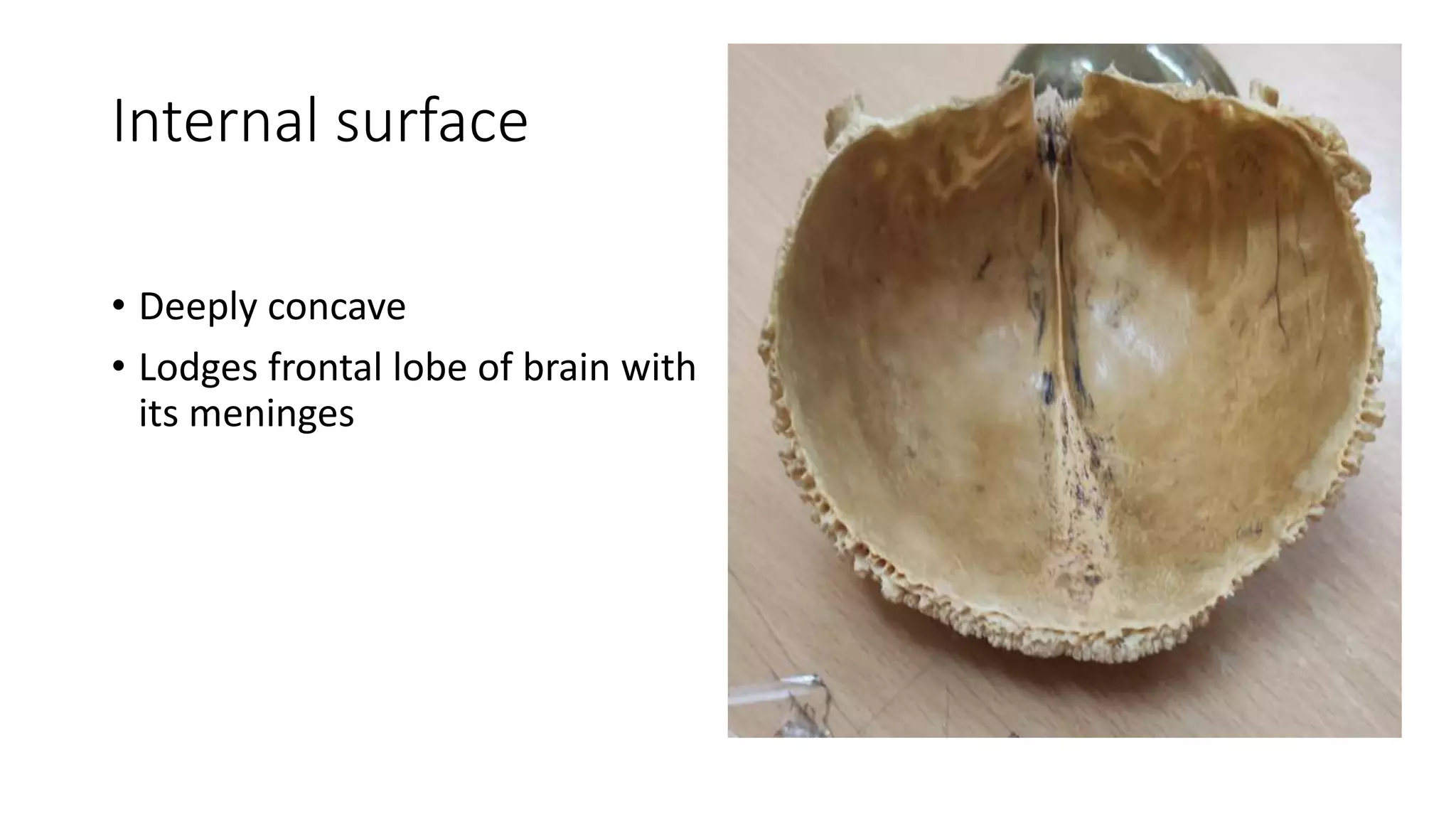
The frontal bone forms the forehead and has several parts including the vertical, orbital, and nasal parts. It has both external and internal surfaces. The external surface is smooth and convex while the internal surface is deeply concave to house the frontal lobe of the brain. Several features are present on both surfaces including the supraorbital margin, notch, foramen, tuberosity, and glabella. The frontal bone articulates with several other bones at the coronal and frontonasal sutures, ethmoid notch, and orbital plates.
Frontal bone
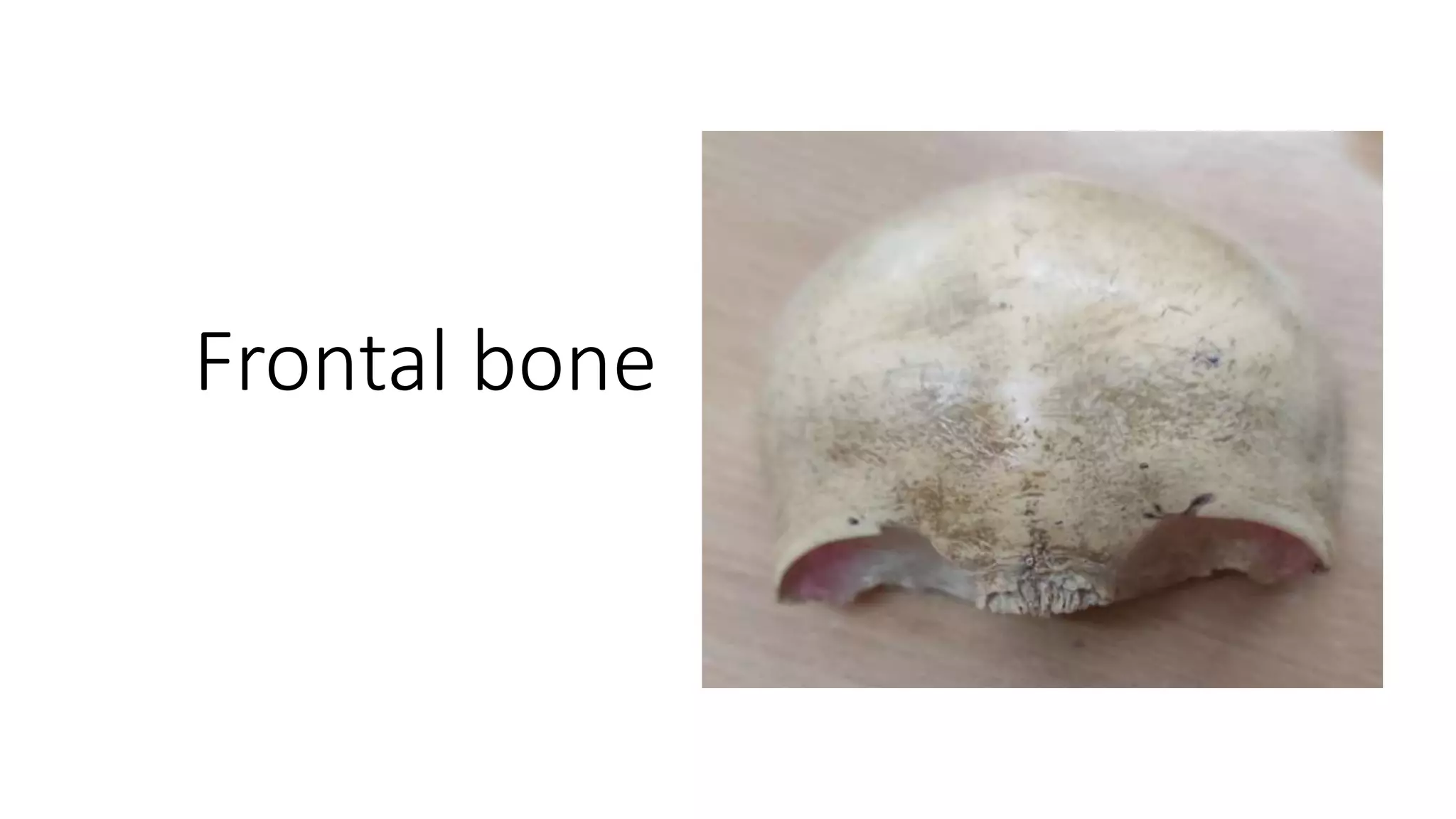
• Formsforehead
• Parts: vertical part, orbital plate,nasal part
• Surfaces: external and internal,rt and left temporal surface
• Rt and lt orbital surface
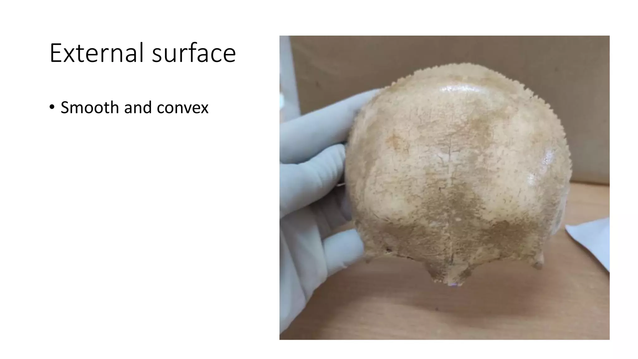
Frontal / metopicsuture
• Present in 9% of individual
• At median plane
• Lower part
• Indicates line of fusion during
development
5.
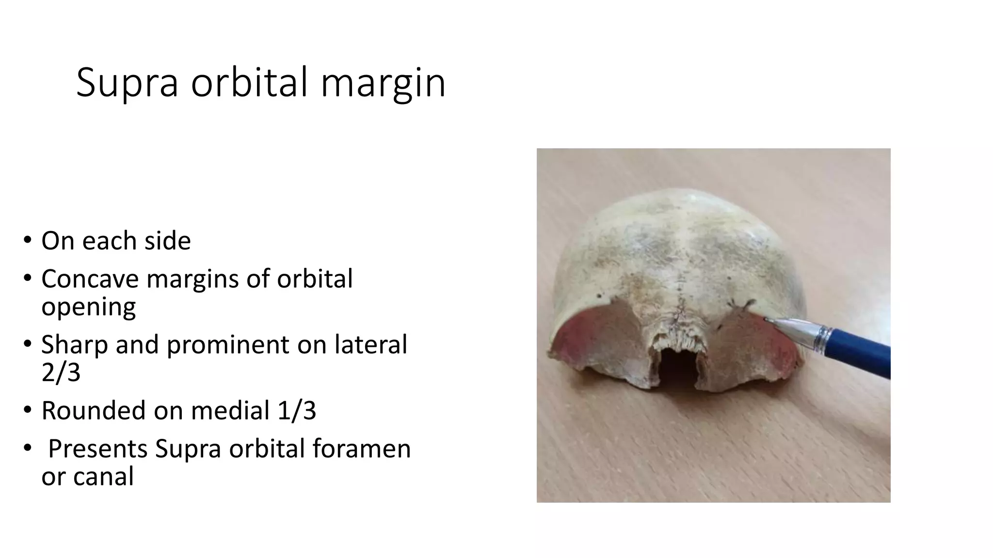
Supra orbital margin
•On each side
• Concave margins of orbital
opening
• Sharp and prominent on lateral
2/3
• Rounded on medial 1/3
• Presents Supra orbital foramen
or canal
6.
Supra orbital foramen/notch&Supra
trochlear foramen
• Present at meeting point of
medial1/3& lateral2/3 of Supra
orbital margin
• Transmitting Supra orbital nerves
and vessels
• Supra trochlear foramen present
occassionally medial to Supra
orbital notch / canal
• Transmitting Supra trochlear
nerves and vessels
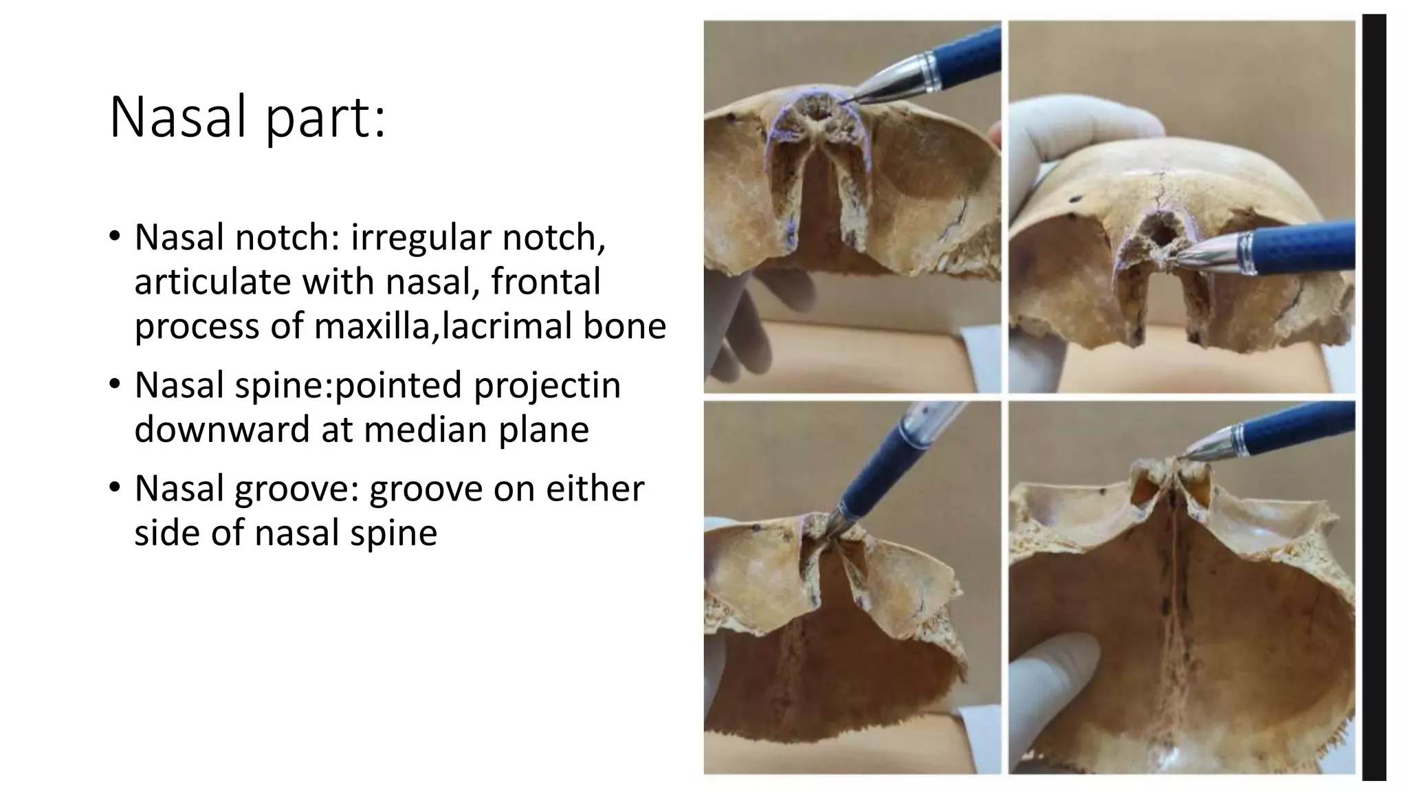
Nasal part:
• Nasalnotch: irregular notch,
articulate with nasal, frontal
process of maxilla,lacrimal bone
• Nasal spine:pointed projectin
downward at median plane
• Nasal groove: groove on either
side of nasal spine
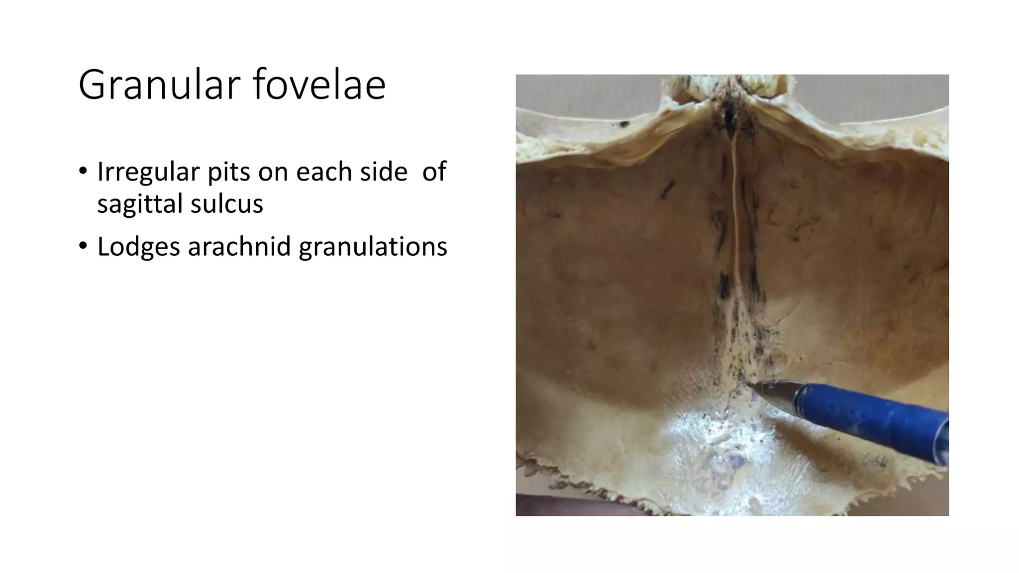
Sagittal sulcus
• Verticalgroove
• At median plane
• Lodges superior sagittal sinus
• Margin receives attachment of
falx cerebri
13.
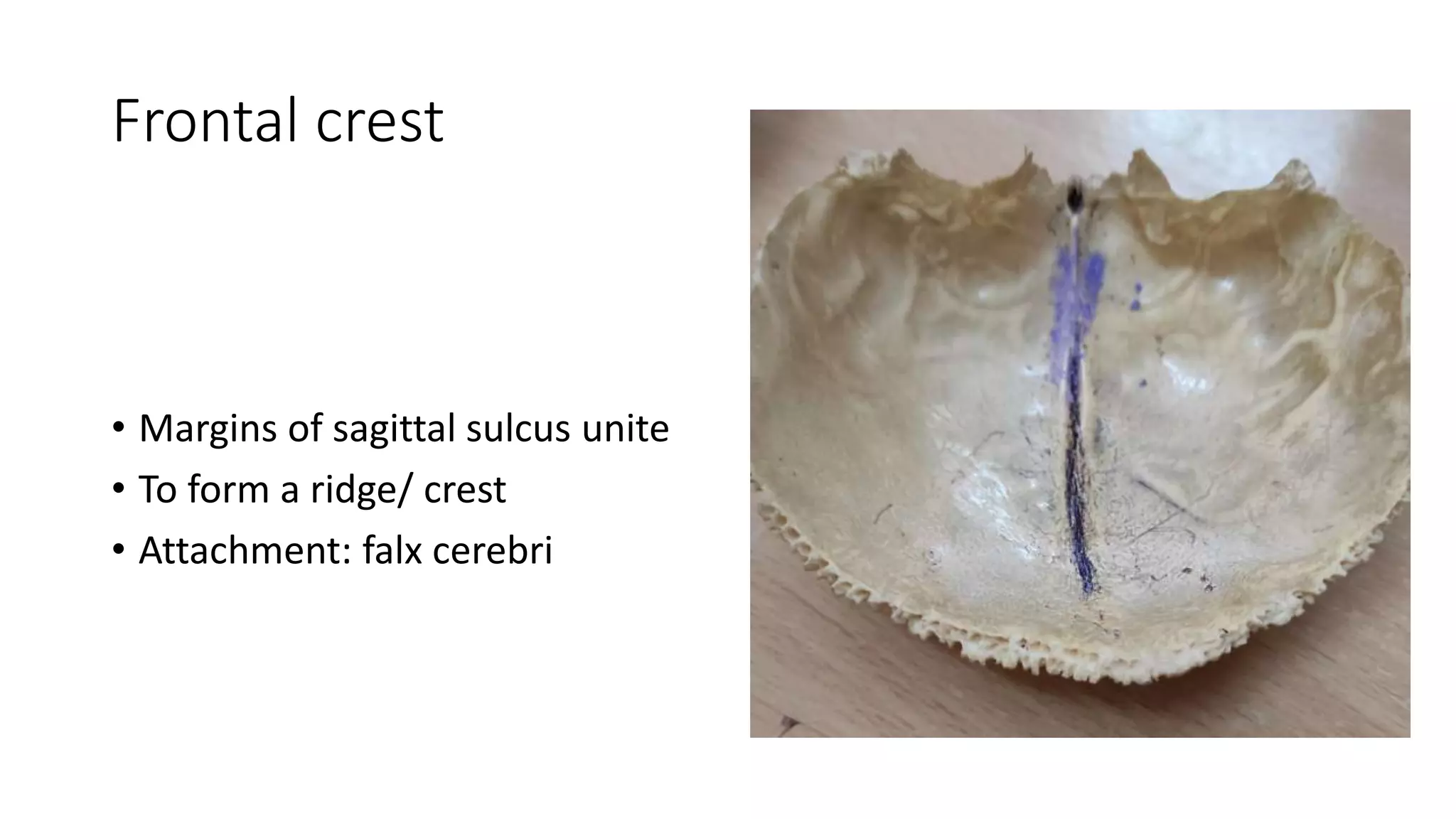
Frontal crest
• Marginsof sagittal sulcus unite
• To form a ridge/ crest
• Attachment: falx cerebri
14.
Foramen caecum
• Frontalcrest ends in a notch
• Convert into foramen
• In articulating with crista galli of
ethmoid bone
• When present transmitts vein
from nasal mucosa_ sup sagittal
sinus
Lacrimal fossa
• Shallowdepression
• Anterolateral part of orbital
plate
• Lodges lacrimal gland
21.
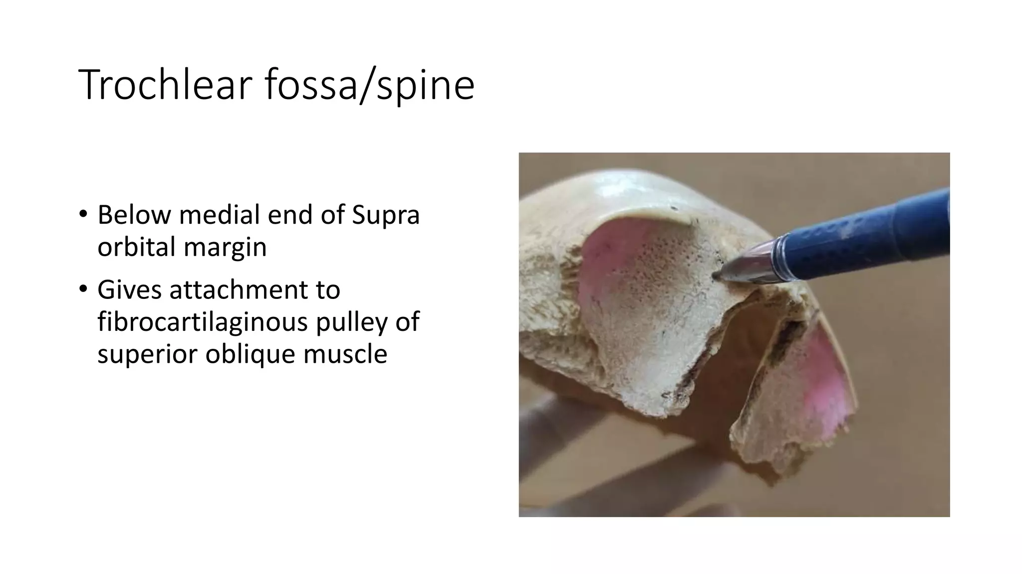
Trochlear fossa/spine
• Belowmedial end of Supra
orbital margin
• Gives attachment to
fibrocartilaginous pulley of
superior oblique muscle
22.
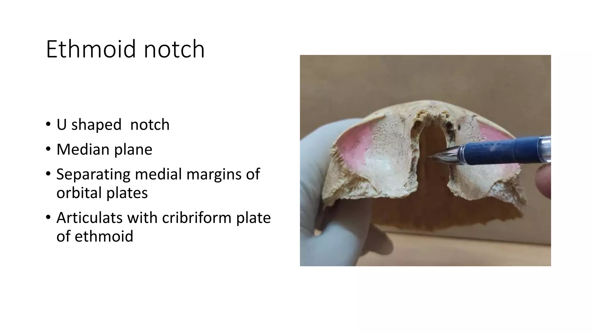
Ethmoid notch
• Ushaped notch
• Median plane
• Separating medial margins of
orbital plates
• Articulats with cribriform plate
of ethmoid
23.
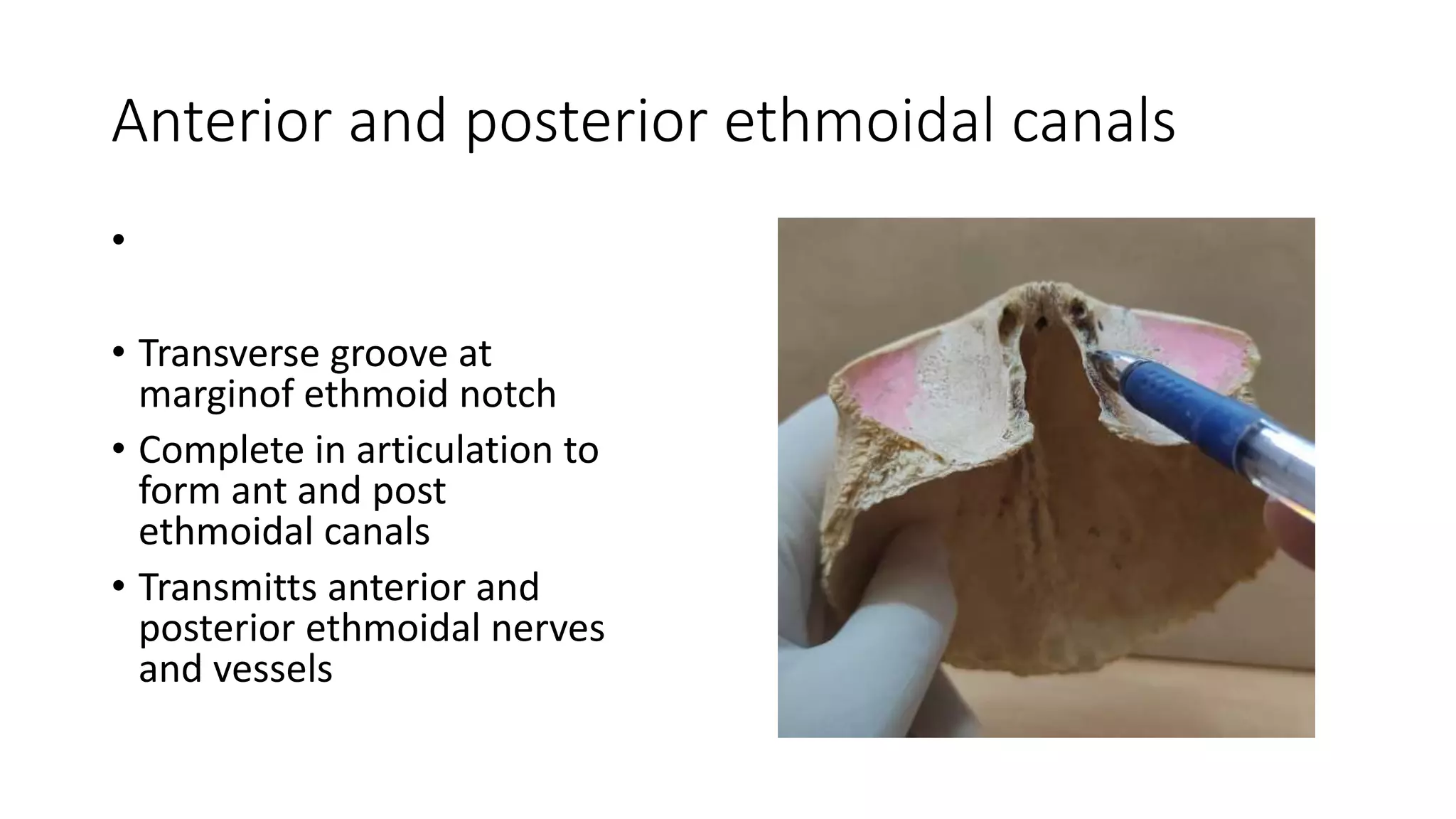
Anterior and posteriorethmoidal canals
•
• Transverse groove at
marginof ethmoid notch
• Complete in articulation to
form ant and post
ethmoidal canals
• Transmitts anterior and
posterior ethmoidal nerves
and vessels
24.
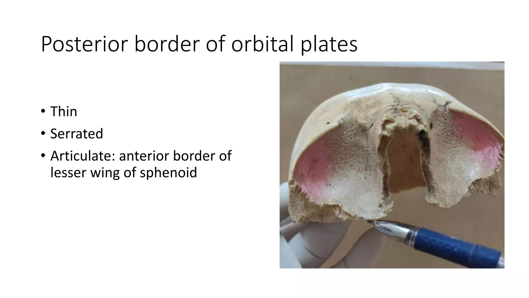
Posterior border oforbital plates
• Thin
• Serrated
• Articulate: anterior border of
lesser wing of sphenoid