


![Lambdoid suture The two parietal bones and the occipital meet at an inverted Y-shaped suture, which is therefore called the lambdoid (resembling the Greek letter lambda [ ]). ](https://image.slidesharecdn.com/skull-thenormas-110913232421-phpapp01/85/Skull-the-normas-21-320.jpg)


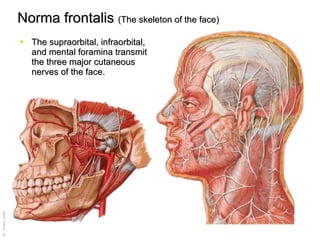
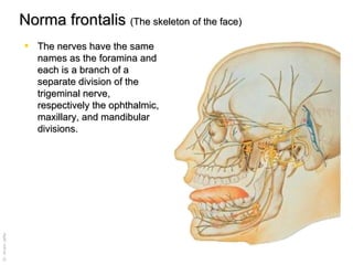
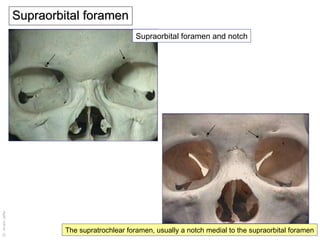


The document discusses the anatomy of the skull, detailing the major bones forming the cranial cavity, the structure and blood supply of flat bones, and the implications of sutural closures that can lead to deformities. It explains ossification processes and provides surface landmarks, with a focus on sutures like the coronal and lambdoid sutures, as well as important foramina associated with facial nerves. Additionally, it describes the classifications of skull views and the developmental features of the infant skull.


![Introduction to skull[1]](https://cdn.slidesharecdn.com/ss_thumbnails/introductiontoskull1-170504174910-thumbnail.jpg?width=640&height=640&fit=bounds)


