Downloaded 1,975 times
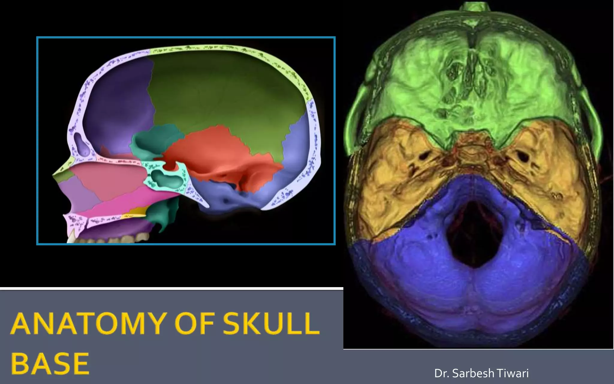
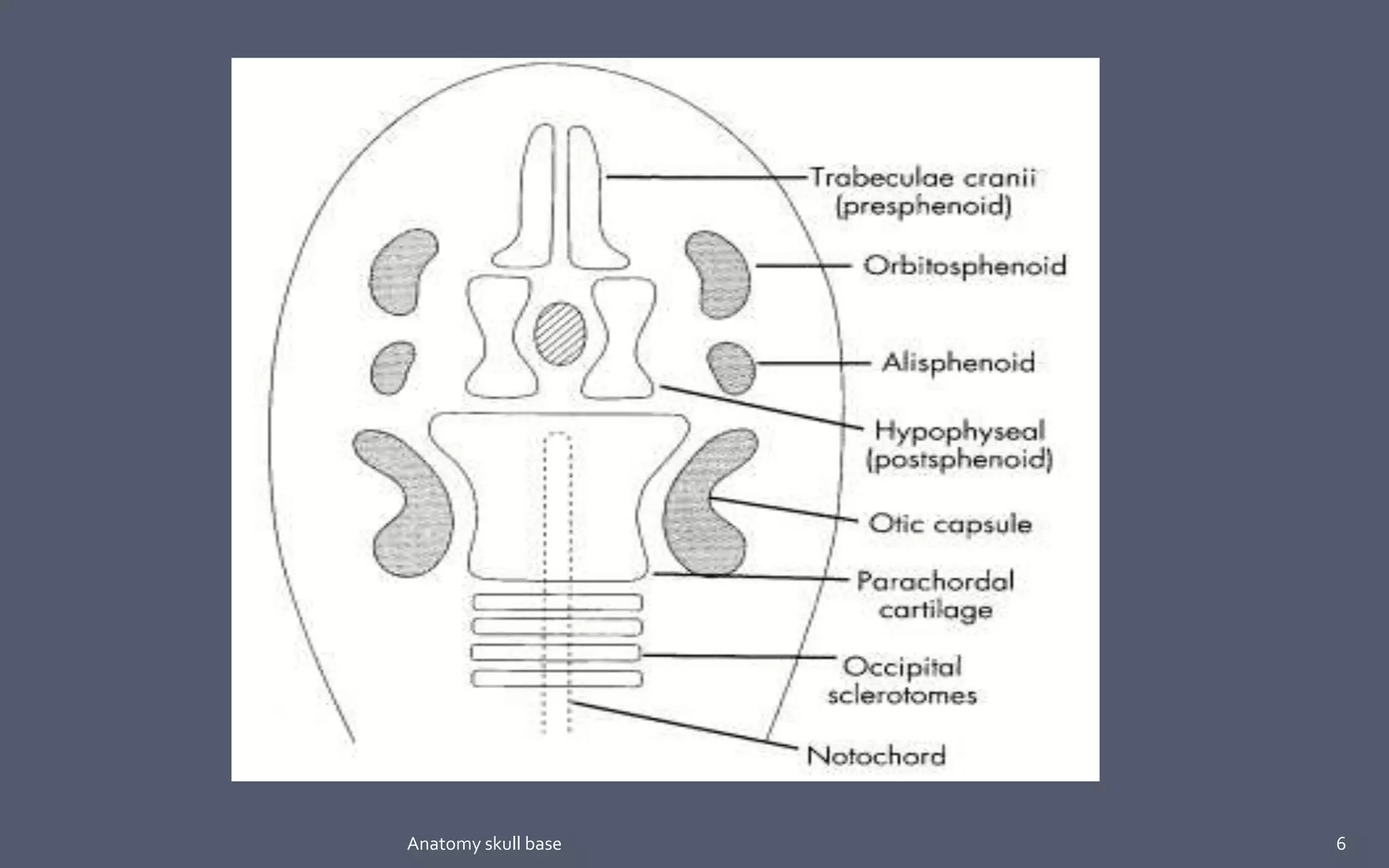

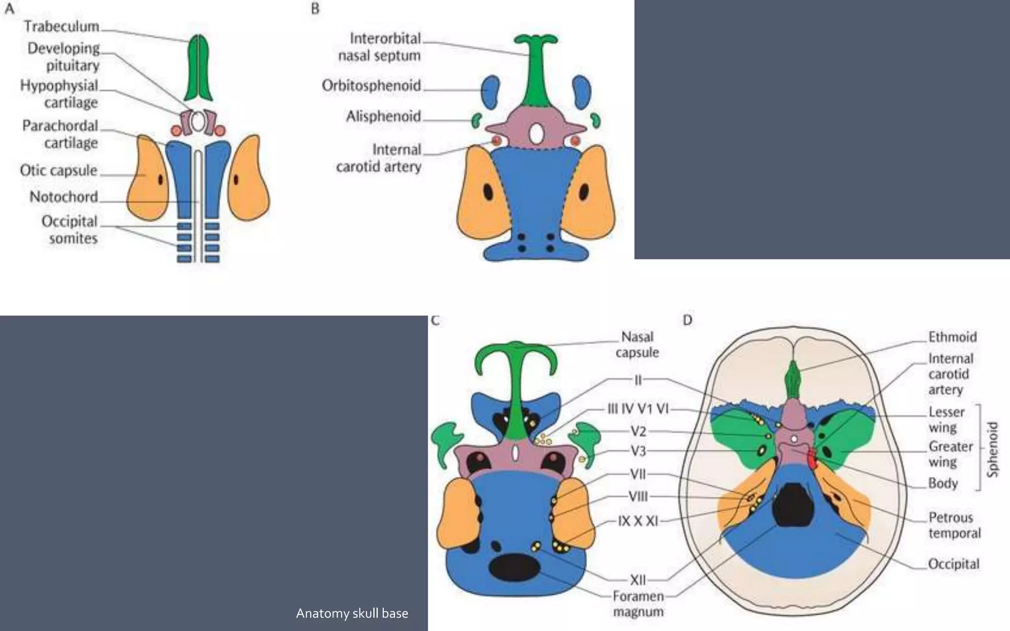
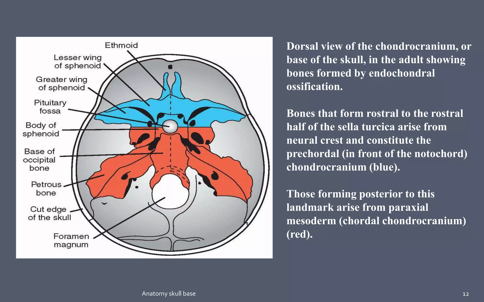
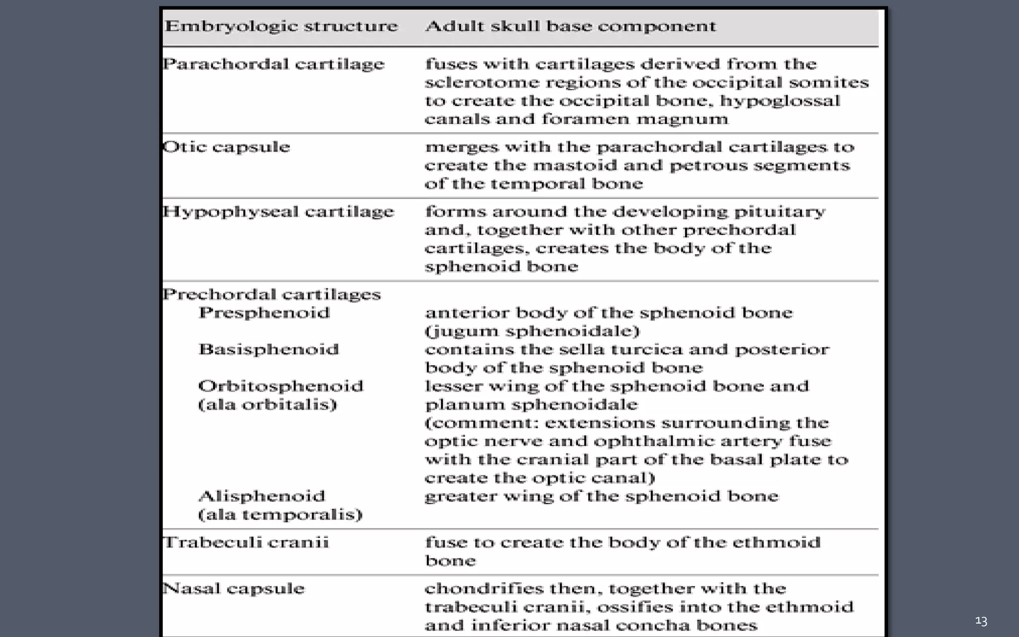
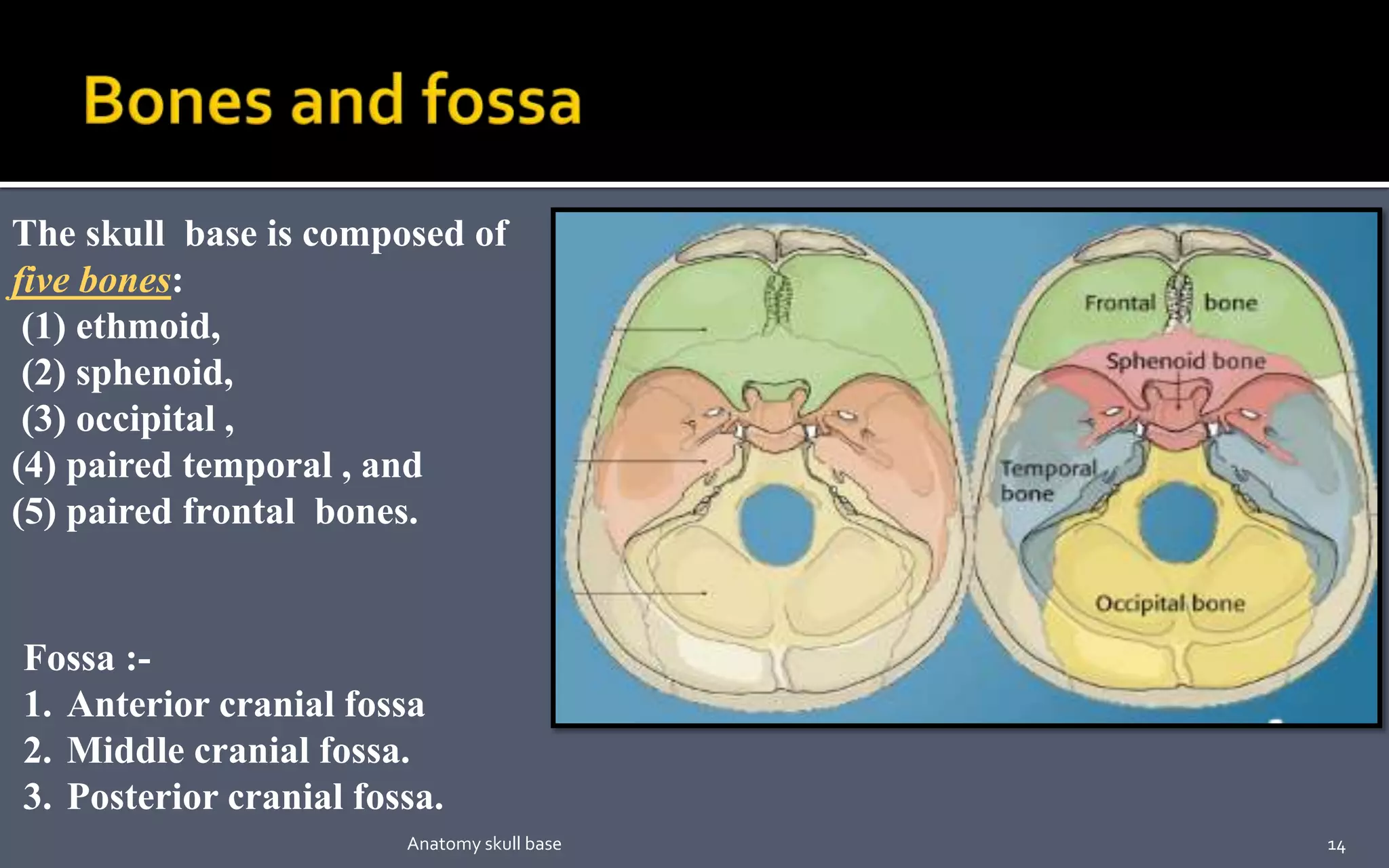
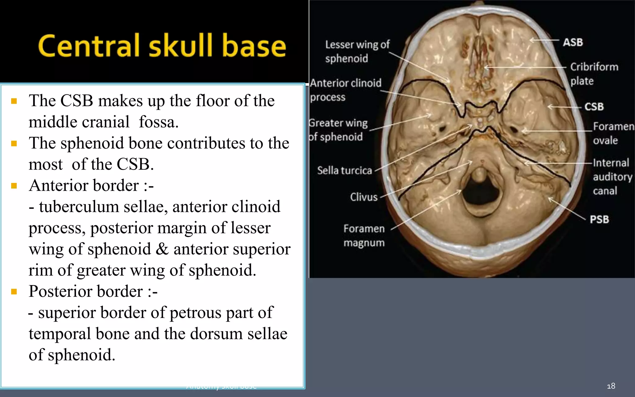
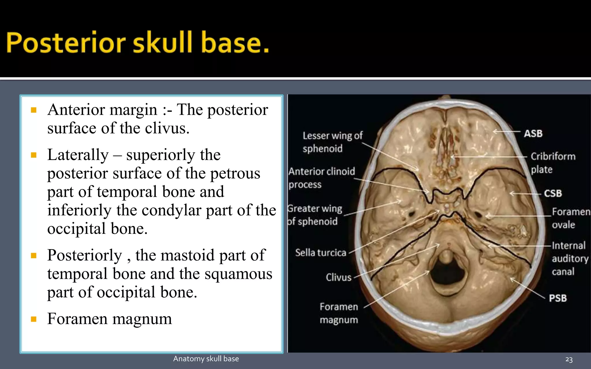
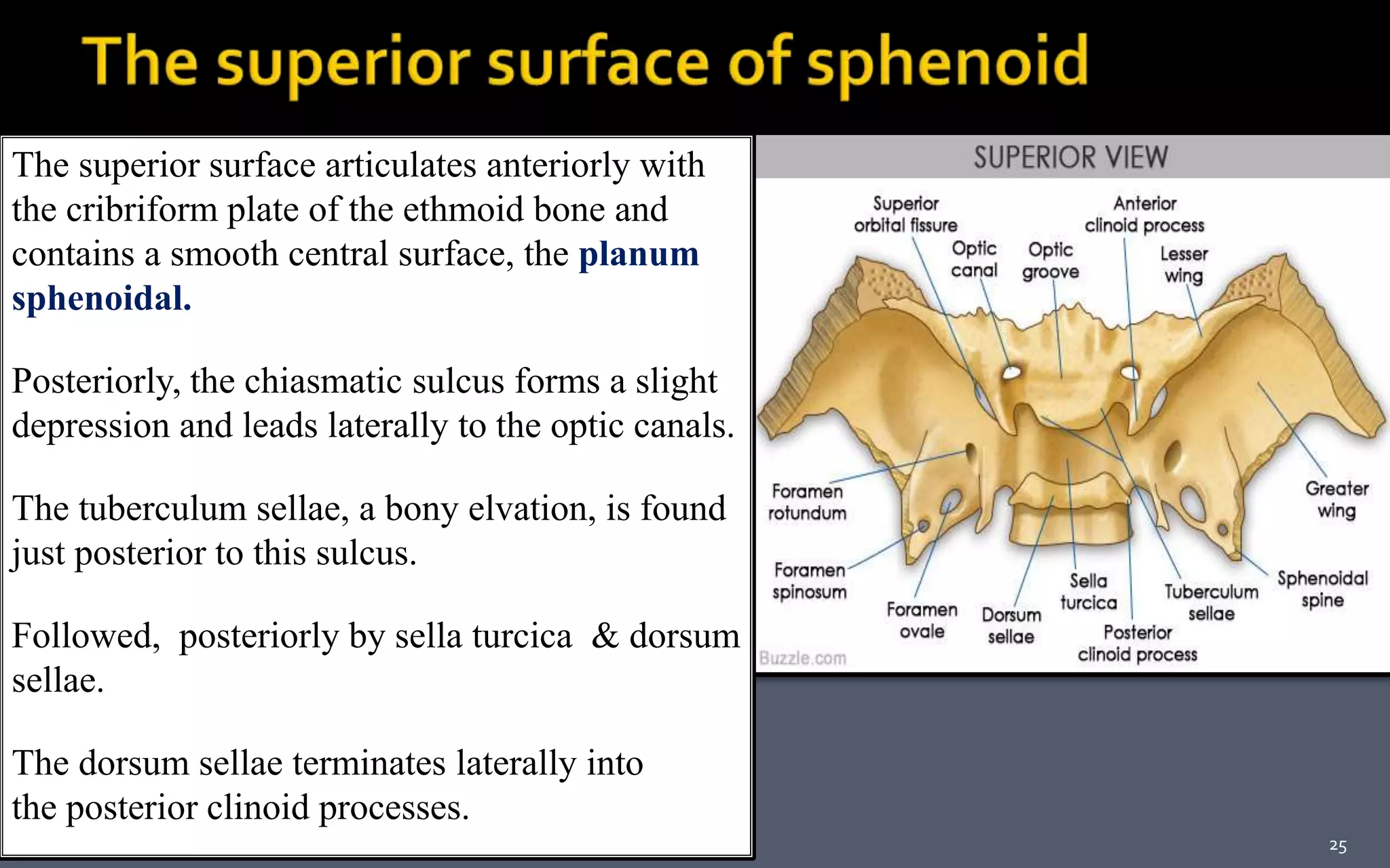
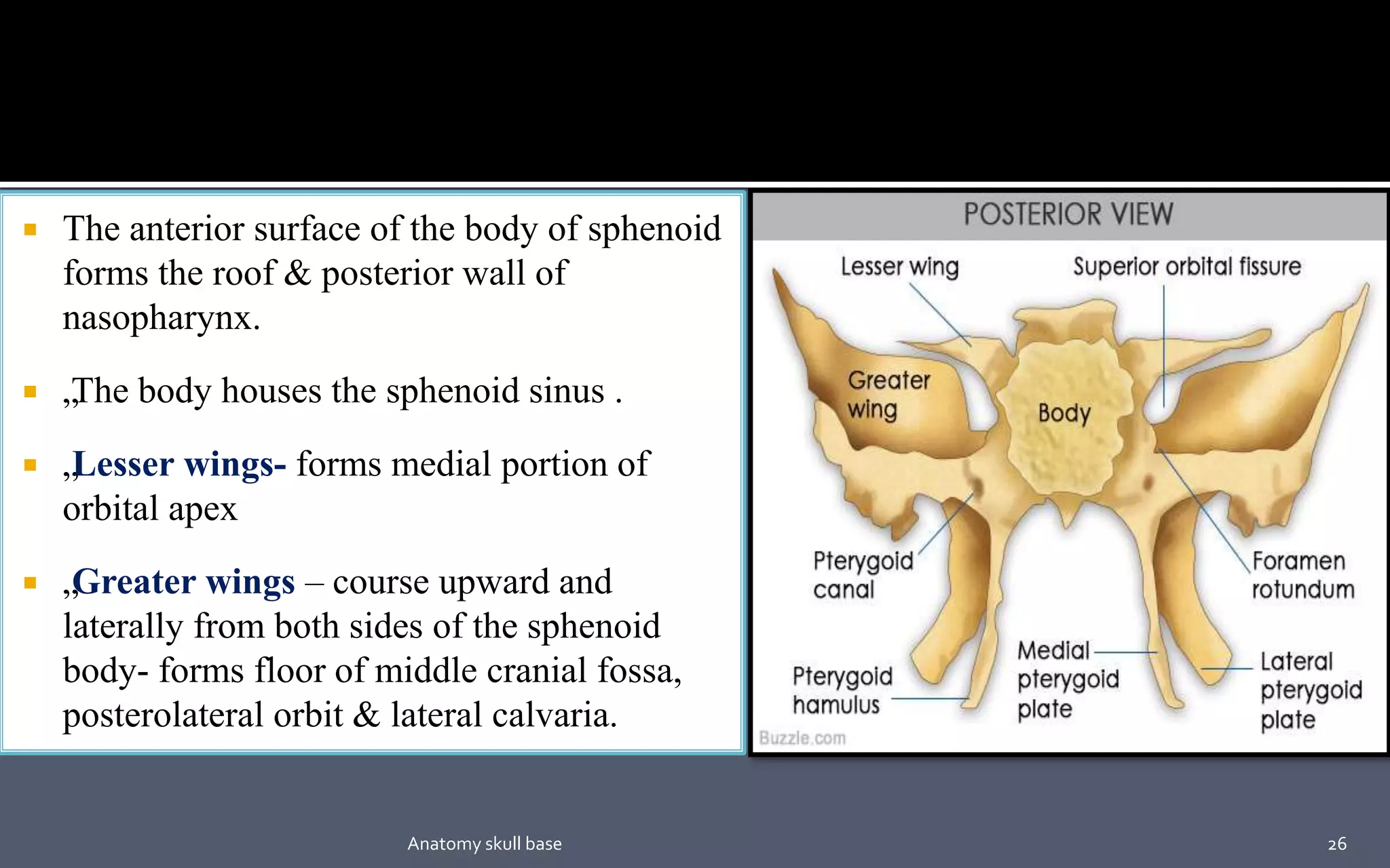
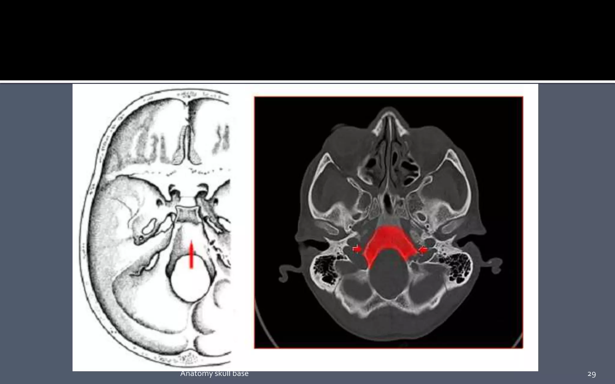
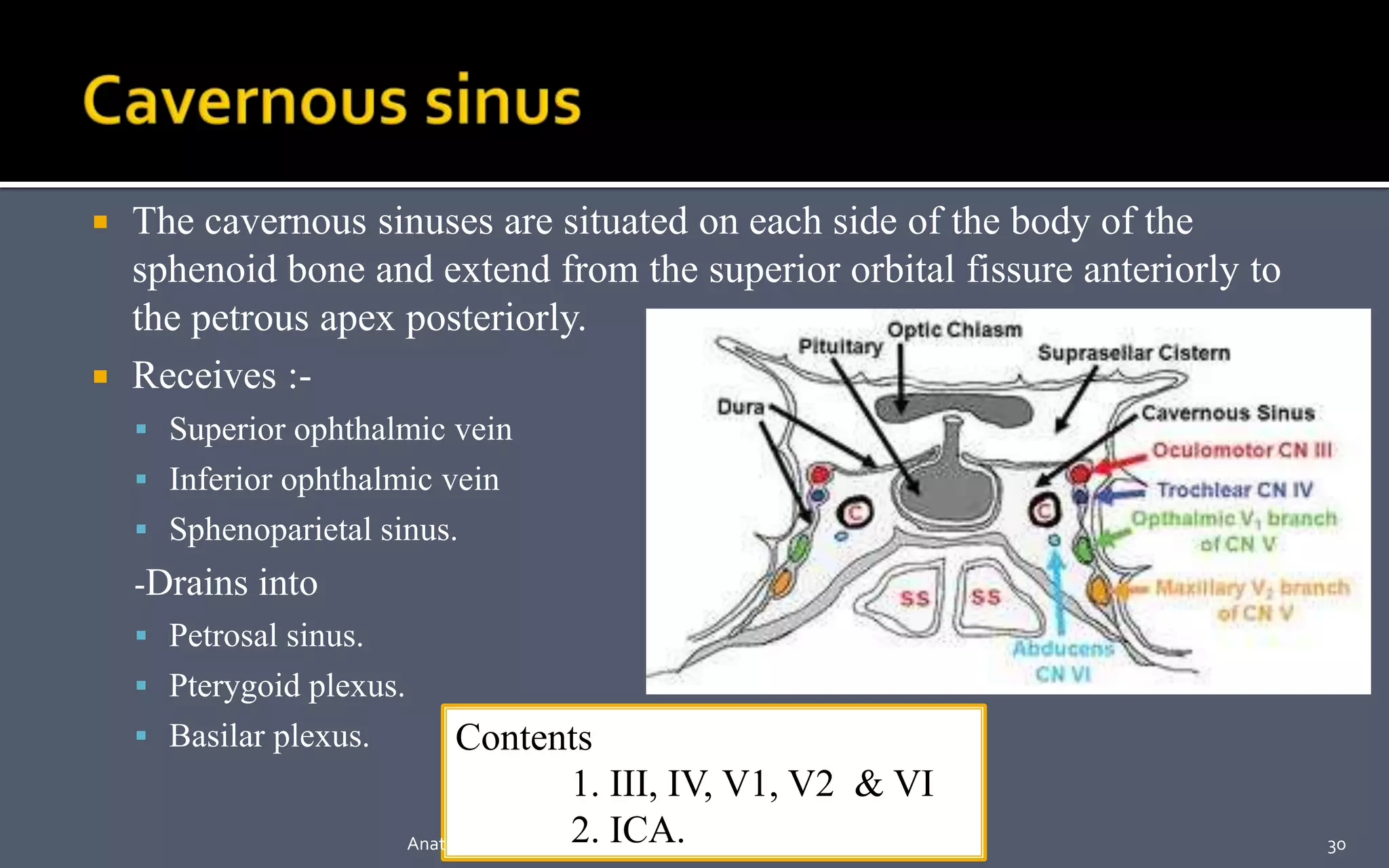
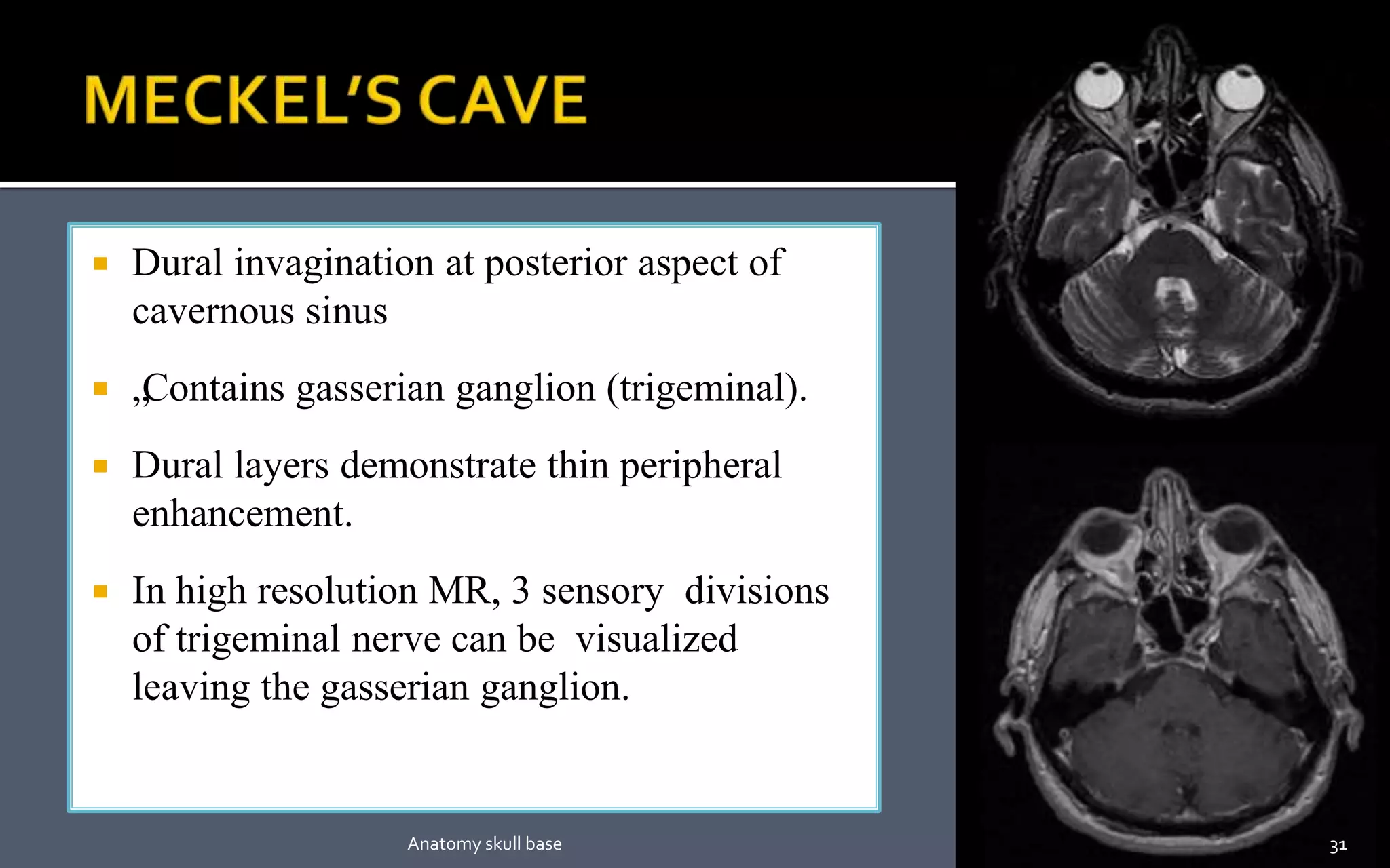
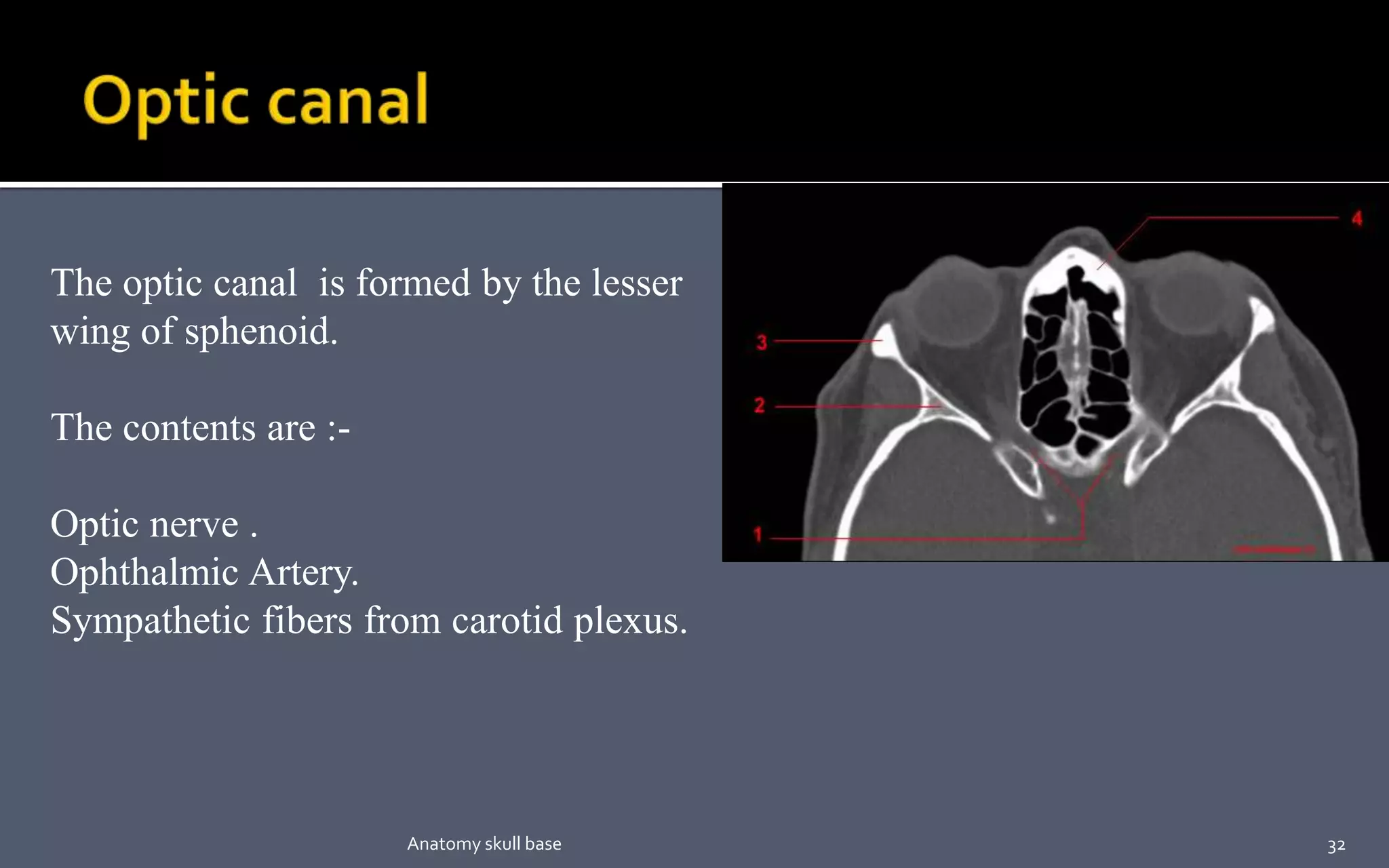
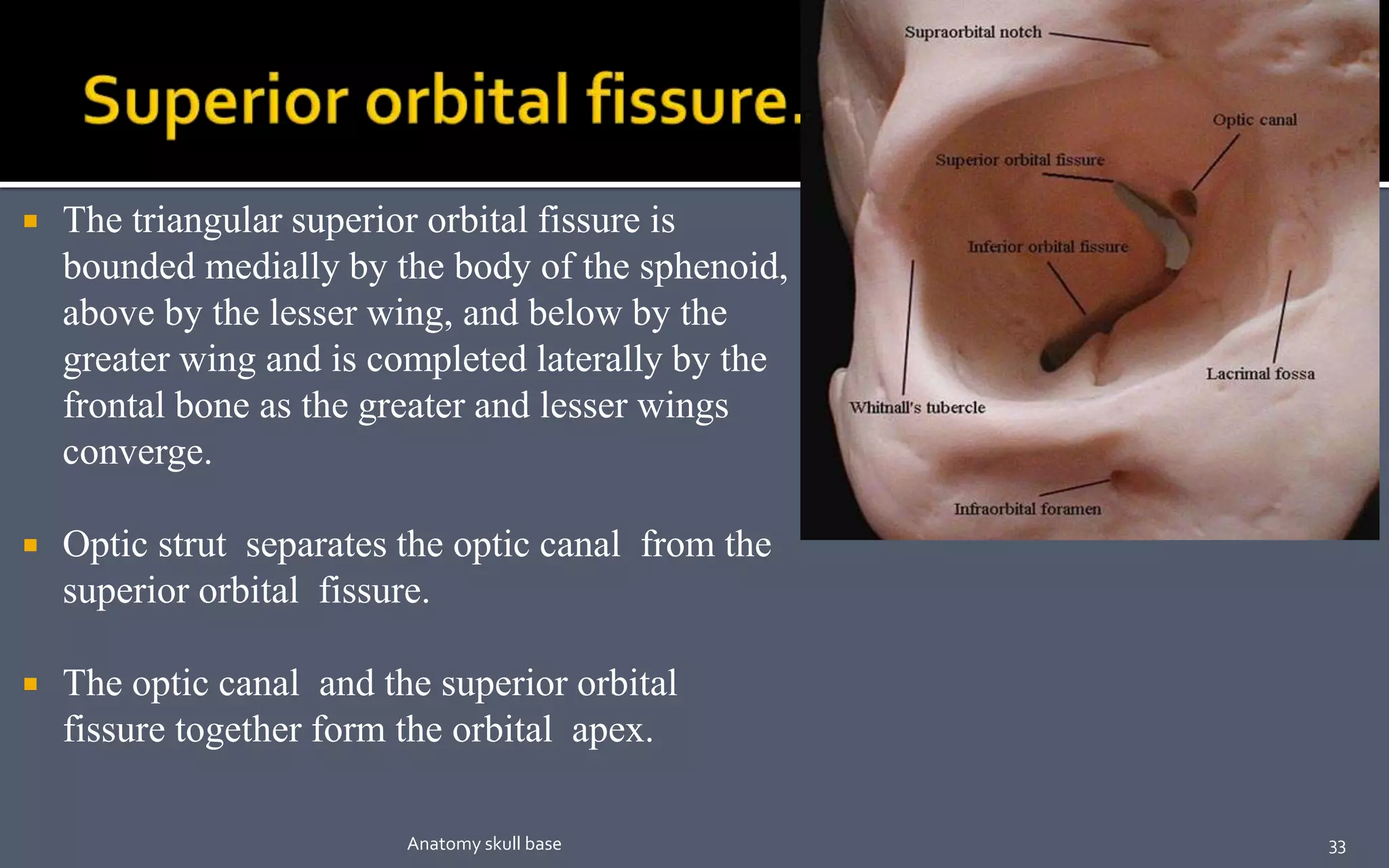
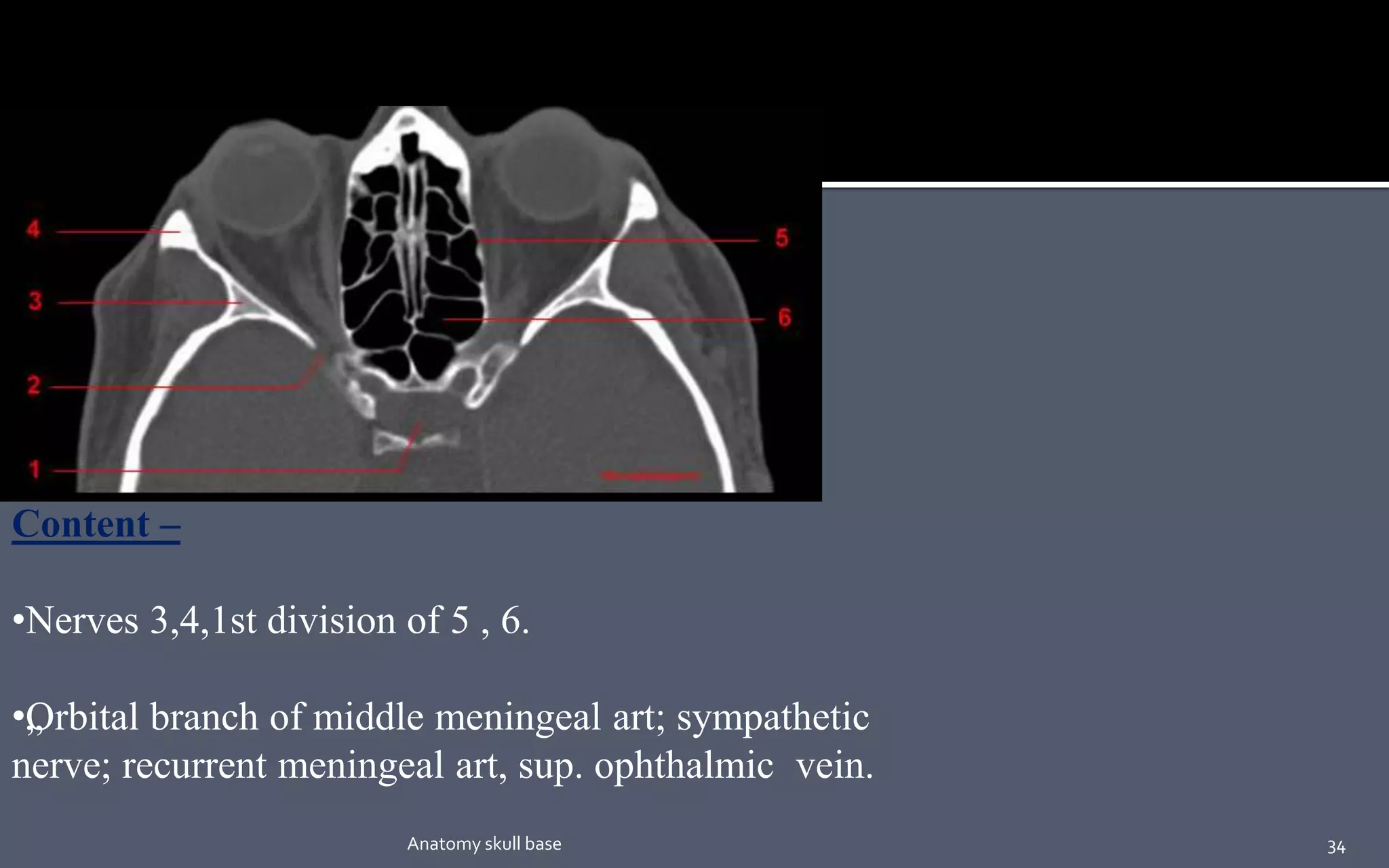


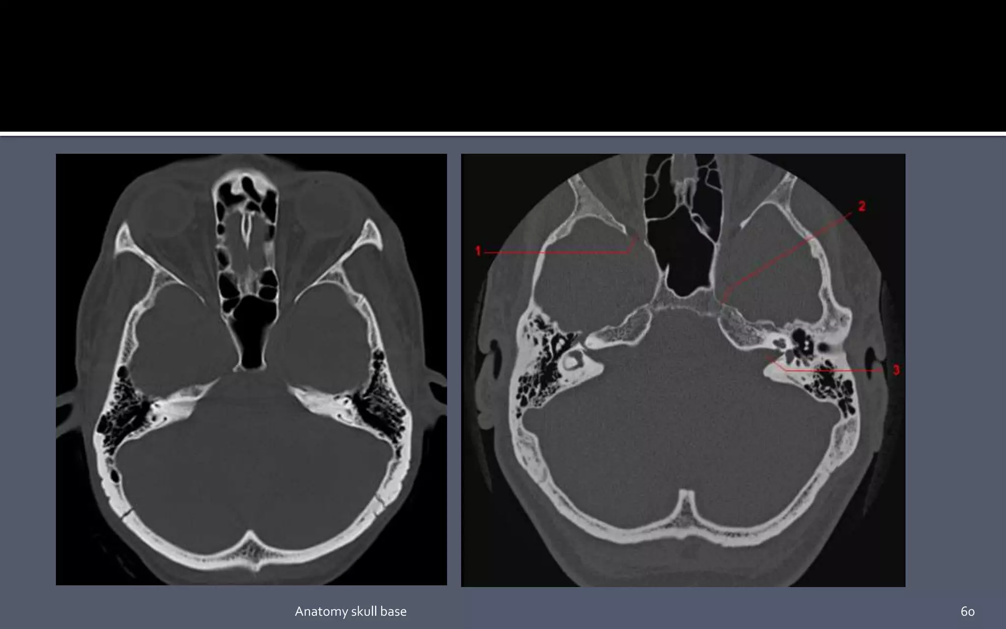


This document discusses the anatomy of the skull base. It begins by explaining that the skull base develops primarily from cartilage precursors and forms the floor of the cranial cavity. It then describes the development of the various cartilage components that fuse together to form bones of the skull base, including the parachordal, sclerotomal, hypophyseal, presphenoid, orbitosphenoid, and alisphenoid cartilages. The document further details the boundaries and structures of the anterior, middle, and posterior skull base, emphasizing the role of the sphenoid bone in forming the central skull base. Key anatomical landmarks and contents of the cavernous sinus, optic canal, and superior orbital fiss


![Radiological anatomy of_temporal_bone[1]](https://cdn.slidesharecdn.com/ss_thumbnails/radiologicalanatomyoftemporalbone1-171112100915-thumbnail.jpg?width=640&height=640&fit=bounds)


