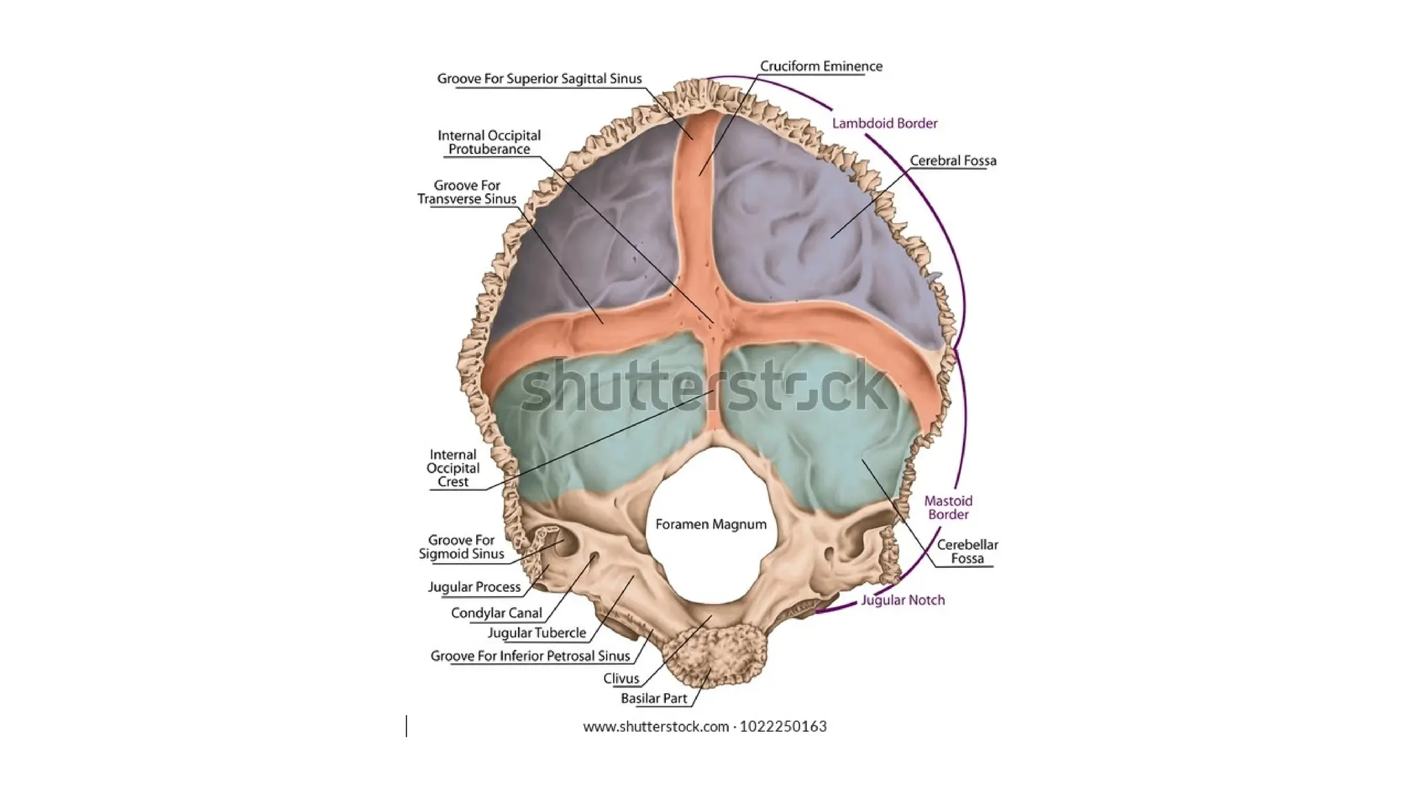
CRANIAL CAVITY
AIZAZ
Demonstrator Surgical
KMU-IPMS Peshawar
Contents
The cranial cavity contains the
• Brain and its surrounding meninges.
• Portions of the cranial nerves
• Arteries, veins, and venous sinuses.
Vault of the Skull
• The internal surface of the vault shows the coronal, sagittal, and lambdoid
sutures.
• In the midline is a shallow sagittal groove that lodges the superior sagittal
sinus.
• On each side of the groove are several small pits, called granular pits, which
lodge the lateral lacunae and arachnoid granulations.
• Several narrow grooves are present for the anterior and posterior divisions of
the middle meningeal vessels as they pass up the side of the skull to the vault.
Base of the Skull
• The interior of the base of the skull is divided into three cranial fossae: anterior,
middle, and posterior.
• The anterior cranial fossa is separated from the middle cranial fossa by the
lesser wing of the sphenoid,
• The middle cranial fossa is separated from the posterior cranial fossa by the
petrous part of the temporal bone.
Anterior Cranial Fossa
• The anterior cranial fossa is the most shallow and superior of the three cranial fossae.
• It lies superiorly over the nasal and orbital cavities.
• The anterior cranial fossa lodges the frontal lobes of the cerebral hemispheres.
• It is bounded anteriorly by the inner surface of the frontal bone.
• In the midline is a Frontal crest for the attachment of the falx cerebri.
• Posterior boundary is the sharp lesser wing of the sphenoid bone.
• The medial end of the lesser wing of the sphenoid forms the anterior clinoid process
on each side, which gives attachment to the tentorium cerebelli.
Anterior Cranial Fossa
• The median part of the anterior cranial fossa is incomplete posteriorly by the groove for the optic
chiasma.
• The floor of the fossa is formed by the ridged orbital plates of the frontal bone laterally and by
the cribriform plate of the ethmoid medially.
• Grista Galli is a sharp upward projection of the ethmoid bone in the midline act as another
attachment point for the falx cerebri.
• Along side the Crista Galli is a narrow slit in the cribriform plate for the passage of the anterior
ethmoidal nerve into the nasal cavity.
• The upper surface of the cribriform plate supports the olfactory bulbs, and the small perforations
in the cribriform plate are for the olfactory nerves.
Anterior Cranial Fossa
• The ethmoid bone in particular contains the main foramina (openings that
transmit vessels and nerves) of the anterior cranial fossa.
• The cribriform plate is a sheet of bone seen either side of the crista Galli which
contains numerous small foramina – these transmit olfactory nerve fibres into
the nasal cavity. It also contains two larger foramen:
Anterior ethmoidal foramen: transmits the anterior ethmoidal artery, nerve and vein.
Posterior ethmoidal foramen: transmits the posterior ethmoidal artery, nerve and vein
Middle Cranial fossa
• Consists of a small median part and expanded lateral parts.
• Median part is raised formed by the body of the sphenoid accommodating the pituitary
gland.
• Lateral parts form concavities on either side accommodating the temporal lobes of the
brain temporal lobes.
• Consist of three bones, sphenoid and two temporal bones
Bounded anteriorly by the lesser wings of the sphenoid
Bounded posteriorly by the superior borders of the petrous parts of the temporal bones.
Laterally lie the squamous parts of the temporal bones, the greater wings of the sphenoid, and
the parietal bones.
The floor of each lateral part of the middle cranial fossa is formed by the greater wing of the
sphenoid and the squamous and petrous parts of the temporal bone.
Middle Cranial fossa
• The sphenoid bone resembles a bat having a centrally placed body with greater and lesser
wings that are outstretched on each side.
• The body of the sphenoid contains the sphenoid air sinuses, which are lined with mucous
membrane and communicate with the nasal cavity; they serve as voice resonators.
• Optic canals are situated anteriorly in the middle cranial fossa which transmit the optic
nerves and ophthalmic arteries into the orbital cavities. They are connected by the chiasmatic
sulcus, (a groove running between the right and left optic canals).
• The superior orbital fissure, which is a slit like opening between the lesser and the greater
wings of the sphenoid, transmits the lacrimal, frontal, trochlear, oculomotor, nasociliary, and
abducent nerves, together with the superior ophthalmic vein.
Middle Cranial fossa
• The foramen rotundum, which is situated behind the medial end of the superior orbital fissure,
perforates the greater wing of the sphenoid and transmits the maxillary nerve from the
trigeminal ganglion to the pterygopalatine fossa.
• The foramen ovale lies posterolateral to the foramen rotundum. It perforates the greater wing
of the sphenoid and transmits the large sensory root and small motor root of the mandibular
nerve to the infratemporal fossa; the lesser petrosal nerve also passes through it.
• The small foramen spinosum lies posterolateral to the foramen ovale and also perforates the
greater wing of the sphenoid. transmits the middle meningeal artery from the infratemporal
fossa into the cranial cavity.
Middle Cranial fossa
• The foramen lacerum is large and irregularly shaped lies between the apex of the
petrous part of the temporal bone and the sphenoid bone.
• The inferior opening of the foramen lacerum in life is filled by cartilage and fibrous
tissue, and only small blood vessels pass through this tissue from the cranial cavity to
the neck.
• The carotid canal opens into the side of the foramen lacerum above the closed inferior
opening. The internal carotid artery enters the foramen through the carotid canal.
• Lateral to the foramen lacerum is an impression on the apex of the petrous part of the
temporal bone for the trigeminal ganglion
Middle Cranial fossa
• On the anterior surface of the petrous bone are two grooves for nerves;
• The largest medial groove is for the greater petrosal nerve, a branch of the facial nerve;
• The smaller lateral groove is for the lesser petrosal nerve, a branch of the tympanic
plexus.
• The greater petrosal nerve enters the foramen lacerum deep to the trigeminal ganglion
and joins the deep petrosal nerve (sympathetic fibers from around the internal carotid
artery), to form the nerve of the pterygoid canal.
• The lesser petrosal nerve passes forward to the foramen ovale.
Middle Cranial fossa
• The arcuate eminence is a rounded eminence found on the anterior surface of
the petrous bone and is caused by the underlying superior semicircular canal.
• The tegmen tympani, also known as the tegmental wall or roof of the
tympanic cavity, is a thin plate of the petrous part of the temporal bone that
separates the intracranial compartment and middle ear
Middle Cranial fossa
• Central part of the fossa is formed by the body of sphenoid contains the sella turcica
a saddle-shaped bony prominence. Which hold and support pituitary gland, and
consists of three parts:
Tuberculum sellae: (horn of the saddle) A vertical elevation of bone. It forms the anterior wall
of the sella turcica, and the posterior aspect of the chiasmatic sulcus
Hypophysial fossa or pituitary fossa: (seat of the saddle) sits in the middle of the sella turcica. It
is a depression in the body of the sphenoid, which holds the pituitary gland.
Dorsum sellae (back of the saddle) forms the posterior wall of the sella turcica. It is a large
square of bone, pointing upwards and forwards. It separates the middle cranial fossa from the
posterior cranial fossa.
Posterior cranial Fossa
• The fossa is deep and lodges the cerebellum, pons, and medulla oblongata.
• Boundaries
Anteriorly: superior border of the petrous part of the temporal bone,
Posteriorly: by the internal surface of the squamous part of the occipital bone
Floor: the basilar, condylar, and squamous parts of the occipital bone and the mastoid
part of the temporal bone.
Roof: formed by a fold of dura, the tentorium cerebelli.
Posterior cranial Fossa
• The foramen magnum occupies the central area of the floor and transmits the
medulla oblongata and its surrounding meninges, the ascending spinal parts of
the accessory nerves, and the two vertebral arteries.
• The hypoglossal canal is situated above the anterolateral boundary of the
foramen magnum and transmits the hypoglossal nerve.
• The jugular foramen lies between the lower border of the petrous part of the
temporal bone and the condylar part of the occipital bone. It transmits the
following structures from before backward:
Inferior petrosal sinus, the 9th, 10th, and 11th cranial nerves, and the large sigmoid
sinus. The inferior petrosal sinus descends in the groove on the lower border of the
petrous part of the temporal bone to reach the foramen. The sigmoid sinus turns down
through the foramen to become the internal jugular vein.
Posterior cranial Fossa
• The internal acoustic meatus pierces the posterior surface of the petrous part
of the temporal bone and transmit vestibulocochlear nerve and the motor
and sensory roots of the facial nerve.
• The internal occipital crest runs upward in the midline posteriorly from the
foramen magnum to the internal occipital protuberance and small falx
cerebelli over the occipital sinus.
Posterior cranial Fossa
• The groove for transverse sinus is a groove which runs along the internal
surface of the occipital bone, running laterally between the superior and
inferior fossae of the cruciform eminence. The transverse sinuses travel along
this groove.
• This groove curves around on either side, on the internal surface of the
occipital bone, to reach the posteroinferior corner of the parietal bone.
• The groove now passes onto the mastoid part of the temporal bone, and here
the transverse sinus becomes the sigmoid sinus
• The superior petrosal sinus runs backward along the upper border of the
petrous bone in a narrow groove and drains into the sigmoid sinus
Cranial cavity (fossas).pptx .  .  .  .  .  .

Cranial cavity (fossas).pptx . . . . . .

  • 1.
  • 2.
    Contents The cranial cavitycontains the • Brain and its surrounding meninges. • Portions of the cranial nerves • Arteries, veins, and venous sinuses.
  • 3.
    Vault of theSkull • The internal surface of the vault shows the coronal, sagittal, and lambdoid sutures. • In the midline is a shallow sagittal groove that lodges the superior sagittal sinus. • On each side of the groove are several small pits, called granular pits, which lodge the lateral lacunae and arachnoid granulations. • Several narrow grooves are present for the anterior and posterior divisions of the middle meningeal vessels as they pass up the side of the skull to the vault.
  • 6.
    Base of theSkull • The interior of the base of the skull is divided into three cranial fossae: anterior, middle, and posterior. • The anterior cranial fossa is separated from the middle cranial fossa by the lesser wing of the sphenoid, • The middle cranial fossa is separated from the posterior cranial fossa by the petrous part of the temporal bone.
  • 8.
    Anterior Cranial Fossa •The anterior cranial fossa is the most shallow and superior of the three cranial fossae. • It lies superiorly over the nasal and orbital cavities. • The anterior cranial fossa lodges the frontal lobes of the cerebral hemispheres. • It is bounded anteriorly by the inner surface of the frontal bone. • In the midline is a Frontal crest for the attachment of the falx cerebri. • Posterior boundary is the sharp lesser wing of the sphenoid bone. • The medial end of the lesser wing of the sphenoid forms the anterior clinoid process on each side, which gives attachment to the tentorium cerebelli.
  • 11.
    Anterior Cranial Fossa •The median part of the anterior cranial fossa is incomplete posteriorly by the groove for the optic chiasma. • The floor of the fossa is formed by the ridged orbital plates of the frontal bone laterally and by the cribriform plate of the ethmoid medially. • Grista Galli is a sharp upward projection of the ethmoid bone in the midline act as another attachment point for the falx cerebri. • Along side the Crista Galli is a narrow slit in the cribriform plate for the passage of the anterior ethmoidal nerve into the nasal cavity. • The upper surface of the cribriform plate supports the olfactory bulbs, and the small perforations in the cribriform plate are for the olfactory nerves.
  • 14.
    Anterior Cranial Fossa •The ethmoid bone in particular contains the main foramina (openings that transmit vessels and nerves) of the anterior cranial fossa. • The cribriform plate is a sheet of bone seen either side of the crista Galli which contains numerous small foramina – these transmit olfactory nerve fibres into the nasal cavity. It also contains two larger foramen: Anterior ethmoidal foramen: transmits the anterior ethmoidal artery, nerve and vein. Posterior ethmoidal foramen: transmits the posterior ethmoidal artery, nerve and vein
  • 15.
    Middle Cranial fossa •Consists of a small median part and expanded lateral parts. • Median part is raised formed by the body of the sphenoid accommodating the pituitary gland. • Lateral parts form concavities on either side accommodating the temporal lobes of the brain temporal lobes. • Consist of three bones, sphenoid and two temporal bones Bounded anteriorly by the lesser wings of the sphenoid Bounded posteriorly by the superior borders of the petrous parts of the temporal bones. Laterally lie the squamous parts of the temporal bones, the greater wings of the sphenoid, and the parietal bones. The floor of each lateral part of the middle cranial fossa is formed by the greater wing of the sphenoid and the squamous and petrous parts of the temporal bone.
  • 20.
    Middle Cranial fossa •The sphenoid bone resembles a bat having a centrally placed body with greater and lesser wings that are outstretched on each side. • The body of the sphenoid contains the sphenoid air sinuses, which are lined with mucous membrane and communicate with the nasal cavity; they serve as voice resonators. • Optic canals are situated anteriorly in the middle cranial fossa which transmit the optic nerves and ophthalmic arteries into the orbital cavities. They are connected by the chiasmatic sulcus, (a groove running between the right and left optic canals). • The superior orbital fissure, which is a slit like opening between the lesser and the greater wings of the sphenoid, transmits the lacrimal, frontal, trochlear, oculomotor, nasociliary, and abducent nerves, together with the superior ophthalmic vein.
  • 21.
    Middle Cranial fossa •The foramen rotundum, which is situated behind the medial end of the superior orbital fissure, perforates the greater wing of the sphenoid and transmits the maxillary nerve from the trigeminal ganglion to the pterygopalatine fossa. • The foramen ovale lies posterolateral to the foramen rotundum. It perforates the greater wing of the sphenoid and transmits the large sensory root and small motor root of the mandibular nerve to the infratemporal fossa; the lesser petrosal nerve also passes through it. • The small foramen spinosum lies posterolateral to the foramen ovale and also perforates the greater wing of the sphenoid. transmits the middle meningeal artery from the infratemporal fossa into the cranial cavity.
  • 24.
    Middle Cranial fossa •The foramen lacerum is large and irregularly shaped lies between the apex of the petrous part of the temporal bone and the sphenoid bone. • The inferior opening of the foramen lacerum in life is filled by cartilage and fibrous tissue, and only small blood vessels pass through this tissue from the cranial cavity to the neck. • The carotid canal opens into the side of the foramen lacerum above the closed inferior opening. The internal carotid artery enters the foramen through the carotid canal. • Lateral to the foramen lacerum is an impression on the apex of the petrous part of the temporal bone for the trigeminal ganglion
  • 25.
    Middle Cranial fossa •On the anterior surface of the petrous bone are two grooves for nerves; • The largest medial groove is for the greater petrosal nerve, a branch of the facial nerve; • The smaller lateral groove is for the lesser petrosal nerve, a branch of the tympanic plexus. • The greater petrosal nerve enters the foramen lacerum deep to the trigeminal ganglion and joins the deep petrosal nerve (sympathetic fibers from around the internal carotid artery), to form the nerve of the pterygoid canal. • The lesser petrosal nerve passes forward to the foramen ovale.
  • 27.
    Middle Cranial fossa •The arcuate eminence is a rounded eminence found on the anterior surface of the petrous bone and is caused by the underlying superior semicircular canal. • The tegmen tympani, also known as the tegmental wall or roof of the tympanic cavity, is a thin plate of the petrous part of the temporal bone that separates the intracranial compartment and middle ear
  • 29.
    Middle Cranial fossa •Central part of the fossa is formed by the body of sphenoid contains the sella turcica a saddle-shaped bony prominence. Which hold and support pituitary gland, and consists of three parts: Tuberculum sellae: (horn of the saddle) A vertical elevation of bone. It forms the anterior wall of the sella turcica, and the posterior aspect of the chiasmatic sulcus Hypophysial fossa or pituitary fossa: (seat of the saddle) sits in the middle of the sella turcica. It is a depression in the body of the sphenoid, which holds the pituitary gland. Dorsum sellae (back of the saddle) forms the posterior wall of the sella turcica. It is a large square of bone, pointing upwards and forwards. It separates the middle cranial fossa from the posterior cranial fossa.
  • 32.
    Posterior cranial Fossa •The fossa is deep and lodges the cerebellum, pons, and medulla oblongata. • Boundaries Anteriorly: superior border of the petrous part of the temporal bone, Posteriorly: by the internal surface of the squamous part of the occipital bone Floor: the basilar, condylar, and squamous parts of the occipital bone and the mastoid part of the temporal bone. Roof: formed by a fold of dura, the tentorium cerebelli.
  • 36.
    Posterior cranial Fossa •The foramen magnum occupies the central area of the floor and transmits the medulla oblongata and its surrounding meninges, the ascending spinal parts of the accessory nerves, and the two vertebral arteries. • The hypoglossal canal is situated above the anterolateral boundary of the foramen magnum and transmits the hypoglossal nerve. • The jugular foramen lies between the lower border of the petrous part of the temporal bone and the condylar part of the occipital bone. It transmits the following structures from before backward: Inferior petrosal sinus, the 9th, 10th, and 11th cranial nerves, and the large sigmoid sinus. The inferior petrosal sinus descends in the groove on the lower border of the petrous part of the temporal bone to reach the foramen. The sigmoid sinus turns down through the foramen to become the internal jugular vein.
  • 38.
    Posterior cranial Fossa •The internal acoustic meatus pierces the posterior surface of the petrous part of the temporal bone and transmit vestibulocochlear nerve and the motor and sensory roots of the facial nerve. • The internal occipital crest runs upward in the midline posteriorly from the foramen magnum to the internal occipital protuberance and small falx cerebelli over the occipital sinus.
  • 44.
    Posterior cranial Fossa •The groove for transverse sinus is a groove which runs along the internal surface of the occipital bone, running laterally between the superior and inferior fossae of the cruciform eminence. The transverse sinuses travel along this groove. • This groove curves around on either side, on the internal surface of the occipital bone, to reach the posteroinferior corner of the parietal bone. • The groove now passes onto the mastoid part of the temporal bone, and here the transverse sinus becomes the sigmoid sinus • The superior petrosal sinus runs backward along the upper border of the petrous bone in a narrow groove and drains into the sigmoid sinus