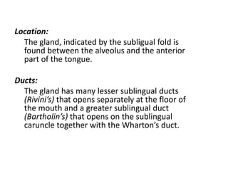
Downloaded 2,159 times
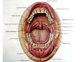
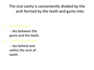
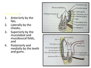


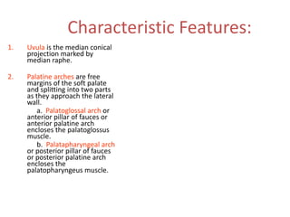
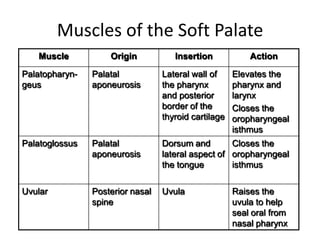
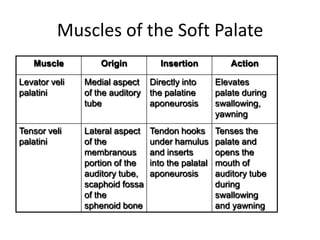

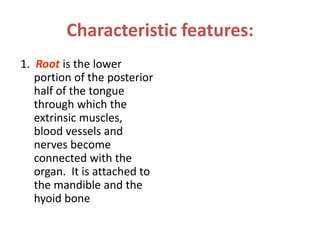

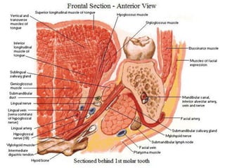


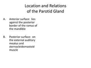
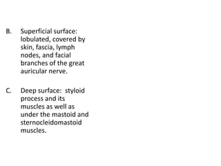
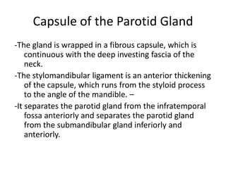
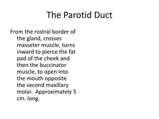


The oral cavity is divided into the oral vestibule anteriorly and the oral cavity proper posteriorly by the arch formed by the teeth and gums. The document describes the boundaries and features of these regions as well as the tongue, palate, lips, cheeks, floor of the mouth, and salivary glands. It provides detailed information on the anatomy, muscles, blood supply, and innervation of structures within the oral cavity.


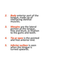
![PERI-PROSTHETIC FRACTURE NAIL-PLATE CONSTRUCT [NPC].pptx](https://cdn.slidesharecdn.com/ss_thumbnails/drarunkumardrmohamedashrafperiprostheticfrasturenail-plateconstructnpc-260209164459-7e9d15a1-thumbnail.jpg?width=640&height=640&fit=bounds)

