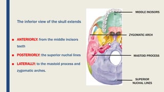
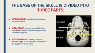
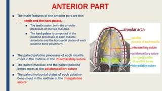
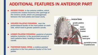
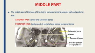
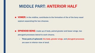
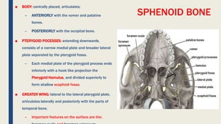
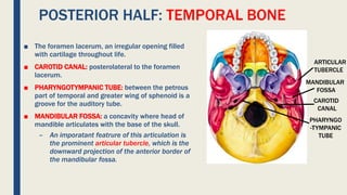
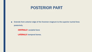
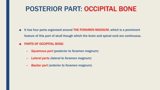
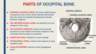
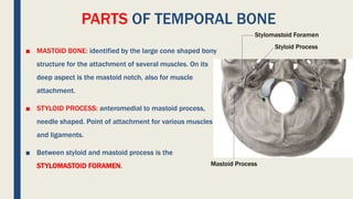
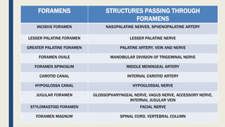
NORMA BASALIS
INFERIOR VIEW OF SKULL
DR. TANYA RASHID
ANATOMY DEPT
The inferior view of the skull extends
โ–  ANTERIORLY: from the middle incisors
teeth
โ–  POSTERIORLY: the superior nuchal lines
โ–  LATERALLY: to the mastoid process and
zygomatic arches.
MIDDLE INCISORS
SUPERIOR
NUCHAL LINES
MASTOID PROCESS
ZYGOMATIC ARCH
THE BASE OF THE SKULL IS DIVIDED INTO
THREE PARTS
โ–  ANTERIOR PART: includes the teeth and
the hard palate
โ–  MIDDLE PART: extends from behind the
hard palate to the anterior margin of the
foramen magnum
โ–  POSTERIOR PART: extends from the
anterior edge of the foramen magnum to
the superior nuchal lines.
ANTERIOR PART
โ–  The main features of the anterior part are the:
โ€“ teeth and the hard palate.
โ–  The teeth project from the alveolar
processes of the two maxillae.
โ–  The hard palate is composed of the
palatine processes of each maxilla
anteriorly and the horizontal plates of each
palatine bone posteriorly.
โ–  The paired palatine processes of each maxilla
meet in the midline at the intermaxillary suture
โ–  The paired maxillae and the paired palatine
bones meet at the palatomaxillary suture
โ–  The paired horizontal plates of each palatine
bone meet in the midline at the interpalatine
suture.
ADDITIONAL FEATURES IN ANTERIOR PART
โ–  INCISIVE FOSSA: in the anterior midline, which
contains the incisive foramina (the openings of
the incisive canals, which are passageways
between the hard palate and nasal cavity.
โ–  GREATER PALATINE FORAMINA: near the
posterolateral border of the hard palate on each
side, which lead to greater palatine canals.
โ–  LESSER PALATINE FORAMINA: posterior of greater
palatine foramina, in the pyramidal process of
each palatine bone, leading to lesser palatine
canals.
โ–  POSTERIOR NASAL SPINE: a midline pointed
projection in the free posterior border of the hard
palate.
MIDDLE PART
โ–  The middle part of the base of the skull is complex forming anterior half and posterior
half.
โ€“ ANTERIOR HALF: vomer and sphenoid bones
โ€“ POSTERIOR HALF: basilar part of occipital and paired temporal bones
MIDDLE PART: ANTERIOR HALF
โ–  VOMER: in the midline, contributes to the formation of the of the bony nasal
septum separating the two choanae.
โ–  SPHENOID BONE: made up of body, paired greater and lesser wings, two
pterygoid processes lateral to each choana.
โ€“ Three parts of sphenoid: the body, greater wings, and pterygoid processes
are seen in inferior view of skull.
SPHENOID BONE
โ–  BODY: centrally placed, articulates;
โ€“ ANTERIORLY with the vomer and palatine
bones.
โ€“ POSTERIORLY with the occipital bone.
โ–  PTERYGOID POCESSES: extending downwards,
consists of a narrow medial plate and broader lateral
plate separated by the pterygoid fossa.
โ€“ Each medial plate of the pterygoid process ends
inferiorly with a hook like projection the
Pterygoid Hamulus, and divided superiorly to
form shallow scaphoid fossa.
โ–  GREATER WING: lateral to the lateral pterygoid plate,
articulates laterally and posteriorly with the parts of
temporal bone.
โ€“ Important features on the surface are the;
MIDDLE PART: POSTERIOR HALF
โ–  CONSISTS OF THE:
occipital and the paired temporal bones.
โ–  OCCIPITAL BONE: more specifically its basilar part.
โ€“ POSTERIORLY: foramen magnum
โ€“ LATERALLY: temporal bones.
โ€“ Prominent structure on the basilar part of the
occipital bone is a PHARYNGEAL TUBERCLE, a
bony prominence for the attachment of the parts
of pharynx to the base of the skull.
POSTERIOR HALF: TEMPORAL BONE
โ–  The foramen lacerum, an irregular opening filled
with cartilage throughout life.
โ–  CAROTID CANAL: posterolateral to the foramen
lacerum.
โ–  PHARYNGOTYMPANIC TUBE: between the petrous
part of temporal and greater wing of sphenoid is a
groove for the auditory tube.
โ–  MANDIBULAR FOSSA: a concavity where head of
mandible articulates with the base of the skull.
โ€“ An imporatant featrure of this articulation is
the prominent articular tubercle, which is the
downward projection of the anterior border of
the mandibular fossa.
CAROTID
CANAL
PHARYNGO
-TYMPANIC
TUBE
MANDIBULAR
FOSSA
ARTICULAR
TUBERCLE
POSTERIOR PART
โ–  Extends from anterior edge of the foramen magnum to the superior nuchal lines
posteriorly.
CENTRALLY: occipital bone
LATERALLY: temporal bones.
POSTERIOR PART: OCCIPITAL BONE
โ–  It has four parts organized around THE FORAMEN MAGNUM, which is a prominent
feature of this part of skull though which the brain and spinal cord are continuous.
โ–  PARTS OF OCCIPITAL BONE:
โ€“ Squamous part (posterior to foramen magnum)
โ€“ Lateral parts (lateral to foramen magnum)
โ€“ Basilar part (anterior to foramen magnum)
PARTS OF OCCIPITAL BONE
โ–  EXTERNAL OCCIPITAL CREST: the most visible feature
of squamous part is the, which extends downwards
from the external occipital protuberance towards
foramen magnum.
โ–  THE INFERIOR NUCHAL LINES: are laterally from the
midpoint of the crest.
โ–  THE OCCIPITAL CONDYLES: which are the paired
structures on the border of foramen magnum. They
articulate with the atlas (c1 vertebra).
โ–  CONDYLAR CANAL: Posterior to each condyle
โ–  HYPOGLOSSAL CANAL: anterior and superior to each
condylar canal.
โ–  JUGULAR FORAMEN: lateral to each hypoglossal
canal
EXTERNAL OCCIPITAL CREST
INFERIOR NUCHAL LINES
OCCIPITAL CONDYLE
CONDYLAR
CANAL
HYPOGLOSSAL
CANAL
JUGULAR
FORAMEN
PARTS OF TEMPORAL BONE
โ–  MASTOID BONE: identified by the large cone shaped bony
structure for the attachment of several muscles. On its
deep aspect is the mastoid notch, also for muscle
attachment.
โ–  STYLOID PROCESS: anteromedial to mastoid process,
needle shaped. Point of attachment for various muscles
and ligaments.
โ–  Between styloid and mastoid process is the
STYLOMASTOID FORAMEN.
Styloid Process
Stylomastoid Foramen
Mastoid Process
FORAMENS STRUCTURES PASSING THROUGH
FORAMENS
INCISIVE FORAMEN NASOPALATINE NERVES, SPHENOPALATINE ARTERY
LESSER PALATINE FORAMEN LESSER PALATINE NERVE
GREATER PALATINE FORAMEN PALATINE ARTERY, VEIN AND NERVE
FORAMEN OVALE MANDIBULAR DIVISION OF TRIGEMINAL NERVE
FORAMEN SPINOSUM MIDDLE MENINGEAL ARTERY
CAROTID CANAL INTERNAL CAROTID ARTERY
HYPOGLOSSA CANAL HYPOGLOSSAL NERVE
JUGULAR FORAMEN GLOSSOPHARYNGEAL NERVE, VAGUS NERVE, ACCESSORY NERVE,
INTERNAL JUGULAR VEIN
STYLOMASTOID FORAMEN FACIAL NERVE
FORAMEN MAGNUM SPINAL CORD, VERTEBRAL COLUMN
MULTIPLE CHOICE QUESTIONS
โ–  WHICH OF THE FOLLOWING IS THE PASSAGEWAY BETWEEN HARD PALATE AND NASAL
CAVITY ?
a) Incisive foramen
b) Palatine foramen
c) Pterygoid fossa
d) Scaphoid fossa
โ–  Pharyngeal tubercle has the attachment for the ?
a) Brainstem
b) Pharynx
c) Pituitary gland
d) Spinal cord
โ–  What structures passes through stylomastoid foramen ?
a) Facial nerve
b) Glossopharyngeal nerve
c) Mandibular division of trigeminal nerve
d) Middle meningeal artery
Norma basalis

Norma basalis

  • 1.
    NORMA BASALIS INFERIOR VIEWOF SKULL DR. TANYA RASHID ANATOMY DEPT
  • 2.
    The inferior viewof the skull extends โ–  ANTERIORLY: from the middle incisors teeth โ–  POSTERIORLY: the superior nuchal lines โ–  LATERALLY: to the mastoid process and zygomatic arches. MIDDLE INCISORS SUPERIOR NUCHAL LINES MASTOID PROCESS ZYGOMATIC ARCH
  • 3.
    THE BASE OFTHE SKULL IS DIVIDED INTO THREE PARTS โ–  ANTERIOR PART: includes the teeth and the hard palate โ–  MIDDLE PART: extends from behind the hard palate to the anterior margin of the foramen magnum โ–  POSTERIOR PART: extends from the anterior edge of the foramen magnum to the superior nuchal lines.
  • 4.
    ANTERIOR PART โ–  Themain features of the anterior part are the: โ€“ teeth and the hard palate. โ–  The teeth project from the alveolar processes of the two maxillae. โ–  The hard palate is composed of the palatine processes of each maxilla anteriorly and the horizontal plates of each palatine bone posteriorly. โ–  The paired palatine processes of each maxilla meet in the midline at the intermaxillary suture โ–  The paired maxillae and the paired palatine bones meet at the palatomaxillary suture โ–  The paired horizontal plates of each palatine bone meet in the midline at the interpalatine suture.
  • 5.
    ADDITIONAL FEATURES INANTERIOR PART โ–  INCISIVE FOSSA: in the anterior midline, which contains the incisive foramina (the openings of the incisive canals, which are passageways between the hard palate and nasal cavity. โ–  GREATER PALATINE FORAMINA: near the posterolateral border of the hard palate on each side, which lead to greater palatine canals. โ–  LESSER PALATINE FORAMINA: posterior of greater palatine foramina, in the pyramidal process of each palatine bone, leading to lesser palatine canals. โ–  POSTERIOR NASAL SPINE: a midline pointed projection in the free posterior border of the hard palate.
  • 6.
    MIDDLE PART โ–  Themiddle part of the base of the skull is complex forming anterior half and posterior half. โ€“ ANTERIOR HALF: vomer and sphenoid bones โ€“ POSTERIOR HALF: basilar part of occipital and paired temporal bones
  • 7.
    MIDDLE PART: ANTERIORHALF โ–  VOMER: in the midline, contributes to the formation of the of the bony nasal septum separating the two choanae. โ–  SPHENOID BONE: made up of body, paired greater and lesser wings, two pterygoid processes lateral to each choana. โ€“ Three parts of sphenoid: the body, greater wings, and pterygoid processes are seen in inferior view of skull.
  • 8.
    SPHENOID BONE โ–  BODY:centrally placed, articulates; โ€“ ANTERIORLY with the vomer and palatine bones. โ€“ POSTERIORLY with the occipital bone. โ–  PTERYGOID POCESSES: extending downwards, consists of a narrow medial plate and broader lateral plate separated by the pterygoid fossa. โ€“ Each medial plate of the pterygoid process ends inferiorly with a hook like projection the Pterygoid Hamulus, and divided superiorly to form shallow scaphoid fossa. โ–  GREATER WING: lateral to the lateral pterygoid plate, articulates laterally and posteriorly with the parts of temporal bone. โ€“ Important features on the surface are the;
  • 9.
    MIDDLE PART: POSTERIORHALF โ–  CONSISTS OF THE: occipital and the paired temporal bones. โ–  OCCIPITAL BONE: more specifically its basilar part. โ€“ POSTERIORLY: foramen magnum โ€“ LATERALLY: temporal bones. โ€“ Prominent structure on the basilar part of the occipital bone is a PHARYNGEAL TUBERCLE, a bony prominence for the attachment of the parts of pharynx to the base of the skull.
  • 10.
    POSTERIOR HALF: TEMPORALBONE โ–  The foramen lacerum, an irregular opening filled with cartilage throughout life. โ–  CAROTID CANAL: posterolateral to the foramen lacerum. โ–  PHARYNGOTYMPANIC TUBE: between the petrous part of temporal and greater wing of sphenoid is a groove for the auditory tube. โ–  MANDIBULAR FOSSA: a concavity where head of mandible articulates with the base of the skull. โ€“ An imporatant featrure of this articulation is the prominent articular tubercle, which is the downward projection of the anterior border of the mandibular fossa. CAROTID CANAL PHARYNGO -TYMPANIC TUBE MANDIBULAR FOSSA ARTICULAR TUBERCLE
  • 11.
    POSTERIOR PART โ–  Extendsfrom anterior edge of the foramen magnum to the superior nuchal lines posteriorly. CENTRALLY: occipital bone LATERALLY: temporal bones.
  • 12.
    POSTERIOR PART: OCCIPITALBONE โ–  It has four parts organized around THE FORAMEN MAGNUM, which is a prominent feature of this part of skull though which the brain and spinal cord are continuous. โ–  PARTS OF OCCIPITAL BONE: โ€“ Squamous part (posterior to foramen magnum) โ€“ Lateral parts (lateral to foramen magnum) โ€“ Basilar part (anterior to foramen magnum)
  • 13.
    PARTS OF OCCIPITALBONE โ–  EXTERNAL OCCIPITAL CREST: the most visible feature of squamous part is the, which extends downwards from the external occipital protuberance towards foramen magnum. โ–  THE INFERIOR NUCHAL LINES: are laterally from the midpoint of the crest. โ–  THE OCCIPITAL CONDYLES: which are the paired structures on the border of foramen magnum. They articulate with the atlas (c1 vertebra). โ–  CONDYLAR CANAL: Posterior to each condyle โ–  HYPOGLOSSAL CANAL: anterior and superior to each condylar canal. โ–  JUGULAR FORAMEN: lateral to each hypoglossal canal EXTERNAL OCCIPITAL CREST INFERIOR NUCHAL LINES OCCIPITAL CONDYLE CONDYLAR CANAL HYPOGLOSSAL CANAL JUGULAR FORAMEN
  • 14.
    PARTS OF TEMPORALBONE โ–  MASTOID BONE: identified by the large cone shaped bony structure for the attachment of several muscles. On its deep aspect is the mastoid notch, also for muscle attachment. โ–  STYLOID PROCESS: anteromedial to mastoid process, needle shaped. Point of attachment for various muscles and ligaments. โ–  Between styloid and mastoid process is the STYLOMASTOID FORAMEN. Styloid Process Stylomastoid Foramen Mastoid Process
  • 15.
    FORAMENS STRUCTURES PASSINGTHROUGH FORAMENS INCISIVE FORAMEN NASOPALATINE NERVES, SPHENOPALATINE ARTERY LESSER PALATINE FORAMEN LESSER PALATINE NERVE GREATER PALATINE FORAMEN PALATINE ARTERY, VEIN AND NERVE FORAMEN OVALE MANDIBULAR DIVISION OF TRIGEMINAL NERVE FORAMEN SPINOSUM MIDDLE MENINGEAL ARTERY CAROTID CANAL INTERNAL CAROTID ARTERY HYPOGLOSSA CANAL HYPOGLOSSAL NERVE JUGULAR FORAMEN GLOSSOPHARYNGEAL NERVE, VAGUS NERVE, ACCESSORY NERVE, INTERNAL JUGULAR VEIN STYLOMASTOID FORAMEN FACIAL NERVE FORAMEN MAGNUM SPINAL CORD, VERTEBRAL COLUMN
  • 16.
    MULTIPLE CHOICE QUESTIONS โ– WHICH OF THE FOLLOWING IS THE PASSAGEWAY BETWEEN HARD PALATE AND NASAL CAVITY ? a) Incisive foramen b) Palatine foramen c) Pterygoid fossa d) Scaphoid fossa
  • 17.
    โ–  Pharyngeal tuberclehas the attachment for the ? a) Brainstem b) Pharynx c) Pituitary gland d) Spinal cord
  • 18.
    โ–  What structurespasses through stylomastoid foramen ? a) Facial nerve b) Glossopharyngeal nerve c) Mandibular division of trigeminal nerve d) Middle meningeal artery