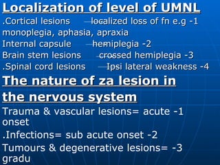
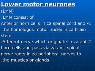
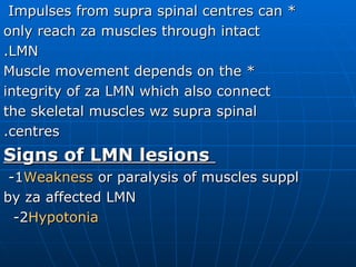
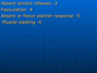
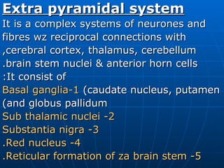
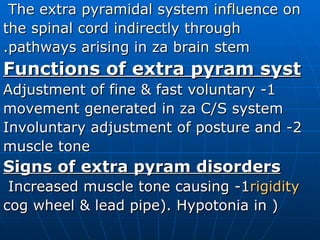
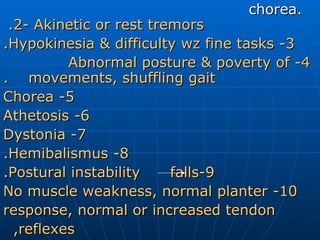
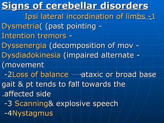
Neurophysiology By: Dr. Osman Sadig
Motor system Consists of: 1-  Pyramidal  OR Corticospinal system (UMN) 2-  Lower motor neurone  (LMN) 3-  Extra pyramidal system 4- Cerebellum .
Corticospinal (pyramidal) system---(UMN) Composed of: 1-  Motor cortex 2-  Corticospinal tracts . 1-  Motor cortex: - Occupies za pre central gyrus - The body is represented upside down,  the area represented depends on the functional importance - Controls za opposite side of za body
2-  Pyramidal tracts (C/S) -  It passes downwards from motor cortex as za corona radiata to za internal capsul occupying the posterior 2/3 of za post.  limb, descends downwards to occupy  za peduncles of mid brain & passes  through za pons to form za pyramids  of za medulla. In za medulla za  majority of C/S tracts decussate wz  those of za opposite side and course  down in za spinal cord in the lateral  column as indirect C/S tracts
 
 
and terminate in za LMN.  The undecussated C/S fibres descend in the anterior column as direct C/S tracts decussate at segmental level & terminate in za LMN. - At different levels of CNS, The C/S  tracts give fibres to za brain stem nuclei and za LMN in za anterior horn of the spinal cord.
Function of C/S: - Motor cortex is responsible for  programming & initiation of skilled  motor acts, specially fine distal  movements, and is influenced by connection from other parts of motor system that controls muscle tone & body posture. - Accurately targeted & coordinated  actions require intact functioning cerebellum.
- Lesions of C/S system above za level of decussation cause contra lateral signs, while below that cause ipsi lateral signs. Signs of UMNL: 1-  Muscle weakness . 2-  Hypertonia  resulting in spasticity. 3-  Hyper- reflexia  (increased tendon  reflex) +/- clonus. 4-  Extensor planter reflex 5-Absent abdominal & cremasteric reflexes 6- In  long standing cases there is wasting .
Localization of level of UMNL 1- Cortical lesions  localized loss of fn e.g. monoplegia, aphasia, apraxia 2- Internal capsule  hemiplegia 3- Brain stem lesions  crossed hemiplegia 4- Spinal cord lesions  Ipsi lateral weakness. The nature of za lesion in the nervous system 1- Trauma & vascular lesions= acute onset 2- Infections= sub acute onset. 3- Tumours & degenerative lesions= gradu onset & progressive course.
4- Demyelinating lesions= remission  and  relapses. 5- Epilepsy, migraine & TIA= paroxysmal. NB: destructive lesion  weakness irritative lesions  seizures
Lower motor neurones (LMN) LMN consist of: 1- Anterior horn cells in za spinal cord and the homologus motor nuclei in za brain  stem 2 Afferent nerve which originate in za ant. horn cells and pass via za ant. spinal nerve roots in za peripheral nerves to the muscles or glands.
* Impulses from supra spinal centres can  only reach za muscles through intact LMN. * Muscle movement depends on the integrity of za LMN which also connect the skeletal muscles wz supra spinal centres. Signs of LMN lesions 1-  Weakness  or paralysis of muscles suppl by za affected LMN 2-  Hypotonia
3- Absent tendon reflexes 4- Fasiculation 5- Absent or flexor planter response 6- Muscle wasting.
Extra pyramidal system It is a complex systems of neurones and fibres wz reciprocal connections with cerebral cortex, thalamus, cerebellum, brain stem nuclei & anterior horn cells. It consist of: 1-Basal ganglia  (caudate nucleus, putamen and globus pallidum) 2- Sub thalamic nuclei 3- Substantia nigra 4- Red nucleus. 5- Reticular formation of za brain stem.
The extra pyramidal system influence on  the spinal cord indirectly through pathways arising in za brain stem. Functions of extra pyram syst 1- Adjustment of fine & fast voluntary movement generated in za C/S system 2- Involuntary adjustment of posture and muscle tone Signs of extra pyram disorders 1- Increased muscle tone causing  rigidity ( cog wheel & lead pipe). Hypotonia in
chorea.  2- Akinetic or rest tremors. 3- Hypokinesia & difficulty wz fine tasks. 4- Abnormal posture & poverty of  movements, shuffling gait. 5- Chorea 6- Athetosis 7- Dystonia 8- Hemibalismus. 9-Postural instability  falls. 10- No muscle weakness, normal planter response, normal or increased tendon reflexes,
The cerebellum -The cerebellum  modulates muscle tone , controls  co ordination of voluntary movements  & is important for  maintainanc   of posture & balance  through  its  connections  with the thalamus,  basal  ganglia, cerebral cortex,  vestibular syst  and za spinal cord - It is closely connected wz za vestibular syst & recieves proprioceptive inputs  from the spinal cord - Cerebellar lesions & its connections  cause  ipsi lateral signs .
Signs of cerebellar disorders 1- Ipsi lateral incordination of limbs   -  Dysmetria  (past pointing) -  Intention tremors -  Dyssenergia  (decomposition of mov -  Dysdiadokinesia  (impaired alternate movement) 2-  Loss of balance   ataxic or broad base gait & pt tends to fall towards the affected side. 3-  Scanning  & explosive speech 4-  Nystagmus
5-  Hypotonia 6- Normal but pendular tendon reflexes 7- Head tilted to za side of lesion 8- Titubation.
The sensory system -The sensory input reaches za nervous sys from specialized receptors in za skin, muscles and joints & through za  autonomic nervous syst from za internal  organs and viscera. - The sensory system comprises: 1- Receptors 2- Peripheral nerves 3- Spinothalamic tracts 4- Posterior column tracts
5-  Medial lemniscus 6-  Main sensory nucleus of za thalamus 7-  Sensory cortex  in za pre central gyrus -  Superficial (exteroceptive ) sensations include pain, temp & touch -  Deep (proprioceptive ) sensations include deep pain, pressure pain, position and vibration sense. -  Interoceptive sensations  arise from the internal organs & viscera. * All sensory impulses arise in za sensory receptors & pass along za 1 st  order
neurone to za spinal cord. These neurone have their cell bodies in za dorsal root  ganglia. 1- Fibres sub serving superficial sensation synapse wz za 2 nd  order neurone in the post. horn of za spinal cord, most of which cross to za opposite site to  ascend in za lateral S/T tract in za dors lateral column to za sensory nucleus of the thalamus, thence za 3 rd  order neuro  relays to za sensory cortex. Some  ascend in za ipsi lateral S/T tract.
2-Fibres sub serving proprioception ascend in za ipsi lateral post. column to synaps with za Gracil & Cuneate nuclei in the lower part of za medulla, thence za 2 nd   order neurone cross za mid line and  ascend in za contra lateral medial  lemniscus to za main sensory nucleus of the thalamus from which the 3 rd  order neurone relays in sensory cortex. 3- Some afferent fibres sub serve spinal reflexes 4- Some fibres arising in za muscle  receptors synapse in za post. horn
 
 
cells & ascend in za ant. & post. spino - cerebellar tracts to za cerebellum sub serving proprioception. 5- Collateral branches from S/T tracts and  from special sensory pathways to the reticular formation of za brain stem are important for arousal & maintainanc of consciousness. 6- Non dominent parietal cortex is  concerned wz asteriognosis, two point  discrimnation and spatial orientation  of za body.
7- The dominant parietal cortex is  concerned wz reception of speech * The body is represented upside down in  motor cortex. * Disorders of sensory syst cause pain, paraethesiae, anaethesia, analgesia and loss of sensory discrimination.
Signs of sensory dysfunction 1- Peripheral nerve lesion  loss of all  sensations & spinal reflexes sub served by za nerve.  2- Post. nerve roots  Dermatomal loss of all sensations & reflexes sub served by za nerve root (+ root pain) 3- Spinothalamic tracts  Contra lateral loss of pain & temp below za lesion 4- Post. Columns  Ipsi lateral loss of  vibration & position sense wz sensory ataxia & +ve Romberg sign.
5- Brain stem  loss of all contra lateral sensations. With mid brain lesions the face is also involved. Pontine & medulla lesions cause ipsi lateral sensory loss. 6- Thalamic lesions  Spontaneous  unpleasant pain & contra lat loss of all sensations. Pain threshold is increased 7- Internal capsule  Contra lat loss of all sensations 8- Sensory cortex  Increased threshold  of sensations, astereognosis, sensory inattention (agnosia) & receptive  dysphasia
 
 
Reflex activities Reflex act represents za simplest integrated activity of nervous system. 1- Stretch or tendon reflexes: they are mono synaptic postural reflexes e.g. knee, ankle, jaw & corneal reflexes. 2- Superficial reflexes: they are poly  synaptic protective reflexes e.g. abdom cremasteric and planter reflexes.
Control of micturition Micturition is controlled by: 1- Higher centres for voluntary control of mict. 2- Parasympath (S2,3,4) causes contractio of detrusal muscles & relaxation of internal sphincter. 3- Sympath (T12 &L1,2 ) causes relaxatio of detrusal muscles & contraction of internal sphincter, 4- Pudendal nerve (S1,2,3) controls  external sphincter.
Bladder dysfunction 1- Cortical lesions: - Post. Central lesions  loss of  awareness of bladder fullness & incont - Pre central lesions  difficulty in initiation of mict & retention of urine. - frontal lesions  inappropriate mict  due to social dis inhibition. 2- Spinal cord lesions: - Above mict centre (pyramidal)  urgency & incontinence (hypertonic) 3- LMN lesions  Atonic bladder  retention with overflow & later autonomic bladder.

Neurophysiology

  • 1.
  • 2.
    Motor system Consistsof: 1- Pyramidal OR Corticospinal system (UMN) 2- Lower motor neurone (LMN) 3- Extra pyramidal system 4- Cerebellum .
  • 3.
    Corticospinal (pyramidal) system---(UMN)Composed of: 1- Motor cortex 2- Corticospinal tracts . 1- Motor cortex: - Occupies za pre central gyrus - The body is represented upside down, the area represented depends on the functional importance - Controls za opposite side of za body
  • 4.
    2- Pyramidaltracts (C/S) - It passes downwards from motor cortex as za corona radiata to za internal capsul occupying the posterior 2/3 of za post. limb, descends downwards to occupy za peduncles of mid brain & passes through za pons to form za pyramids of za medulla. In za medulla za majority of C/S tracts decussate wz those of za opposite side and course down in za spinal cord in the lateral column as indirect C/S tracts
  • 5.
  • 6.
  • 7.
    and terminate inza LMN. The undecussated C/S fibres descend in the anterior column as direct C/S tracts decussate at segmental level & terminate in za LMN. - At different levels of CNS, The C/S tracts give fibres to za brain stem nuclei and za LMN in za anterior horn of the spinal cord.
  • 8.
    Function of C/S:- Motor cortex is responsible for programming & initiation of skilled motor acts, specially fine distal movements, and is influenced by connection from other parts of motor system that controls muscle tone & body posture. - Accurately targeted & coordinated actions require intact functioning cerebellum.
  • 9.
    - Lesions ofC/S system above za level of decussation cause contra lateral signs, while below that cause ipsi lateral signs. Signs of UMNL: 1- Muscle weakness . 2- Hypertonia resulting in spasticity. 3- Hyper- reflexia (increased tendon reflex) +/- clonus. 4- Extensor planter reflex 5-Absent abdominal & cremasteric reflexes 6- In long standing cases there is wasting .
  • 10.
    Localization of levelof UMNL 1- Cortical lesions localized loss of fn e.g. monoplegia, aphasia, apraxia 2- Internal capsule hemiplegia 3- Brain stem lesions crossed hemiplegia 4- Spinal cord lesions Ipsi lateral weakness. The nature of za lesion in the nervous system 1- Trauma & vascular lesions= acute onset 2- Infections= sub acute onset. 3- Tumours & degenerative lesions= gradu onset & progressive course.
  • 11.
    4- Demyelinating lesions=remission and relapses. 5- Epilepsy, migraine & TIA= paroxysmal. NB: destructive lesion weakness irritative lesions seizures
  • 12.
    Lower motor neurones(LMN) LMN consist of: 1- Anterior horn cells in za spinal cord and the homologus motor nuclei in za brain stem 2 Afferent nerve which originate in za ant. horn cells and pass via za ant. spinal nerve roots in za peripheral nerves to the muscles or glands.
  • 13.
    * Impulses fromsupra spinal centres can only reach za muscles through intact LMN. * Muscle movement depends on the integrity of za LMN which also connect the skeletal muscles wz supra spinal centres. Signs of LMN lesions 1- Weakness or paralysis of muscles suppl by za affected LMN 2- Hypotonia
  • 14.
    3- Absent tendonreflexes 4- Fasiculation 5- Absent or flexor planter response 6- Muscle wasting.
  • 15.
    Extra pyramidal systemIt is a complex systems of neurones and fibres wz reciprocal connections with cerebral cortex, thalamus, cerebellum, brain stem nuclei & anterior horn cells. It consist of: 1-Basal ganglia (caudate nucleus, putamen and globus pallidum) 2- Sub thalamic nuclei 3- Substantia nigra 4- Red nucleus. 5- Reticular formation of za brain stem.
  • 16.
    The extra pyramidalsystem influence on the spinal cord indirectly through pathways arising in za brain stem. Functions of extra pyram syst 1- Adjustment of fine & fast voluntary movement generated in za C/S system 2- Involuntary adjustment of posture and muscle tone Signs of extra pyram disorders 1- Increased muscle tone causing rigidity ( cog wheel & lead pipe). Hypotonia in
  • 17.
    chorea. 2-Akinetic or rest tremors. 3- Hypokinesia & difficulty wz fine tasks. 4- Abnormal posture & poverty of movements, shuffling gait. 5- Chorea 6- Athetosis 7- Dystonia 8- Hemibalismus. 9-Postural instability falls. 10- No muscle weakness, normal planter response, normal or increased tendon reflexes,
  • 18.
    The cerebellum -Thecerebellum modulates muscle tone , controls co ordination of voluntary movements & is important for maintainanc of posture & balance through its connections with the thalamus, basal ganglia, cerebral cortex, vestibular syst and za spinal cord - It is closely connected wz za vestibular syst & recieves proprioceptive inputs from the spinal cord - Cerebellar lesions & its connections cause ipsi lateral signs .
  • 19.
    Signs of cerebellardisorders 1- Ipsi lateral incordination of limbs - Dysmetria (past pointing) - Intention tremors - Dyssenergia (decomposition of mov - Dysdiadokinesia (impaired alternate movement) 2- Loss of balance ataxic or broad base gait & pt tends to fall towards the affected side. 3- Scanning & explosive speech 4- Nystagmus
  • 20.
    5- Hypotonia6- Normal but pendular tendon reflexes 7- Head tilted to za side of lesion 8- Titubation.
  • 21.
    The sensory system-The sensory input reaches za nervous sys from specialized receptors in za skin, muscles and joints & through za autonomic nervous syst from za internal organs and viscera. - The sensory system comprises: 1- Receptors 2- Peripheral nerves 3- Spinothalamic tracts 4- Posterior column tracts
  • 22.
    5- Mediallemniscus 6- Main sensory nucleus of za thalamus 7- Sensory cortex in za pre central gyrus - Superficial (exteroceptive ) sensations include pain, temp & touch - Deep (proprioceptive ) sensations include deep pain, pressure pain, position and vibration sense. - Interoceptive sensations arise from the internal organs & viscera. * All sensory impulses arise in za sensory receptors & pass along za 1 st order
  • 23.
    neurone to zaspinal cord. These neurone have their cell bodies in za dorsal root ganglia. 1- Fibres sub serving superficial sensation synapse wz za 2 nd order neurone in the post. horn of za spinal cord, most of which cross to za opposite site to ascend in za lateral S/T tract in za dors lateral column to za sensory nucleus of the thalamus, thence za 3 rd order neuro relays to za sensory cortex. Some ascend in za ipsi lateral S/T tract.
  • 24.
    2-Fibres sub servingproprioception ascend in za ipsi lateral post. column to synaps with za Gracil & Cuneate nuclei in the lower part of za medulla, thence za 2 nd order neurone cross za mid line and ascend in za contra lateral medial lemniscus to za main sensory nucleus of the thalamus from which the 3 rd order neurone relays in sensory cortex. 3- Some afferent fibres sub serve spinal reflexes 4- Some fibres arising in za muscle receptors synapse in za post. horn
  • 25.
  • 26.
  • 27.
    cells & ascendin za ant. & post. spino - cerebellar tracts to za cerebellum sub serving proprioception. 5- Collateral branches from S/T tracts and from special sensory pathways to the reticular formation of za brain stem are important for arousal & maintainanc of consciousness. 6- Non dominent parietal cortex is concerned wz asteriognosis, two point discrimnation and spatial orientation of za body.
  • 28.
    7- The dominantparietal cortex is concerned wz reception of speech * The body is represented upside down in motor cortex. * Disorders of sensory syst cause pain, paraethesiae, anaethesia, analgesia and loss of sensory discrimination.
  • 29.
    Signs of sensorydysfunction 1- Peripheral nerve lesion loss of all sensations & spinal reflexes sub served by za nerve. 2- Post. nerve roots Dermatomal loss of all sensations & reflexes sub served by za nerve root (+ root pain) 3- Spinothalamic tracts Contra lateral loss of pain & temp below za lesion 4- Post. Columns Ipsi lateral loss of vibration & position sense wz sensory ataxia & +ve Romberg sign.
  • 30.
    5- Brain stem loss of all contra lateral sensations. With mid brain lesions the face is also involved. Pontine & medulla lesions cause ipsi lateral sensory loss. 6- Thalamic lesions Spontaneous unpleasant pain & contra lat loss of all sensations. Pain threshold is increased 7- Internal capsule Contra lat loss of all sensations 8- Sensory cortex Increased threshold of sensations, astereognosis, sensory inattention (agnosia) & receptive dysphasia
  • 31.
  • 32.
  • 33.
    Reflex activities Reflexact represents za simplest integrated activity of nervous system. 1- Stretch or tendon reflexes: they are mono synaptic postural reflexes e.g. knee, ankle, jaw & corneal reflexes. 2- Superficial reflexes: they are poly synaptic protective reflexes e.g. abdom cremasteric and planter reflexes.
  • 34.
    Control of micturitionMicturition is controlled by: 1- Higher centres for voluntary control of mict. 2- Parasympath (S2,3,4) causes contractio of detrusal muscles & relaxation of internal sphincter. 3- Sympath (T12 &L1,2 ) causes relaxatio of detrusal muscles & contraction of internal sphincter, 4- Pudendal nerve (S1,2,3) controls external sphincter.
  • 35.
    Bladder dysfunction 1-Cortical lesions: - Post. Central lesions loss of awareness of bladder fullness & incont - Pre central lesions difficulty in initiation of mict & retention of urine. - frontal lesions inappropriate mict due to social dis inhibition. 2- Spinal cord lesions: - Above mict centre (pyramidal) urgency & incontinence (hypertonic) 3- LMN lesions Atonic bladder retention with overflow & later autonomic bladder.