I. Cerebrum
II. Brain Stem
III. Cerebellum.
The Cerebral Cortex
A. Frontal lobe
1) Motor area (area 4):
Frontal lobe
parietal lobe
temporal lobe
occipital lobe
Most people have difficulty differentiating between seizure and convulsion. This presentation also highlights the differences between hysterical fit and grand mal seizure.
How to manage the client is briefly discussed.
The thalamus is the large mass of gray matter in the dorsal part of the diencephalon of the brain with several functions such as relaying of sensory signals, including motor signals, to the cerebral cortex and the regulation of consciousness, sleep, and alertness.
Largest part of hind brain.
Called “ silent area/Little Brain ”
Weight- 150 gms.
Cerebellar cortex is a large folded sheet, each fold is called Folium.
Connected to brain stem by 3 pairs of peduncles- Superior (Brachium conjunctiva), Middle (Brachium Pontis) & Inferior (Restiform body) peduncle.
Electrophysiological assessment of neuromuscular transmissionRahul Kumar
The Presentation discusses the detailed aspects of the Electrophysiological Aspects of Neuromuscular transmission, as well as the diagnostic features of the various types of NMJ Disorders.
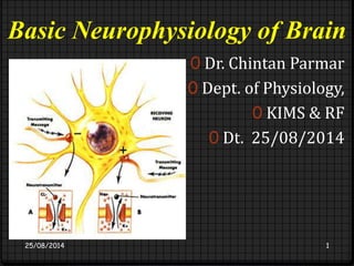
Talks about Neuromuscular transmission in NMJ. It explains how Acetylcholine at a pre synaptic terminal transmits an impulse to the post synaptic terminal
neurohumoral transmission refers to the transmission of impulse through synapse and neuroeffector junction by the release of chemical (humoral) substance.
3. Definitions
0A seizure is the clinical manifestation of an abnormal,
excessive, hyper synchronous discharge of a population
of cortical neurons.
0Epilepsy is a disorder of the CNS characterized by
recurrent seizures unprovoked by an acute systemic
or neurologic insult.
0Epileptogenesis is the sequence of events that turns a
normal neuronal network into a hyper excitable
network.
25/08/2014 KIMS & RF, Symposium on Epilepsy 3
4. Basic Anatomy of Cortex
0The human cerebral cortex consists of 3 to 6 layers of
neurons.
0The phylogenetically oldest part of the cortex
(archipallium) has 3 distinct neuronal layers, and is
represented by the hippocampus, which is found in the
medial temporal lobe.
0The majority of the cortex (neocortex or neopallium)
has 6 distinct cell layers and covers most of the
surface of the cerebral hemispheres.
25/08/2014 KIMS & RF, Symposium on Epilepsy 4
6. Basic Anatomy of Cortex
0The hippocampus consists of three major regions:
subiculum, hippocampus proper and dentate gyrus.
0The hippocampus and dentate gyrus have a 3 layered
cortex.
0The subiculum is the transition zone from the 3 to the 6
layered cortex.
0Important regions of the hippocampus proper include
CA1, CA 2, CA 3 & CA 4.
25/08/2014 KIMS & RF, Symposium on Epilepsy 6
10. 0 The basic mechanism of neuronal excitability is the action
potential.
25/08/2014 KIMS & RF, Symposium on Epilepsy 10
11. Action Potential
0A hyperexcitable state can result from;
0increased excitatory synaptic neurotransmission,
0decreased inhibitory neurotransmission,
0an alteration in voltage-gated ion channels,
0an alteration of intra- or extra-cellular ion
concentrations in favor of membrane
depolarization.
0A hyperexcitable state can also result when several
synchronous subthreshold excitatory stimuli occur,
allowing their temporal summation in the post
synaptic neurons.
25/08/2014 KIMS & RF, Symposium on Epilepsy 11
12. Cellular Mechanisms
0 Neuronal (Intrinsic) Factors Modifying Neuronal Excitability
0 The type, number and distribution of voltage and ligand gated
channels
0 Such channels determine the direction, degree, and rate of
changes in the transmembrane potential, which in turn
determine whether an action potential occurs or not
0 Biochemical modification of receptors
0 Activation of second-messenger systems
0 Modulating gene expression by RNA editing
25/08/2014 KIMS & RF, Symposium on Epilepsy 12
13. Cellular Mechanisms
0Extra-Neuronal (Extrinsic) Factors Modifying
Neuronal Excitability
0Changes in extracellular ion concentration due to
variations in the volume of the extracellular space
0Remodeling of synaptic contacts
0Modulating transmitter metabolism by glial cells
25/08/2014 KIMS & RF, Symposium on Epilepsy 13
14. Cellular Mechanisms
0 The cortex includes two general classes of neurons.
0 The projection, or principal neurons (e.g., pyramidal neurons)
are cells that "project" or send information to neurons located in
distant areas of the brain.
0 Interneurons (e.g., basket cells) are generally considered to be
local-circuit cells which influence the activity of nearby neurons.
0 Most principal neurons form excitatory synapses on post-synaptic
neurons, while most interneurons form inhibitory
synapses on principal cells or other inhibitory neurons.
25/08/2014 KIMS & RF, Symposium on Epilepsy 14
15. Cellular Mechanisms
0Network Organization Influences Neuronal Excitability
0In the dentate gyrus, afferent connections to the network
can directly activate the projection cell (e.g., pyramidal
cells).
0The input can also directly activate local interneurons
(bipolar and basket cells),
0These cells may inhibit projection cells in the vicinity (feed-forward
inhibition).
25/08/2014 KIMS & RF, Symposium on Epilepsy 15
16. Cellular Mechanisms
0Network Organization Influences Neuronal
Excitability
0The projection neuron may in turn activate the
interneurons which in turn act on the projection
neurons (feedback inhibition).
0Sprouting of excitatory axons to make more
numerous connections can increase excitability of
the network of connected neurons
25/08/2014 KIMS & RF, Symposium on Epilepsy 16
17. Focal Seizure Initiation
0 The hypersynchronous discharges that occur during a seizure
may begin in a very discrete region of cortex and then spread to
neighboring regions.
0 Seizure initiation is characterized by two concurrent events:
0 1) high-frequency bursts of action potentials, and
0 2) hypersynchronization of a neuronal population
0 Paroxysmal depolarizing shift - sustained neuronal
depolarization resulting in a burst of action potentials, a plateau-like
depolarization associated with completion of the action
potential burst, and then a rapid repolarization followed by
hyperpolarization
25/08/2014 KIMS & RF, Symposium on Epilepsy 17
19. Seizure Propagation
0 The propagation of bursting activity is normally prevented by intact
hyperpolarization and a region of surrounding inhibition created
by inhibitory neurons.
0 With sufficient activation there is a recruitment of surrounding
neurons.
0 Repetitive discharges lead to:
0 1) an increase in extracellular K+, which blunts the extent of
hyperpolarizing outward K+ currents, tending to depolarize
neighboring neurons;
0 2) accumulation of Ca++ in presynaptic terminals, leading to enhanced
neurotransmitter release; and
0 3) depolarization-induced activation of the NMDA subtype of the
excitatory amino acid receptor, which causes more Ca++ influx and
neuronal activation.
25/08/2014 KIMS & RF, Symposium on Epilepsy 19
20. Epileptogenesis
0 Approximately 50% of patients who suffer a severe head injury will
develop a seizure disorder.
0 In a significant number of these patients, the seizures will not become
clinically evident for months or years.
0 This "silent period" after the initial injury indicates that in some cases
the epileptogenic process involves a gradual transformation of the
neural network over time.
0 Changes occurring during this period could include delayed necrosis
of inhibitory interneurons (or the excitatory interneurons driving
them), or sprouting of axonal collaterals leading to reverberating, or
self-reinforcing, circuits.
25/08/2014 KIMS & RF, Symposium on Epilepsy 20
21. Epileptogenesis
0 An important experimental model of Epileptogenesis is kindling
0 Daily, subconvulsive stimulation (electrical or chemical) of certain
brain regions such as the hippocampus or amygdala result in
electrical afterdischarges, eventually leading to stimulation-induced
clinical seizures, and in some instances, spontaneous
seizures.
0 This change in excitability is permanent and presumably involves
long-lasting biochemical and/or structural changes in the CNS.
0 Alterations in glutamate channel properties, selective loss of
neurons, and axonal reorganization.
25/08/2014 KIMS & RF, Symposium on Epilepsy 21