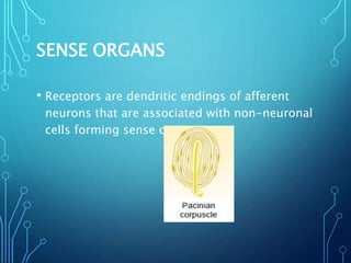
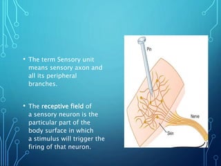
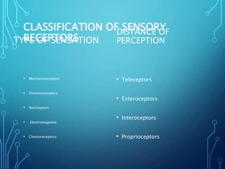
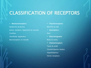
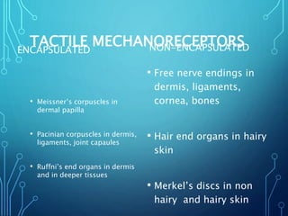
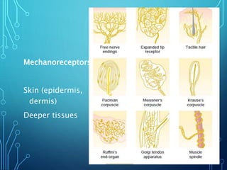
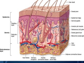
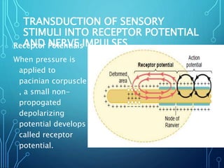
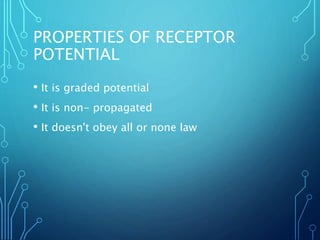
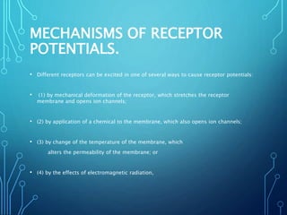
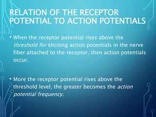
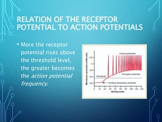
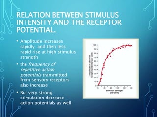
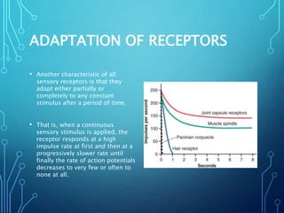
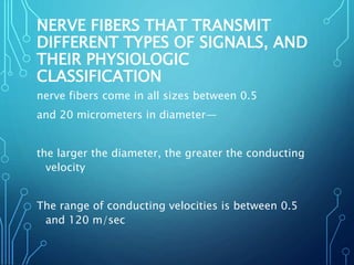
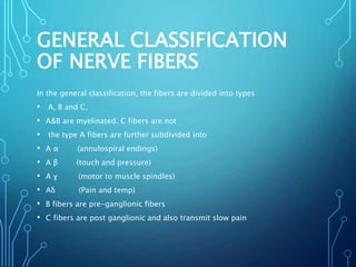
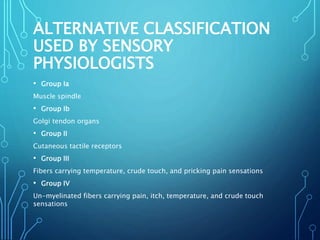
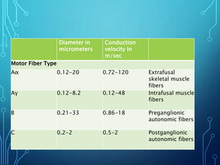
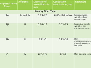
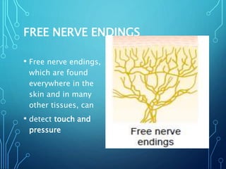
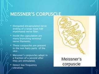
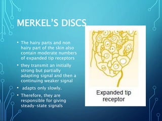
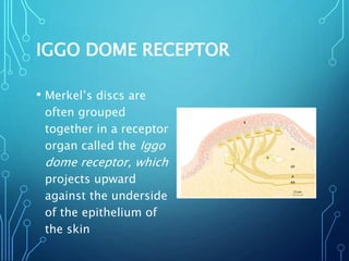
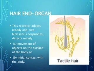
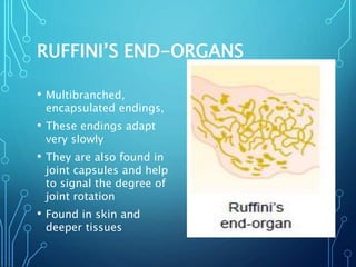
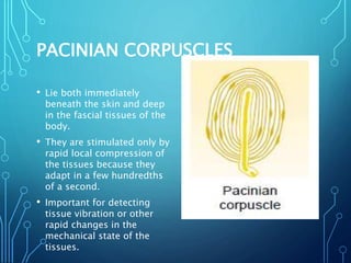
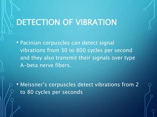
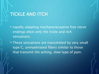
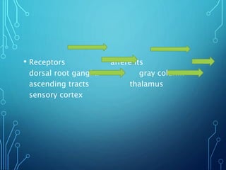
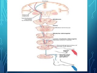
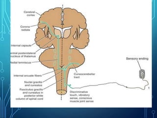
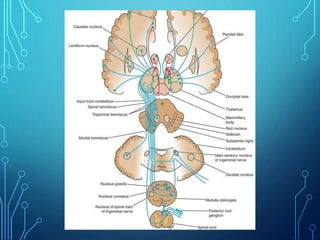
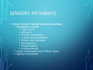
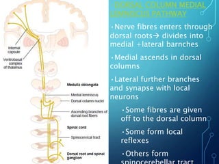
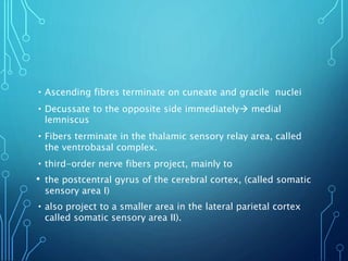
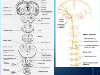
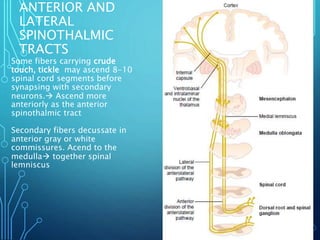
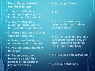
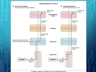
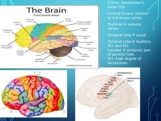
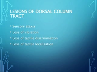
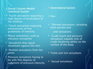
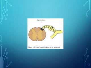
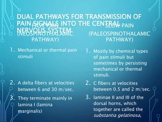
Sensory receptors detect stimuli from the internal and external environment and transmit this information to the central nervous system via sensory neurons. There are several types of sensory receptors that detect different modalities like touch, pressure, vibration, temperature, and pain. Mechanoreceptors include receptors that detect touch and pressure like free nerve endings, Meissner's corpuscles, Merkel's discs, hair receptors, Ruffini endings, and Pacinian corpuscles. Tactile signals are transmitted via myelinated A-beta fibers while pain and itch signals use small diameter C fibers. The dorsal column medial lemniscus pathway transmits tactile, proprioceptive, and vibratory signals from the periphery to the thalamus and som
Receptor by Pandian M, Tutor, Dept of Physiology, DYPMCKOP, MH. This PPT for ...Pandian M
Introduction
SENSORY RECEPTORS
Structurally 3 types of receptors
Transducers
CLASSIFICATION OF RECEPTORS
A. Depending on the source of stimulus(Sherrington’s classification)
B. Depending upon type of stimulus
C. Clinical or anatomical classification of receptors
Production of receptor potential
Properties of receptors
Properties of receptor potential
this ppt shares what synapses are and how information of one neuron is transmitted to other through the synapses. it also includes the properties and plasticity of synaptic transmission
Synapse – Greek word –synaptein. Syn –together; aptein –clasp.
Synapse – Clasping of hands (as in hand shaking between two friends).
Site of functional continuity (transneuronal junctional complex) between two neurons.
Why need of synapse?
A brief overview of the physiology of the neuromuscular junction.It includes a video towards the end sourced from the internet with the copyright watermarks intact.
Nerve conduction studies (NCSs) have become a simple and reliable test of peripheral nerve function.
With adequate standardization, the method now provides a means of not only objectively identifying the lesion but also precisely localizing the site of maximal involvement.
Electrical stimulation of the nerve initiates an impulse that travels along motor or sensory nerve fibers.
The assessment of conduction characteristics depends on the analysis of compound evoked potentials recorded from the muscle in the study of motor fibers and from the nerve itself in the case of sensory fiber
ELECTRICAL STIMULation of the nerve CATHODE AND ANODE: Surface electrodes, usually made of silver plate, come in different sizes, commonly in the range of 0.5 to 1.0 cm in diameter.
Stimulating electrodes consist of a cathode, or negative pole, and an anode, or positive pole, so called because they attract cations and anions.
As the current flows between them, negative charges that accumulate under the cathode, by making inside the axon relatively more positive than outside, depolarize the nerve or cathodal depolarization.
Conversely, positive charges under the anode hyperpolarize the nerve
TYPES OF STIMULATOR
Most commercially available stimulators provide a probe that mounts the cathode and the anode at a fixed distance, usually 2 to 3 cm apart.
The intensity control located in the insulated handle, though bulky, simplifies the operation for a single examiner.
The ordinary banana plugs connected by shielded cable also serve well as stimulating electrodes.
The use of a large diameter electrode for stimulation lowers current density in the skin, causing less pain, although the exact site of nerve activation becomes uncertain.
A monopolar stimulation with a small cathode placed on the nerve trunk and a large anode over the opposite surface in the same limb.
The use of a subcutaneously inserted needle as the cathode reduces the current necessary to excite the nerve compared to surface stimulation.
A surface electrode located on the skin nearby or a second needle electrode inserted in the vicinity of the cathode serves as the anode.
The maximum current during such stimulation causes neither electric nor heat damage to the tissue.
RECORDING OF MUSCLE AND NERVE POTENTIAL
Surface and Needle Electrodes:
Surface electrodes with a larger recording radius serve better than needle electrodes in assessing a compound muscle action potential (CMAP).
Its onset latency indicates the conduction time of the fastest motor fibers, and amplitude, the number of available motor axons.
A needle electrode, despite its small recording radius has its place in identifying the activity from a small muscle when surface recording fails.
Its use also improves segregation of a target activity from neighboring discharges after proximal stimulation, which tends to excite unwanted neighboring nerves simultaneously
Amplifier system
The electrodes convert bioelectric signal resulting from muscle or
The Importance of Community Nursing Care.pdfAD Healthcare
NDIS and Community 24/7 Nursing Care is a specific type of support that may be provided under the NDIS for individuals with complex medical needs who require ongoing nursing care in a community setting, such as their home or a supported accommodation facility.
India Clinical Trials Market: Industry Size and Growth Trends [2030] Analyzed...Kumar Satyam
According to TechSci Research report, "India Clinical Trials Market- By Region, Competition, Forecast & Opportunities, 2030F," the India Clinical Trials Market was valued at USD 2.05 billion in 2024 and is projected to grow at a compound annual growth rate (CAGR) of 8.64% through 2030. The market is driven by a variety of factors, making India an attractive destination for pharmaceutical companies and researchers. India's vast and diverse patient population, cost-effective operational environment, and a large pool of skilled medical professionals contribute significantly to the market's growth. Additionally, increasing government support in streamlining regulations and the growing prevalence of lifestyle diseases further propel the clinical trials market.
Growing Prevalence of Lifestyle Diseases
The rising incidence of lifestyle diseases such as diabetes, cardiovascular diseases, and cancer is a major trend driving the clinical trials market in India. These conditions necessitate the development and testing of new treatment methods, creating a robust demand for clinical trials. The increasing burden of these diseases highlights the need for innovative therapies and underscores the importance of India as a key player in global clinical research.
Defecation
Normal defecation begins with movement in the left colon, moving stool toward the anus. When stool reaches the rectum, the distention causes relaxation of the internal sphincter and an awareness of the need to defecate. At the time of defecation, the external sphincter relaxes, and abdominal muscles contract, increasing intrarectal pressure and forcing the stool out
The Valsalva maneuver exerts pressure to expel faeces through a voluntary contraction of the abdominal muscles while maintaining forced expiration against a closed airway. Patients with cardiovascular disease, glaucoma, increased intracranial pressure, or a new surgical wound are at greater risk for cardiac dysrhythmias and elevated blood pressure with the Valsalva maneuver and need to avoid straining to pass the stool.
Normal defecation is painless, resulting in passage of soft, formed stool
CONSTIPATION
Constipation is a symptom, not a disease. Improper diet, reduced fluid intake, lack of exercise, and certain medications can cause constipation. For example, patients receiving opiates for pain after surgery often require a stool softener or laxative to prevent constipation. The signs of constipation include infrequent bowel movements (less than every 3 days), difficulty passing stools, excessive straining, inability to defecate at will, and hard feaces
IMPACTION
Fecal impaction results from unrelieved constipation. It is a collection of hardened feces wedged in the rectum that a person cannot expel. In cases of severe impaction the mass extends up into the sigmoid colon.
DIARRHEA
Diarrhea is an increase in the number of stools and the passage of liquid, unformed feces. It is associated with disorders affecting digestion, absorption, and secretion in the GI tract. Intestinal contents pass through the small and large intestine too quickly to allow for the usual absorption of fluid and nutrients. Irritation within the colon results in increased mucus secretion. As a result, feces become watery, and the patient is unable to control the urge to defecate. Normally an anal bag is safe and effective in long-term treatment of patients with fecal incontinence at home, in hospice, or in the hospital. Fecal incontinence is expensive and a potentially dangerous condition in terms of contamination and risk of skin ulceration
HEMORRHOIDS
Hemorrhoids are dilated, engorged veins in the lining of the rectum. They are either external or internal.
FLATULENCE
As gas accumulates in the lumen of the intestines, the bowel wall stretches and distends (flatulence). It is a common cause of abdominal fullness, pain, and cramping. Normally intestinal gas escapes through the mouth (belching) or the anus (passing of flatus)
FECAL INCONTINENCE
Fecal incontinence is the inability to control passage of feces and gas from the anus. Incontinence harms a patient’s body image
PREPARATION AND GIVING OF LAXATIVESACCORDING TO POTTER AND PERRY,
An enema is the instillation of a solution into the rectum and sig
One of the most developed cities of India, the city of Chennai is the capital of Tamilnadu and many people from different parts of India come here to earn their bread and butter. Being a metropolitan, the city is filled with towering building and beaches but the sad part as with almost every Indian city
Deep Leg Vein Thrombosis (DVT): Meaning, Causes, Symptoms, Treatment, and Mor...The Lifesciences Magazine
Deep Leg Vein Thrombosis occurs when a blood clot forms in one or more of the deep veins in the legs. These clots can impede blood flow, leading to severe complications.
Telehealth Psychology Building Trust with Clients.pptxThe Harvest Clinic
Telehealth psychology is a digital approach that offers psychological services and mental health care to clients remotely, using technologies like video conferencing, phone calls, text messaging, and mobile apps for communication.
ICH Guidelines for Pharmacovigilance.pdfNEHA GUPTA
The "ICH Guidelines for Pharmacovigilance" PDF provides a comprehensive overview of the International Council for Harmonisation of Technical Requirements for Pharmaceuticals for Human Use (ICH) guidelines related to pharmacovigilance. These guidelines aim to ensure that drugs are safe and effective for patients by monitoring and assessing adverse effects, ensuring proper reporting systems, and improving risk management practices. The document is essential for professionals in the pharmaceutical industry, regulatory authorities, and healthcare providers, offering detailed procedures and standards for pharmacovigilance activities to enhance drug safety and protect public health.
Global launch of the Healthy Ageing and Prevention Index 2nd wave – alongside...ILC- UK
The Healthy Ageing and Prevention Index is an online tool created by ILC that ranks countries on six metrics including, life span, health span, work span, income, environmental performance, and happiness. The Index helps us understand how well countries have adapted to longevity and inform decision makers on what must be done to maximise the economic benefits that comes with living well for longer.
Alongside the 77th World Health Assembly in Geneva on 28 May 2024, we launched the second version of our Index, allowing us to track progress and give new insights into what needs to be done to keep populations healthier for longer.
The speakers included:
Professor Orazio Schillaci, Minister of Health, Italy
Dr Hans Groth, Chairman of the Board, World Demographic & Ageing Forum
Professor Ilona Kickbusch, Founder and Chair, Global Health Centre, Geneva Graduate Institute and co-chair, World Health Summit Council
Dr Natasha Azzopardi Muscat, Director, Country Health Policies and Systems Division, World Health Organisation EURO
Dr Marta Lomazzi, Executive Manager, World Federation of Public Health Associations
Dr Shyam Bishen, Head, Centre for Health and Healthcare and Member of the Executive Committee, World Economic Forum
Dr Karin Tegmark Wisell, Director General, Public Health Agency of Sweden
Antibiotic Stewardship by Anushri Srivastava.pptxAnushriSrivastav
Stewardship is the act of taking good care of something.
Antimicrobial stewardship is a coordinated program that promotes the appropriate use of antimicrobials (including antibiotics), improves patient outcomes, reduces microbial resistance, and decreases the spread of infections caused by multidrug-resistant organisms.
WHO launched the Global Antimicrobial Resistance and Use Surveillance System (GLASS) in 2015 to fill knowledge gaps and inform strategies at all levels.
ACCORDING TO apic.org,
Antimicrobial stewardship is a coordinated program that promotes the appropriate use of antimicrobials (including antibiotics), improves patient outcomes, reduces microbial resistance, and decreases the spread of infections caused by multidrug-resistant organisms.
ACCORDING TO pewtrusts.org,
Antibiotic stewardship refers to efforts in doctors’ offices, hospitals, long term care facilities, and other health care settings to ensure that antibiotics are used only when necessary and appropriate
According to WHO,
Antimicrobial stewardship is a systematic approach to educate and support health care professionals to follow evidence-based guidelines for prescribing and administering antimicrobials
In 1996, John McGowan and Dale Gerding first applied the term antimicrobial stewardship, where they suggested a causal association between antimicrobial agent use and resistance. They also focused on the urgency of large-scale controlled trials of antimicrobial-use regulation employing sophisticated epidemiologic methods, molecular typing, and precise resistance mechanism analysis.
Antimicrobial Stewardship(AMS) refers to the optimal selection, dosing, and duration of antimicrobial treatment resulting in the best clinical outcome with minimal side effects to the patients and minimal impact on subsequent resistance.
According to the 2019 report, in the US, more than 2.8 million antibiotic-resistant infections occur each year, and more than 35000 people die. In addition to this, it also mentioned that 223,900 cases of Clostridoides difficile occurred in 2017, of which 12800 people died. The report did not include viruses or parasites
VISION
Being proactive
Supporting optimal animal and human health
Exploring ways to reduce overall use of antimicrobials
Using the drugs that prevent and treat disease by killing microscopic organisms in a responsible way
GOAL
to prevent the generation and spread of antimicrobial resistance (AMR). Doing so will preserve the effectiveness of these drugs in animals and humans for years to come.
being to preserve human and animal health and the effectiveness of antimicrobial medications.
to implement a multidisciplinary approach in assembling a stewardship team to include an infectious disease physician, a clinical pharmacist with infectious diseases training, infection preventionist, and a close collaboration with the staff in the clinical microbiology laboratory
to prevent antimicrobial overuse, misuse and abuse.
to minimize the developme
Navigating Challenges: Mental Health, Legislation, and the Prison System in B...Guillermo Rivera
This conference will delve into the intricate intersections between mental health, legal frameworks, and the prison system in Bolivia. It aims to provide a comprehensive overview of the current challenges faced by mental health professionals working within the legislative and correctional landscapes. Topics of discussion will include the prevalence and impact of mental health issues among the incarcerated population, the effectiveness of existing mental health policies and legislation, and potential reforms to enhance the mental health support system within prisons.
2. • Student should be able to
• understand the types of sensory receptors
• enumerate and understand the properties of
receptors
3. SENSORY RECEPTORS
• Information about the internal and external environment activates
the CNS via a variety of sensory receptors.
• These receptors are transducers that convert various forms of
energy in the environment into action potentials in neurons.
• Stimulus is an event or particular form of energy that evokes a
specific functional reaction in an organ or receptor. (mechanical,
chemical, EMG, temp)
4. SENSE ORGANS
• Receptors are dendritic endings of afferent
neurons that are associated with non-neuronal
cells forming sense organs.
5. • The term Sensory unit
means sensory axon and
all its peripheral
branches.
• The receptive field of
a sensory neuron is the
particular part of the
body surface in which
a stimulus will trigger the
firing of that neuron.
6. CLASSIFICATION OF SENSORY
RECEPTORSTYPE OF SENSATION
• Mechanoreceptors
• Thermoreceptors
• Nociceptors
• Electromagnetic
• Chemoreceptors
DISTANCE OF
PERCEPTION
• Teleceptors
• Exteroceptors
• Interoceptors
• Proprioceptors
8. TACTILE MECHANORECEPTORSENCAPSULATED
• Meissner’s corpuscles in
dermal papilla
• Pacinian corpuscles in dermis,
ligaments, joint capaules
• Ruffni’s end organs in dermis
and in deeper tissues
NON-ENCAPSULATED
• Free nerve endings in
dermis, ligaments,
cornea, bones
• Hair end organs in hairy
skin
• Merkel’s discs in non
hairy and hairy skin
12. MODALITY OF SENSATION
• Differential Sensitivity of Receptors for particular
stimulus or specific energy for which it is designed
• When nerve fiber from the receptors is stimulated the
perception is that for which the receptor is specialized,
no matter where and how that nerve is stimulated. This
is Muller’s law of specific energy.
• The specificity of nerve fibers for transmitting only one
modality of sensation is called the labeled line principle.
13. EXPLANNATION
• Each nerve tract terminates at a specific point in the
central nervous system, and the type of sensation felt
when a nerve fiber is stimulated is determined by the
point in the nervous system to which the fiber leads.
• For instance if a pain fiber is stimulated, the person
perceives pain regardless of what type of stimulus
excites the fiber.
• Likewise, if a touch fiber is stimulated by electrical
excitation of a touch receptor or in any other way, the
person perceives touch.
14. LAW OF PROJECTION
• No matter which part of a pathway you
stimulate between the receptor and the brain
center, your brain will locate the stimulus
where receptors are (e.g. phantom limb)
• reorganization of brain map in somatosensory
cortex
• Some believe it can be blocked by local
anesthetics in spinal cord.
15. • A phantom limb is the sensation that an amputated
or missing limb is still attached to the body.
• Approximately 60 to 80% of individuals with an
amputation experience phantom sensations in their
amputated limb, and the majority of the sensations
are painful.
• Phantom sensations may also occur after the
removal of body parts e.g. after amputation of the
breast, extraction of a tooth (phantom tooth pain)
or removal of an eye (phantom eye syndrome)
16. DALE’S LAW
• At one type of synapse only one type of
neurotransmitter is released
17.
18. TRANSDUCTION OF SENSORY
STIMULI INTO RECEPTOR POTENTIAL
AND NERVE IMPULSESReceptor Potentials
When pressure is
applied to
pacinian corpuscle
, a small non-
propogated
depolarizing
potential develops
called receptor
potential.
20. MECHANISMS OF RECEPTOR
POTENTIALS.
• Different receptors can be excited in one of several ways to cause receptor potentials:
• (1) by mechanical deformation of the receptor, which stretches the receptor
membrane and opens ion channels;
• (2) by application of a chemical to the membrane, which also opens ion channels;
• (3) by change of the temperature of the membrane, which
alters the permeability of the membrane; or
• (4) by the effects of electromagnetic radiation,
21.
22. RELATION OF THE RECEPTOR
POTENTIAL TO ACTION POTENTIALS
• When the receptor potential rises above the
threshold for eliciting action potentials in the nerve
fiber attached to the receptor, then action potentials
occur.
• More the receptor potential rises above the
threshold level, the greater becomes the action
potential frequency.
23. RELATION OF THE RECEPTOR
POTENTIAL TO ACTION POTENTIALS
• More the receptor
potential rises above
the threshold level,
the greater becomes
the action potential
frequency.
24. RELATION BETWEEN STIMULUS
INTENSITY AND THE RECEPTOR
POTENTIAL.
• Amplitude increases
rapidly and then less
rapid rise at high stimulus
strength
• the frequency of
repetitive action
potentials transmitted
from sensory receptors
also increase
• But very strong
stimulation decrease
action potentials as well
25. ADAPTATION OF RECEPTORS
• Another characteristic of all
sensory receptors is that they
adapt either partially or
completely to any constant
stimulus after a period of time.
• That is, when a continuous
sensory stimulus is applied, the
receptor responds at a high
impulse rate at first and then at a
progressively slower rate until
finally the rate of action potentials
decreases to very few or often to
none at all.
26. MECHANISMS BY WHICH RECEPTORS
ADAPT
• part of the adaptation results from
readjustments in the structure of the receptor
itself,
• and part from an electrical type of
accommodation in the terminal nerve fibril
27. SLOWLY AND RAPIDLY
ADAPTING RECEPTORS
• Slowly Adapting Receptors Detect
Continuous Stimulus Strength—The
“Tonic” Receptors.
• impulses from the muscle spindles and
Golgi tendon apparatuses allow the
nervous system to know the status of
muscle contraction
• Receptors of the macula in the
vestibular apparatuses
• pain receptors
• baroreceptors of the arterial tree
• chemoreceptors of the carotid and
aortic bodies.
• Rapidly Adapting Receptors Detect
Change in Stimulus Strength—The “Rate
Receptors,” “Movement Receptors,” or
“Phasic Receptors.”
• Pacinian and meissner’s
• Importance of the Rate Receptors—
Their Predictive Function.
29. • To classify nerve fibers
• To understand the properties and differences
of nerve fibers
30. NERVE FIBERS THAT TRANSMIT
DIFFERENT TYPES OF SIGNALS, AND
THEIR PHYSIOLOGIC
CLASSIFICATION
nerve fibers come in all sizes between 0.5
and 20 micrometers in diameter—
the larger the diameter, the greater the conducting
velocity
The range of conducting velocities is between 0.5
and 120 m/sec
31. GENERAL CLASSIFICATION
OF NERVE FIBERS
In the general classification, the fibers are divided into types
• A, B and C,
• A&B are myelinated. C fibers are not
• the type A fibers are further subdivided into
• A α (annulospiral endings)
• A β (touch and pressure)
• A ɣ (motor to muscle spindles)
• Aδ (Pain and temp)
• B fibers are pre-ganglionic fibers
• C fibers are post ganglionic and also transmit slow pain
32. ALTERNATIVE CLASSIFICATION
USED BY SENSORY
PHYSIOLOGISTS
• Group Ia
Muscle spindle
• Group Ib
Golgi tendon organs
• Group II
Cutaneous tactile receptors
• Group III
Fibers carrying temperature, crude touch, and pricking pain sensations
• Group IV
Un-myelinated fibers carrying pain, itch, temperature, and crude touch
sensations
33. Diameter in
micrometers
Conduction
velocity in
m/sec
Motor Fiber Type
Aα 0.12-20 0.72-120 Extrafusal
skeletal muscle
fibers
Aγ 0.12-8.2 0.12-48 Intrafusal muscle
fibers
B 0.21-33 0.86-18 Preganglionic
autonomic fibers
C 0.2-2 0.5-2 Postganglionic
autonomic fibers
34. Peripheral nerve
fibers
Afferents Diameter of
nerve fibers in
mm
Conduction
velocity in m/sec
Receptors
Sensory Fiber Type
Aα Ia and Ib 0.13-20 0.80-120 m/sec Primary muscle
spindles, Golgi
tendon organ
Aβ II 0.16-12 0.35-75 Secondary muscle
spindles, skin
mechanoreceptors
Aδ III 0.11-5 0.15-30 Skin
mechanoreceptors,
thermal receptors,
fast pain
C IV 0.2-1.5 0.5-2 Slow pain and temp
38. CONVERGENCE OF SIGNALS
• Convergence means signals from multiple inputs uniting
to excite a single neuron.
• Convergence from a single source. That is, multiple
terminals from a single incoming fiber tract terminate
on the same neuron.
• Convergence can also result from input signals
(excitatory or inhibitory) from multiple sources
41. Classification of Sensations
• The somatic senses are the nervous mechanisms
that collect sensory information from all over the
body.
• Special senses, which mean specifically vision,
hearing, smell, taste and equilibrium.
42. CLASSIFICATION OF SOMATIC
SENSES
• The mechanoreceptive somatic senses,
which include both tactile and position
sensations.
• The thermoreceptive senses, which detect
heat and cold
• The pain sense, which is activated by any
factor that damages the tissues.
43. MECHANORECEPTIVE SOMATIC
SENSES
• The tactile senses include touch, pressure,
vibration, and tickle
• the position senses include static position and rate
of movement
44. DETECTION AND TRANSMISSION OF
TACTILE SENSATIONS
• Touch, pressure, and vibration are all
detected by the same types of receptors
called tactile receptors.
• There are at least six entirely different
types of tactile receptors
45. FREE NERVE ENDINGS
• Free nerve endings,
which are found
everywhere in the
skin and in many
other tissues, can
• detect touch and
pressure
46. MEISSNER’S CORPUSCLE
• Elongated encapsulated nerve
ending of a large (type AB)
myelinated nerve fiber.
• Inside the capsulation are
many branching terminal
nerve filaments.
• These corpuscles are present
in the non hairy parts of the
skin .
• Meissner’s corpuscles adapt in
a fraction of a second after
they are stimulated,
• Detect low frequency
vibration.
47. MERKEL’S DISCS
• The hairy parts and non
hairy part of the skin also
contain moderate numbers
of expanded tip receptors
• they transmit an initially
strong but partially
adapting signal and then a
continuing weaker signal
• adapts only slowly.
• Therefore, they are
responsible for giving
steady-state signals
48. IGGO DOME RECEPTOR
• Merkel’s discs are
often grouped
together in a receptor
organ called the Iggo
dome receptor, which
projects upward
against the underside
of the epithelium of
the skin
49. HAIR END-ORGAN
• This receptor adapts
readily and, like
Meissner’s corpuscles,
detects mainly
• (a) movement of
objects on the surface
of the body or
• (b) initial contact with
the body.
50. RUFFINI’S END-ORGANS
• Multibranched,
encapsulated endings,
• These endings adapt
very slowly
• They are also found in
joint capsules and help
to signal the degree of
joint rotation
• Found in skin and
deeper tissues
51. PACINIAN CORPUSCLES
• Lie both immediately
beneath the skin and deep
in the fascial tissues of the
body.
• They are stimulated only by
rapid local compression of
the tissues because they
adapt in a few hundredths
of a second.
• Important for detecting
tissue vibration or other
rapid changes in the
mechanical state of the
tissues.
52. • Position sense
1. Static
2. Rate of movement called kinesthesia
• Receptors
1. Muscle spindles
2. Golgi tendon organs
3. Ruffni’s endings
4. Pacinian
54. TRANSMISSION OF TACTILE SIGNALS
IN PERIPHERAL NERVE FIBERS
• Transmit their signals in type A-beta nerve fibers
that have transmission velocities ranging from 30
to 70 m/sec.
• free nerve ending tactile receptors transmit signals
mainly by way of the small type A-delta myelinated
fibers that conduct at velocities of only 5 to 30
m/sec.
• Some tactile free nerve endings transmit by way of
type C unmyelinated fibers (up to 2 m/sec;) these
send signals into the spinal cord and lower brain
stem, mainly the sensation of tickle.
55. DETECTION OF VIBRATION
• Pacinian corpuscles can detect signal
vibrations from 30 to 800 cycles per second
and they also transmit their signals over type
A-beta nerve fibers.
• Meissner’s corpuscles detect vibrations from 2
to 80 cycles per seconds
56. TICKLE AND ITCH
• rapidly adapting mechanoreceptive free nerve
endings elicit only the tickle and itch
sensations.
• These sensations are transmitted by very small
type C, unmyelinated fibers similar to those
that transmit the aching, slow type of pain.
62. •DORSAL COLUMN MEDIAL
LEMNISCUS PATHWAY
•Nerve fibres enters through
dorsal roots divides into
medial +lateral barnches
•Medial ascends in dorsal
columns
•Lateral further branches
and synapse with local
neurons
•Some fibres are given
off to the dorsal column
•Some form local
reflexes
•Others form
63. •Ascending fibres terminate on cuneate and gracile nuclei
•Decussate to the opposite side immediately medial
lemniscus
•Fibers terminate in the thalamic sensory relay area, called
the ventrobasal complex.
•third-order nerve fibers project, mainly to
• the postcentral gyrus of the cerebral cortex, (called somatic
sensory area I)
•also project to a smaller area in the lateral parietal cortex
called somatic sensory area II).
64.
65. • the fibers from the lower parts
of the body lie toward the
center of the cord, whereas
new fibres enter laterally.
• In the thalamus, the tail end of
the body represented by the
most lateral portions of the
ventrobasal complex and the
head and face represented by
medial areas of the complex.
• because of the crossing of the
medial lemnisci in the medulla,
the left side of the body is
represented in the right side of
the thalamus, and the right
side of the body in the left
side of the thalamus.
Spatial Orientation
66.
67. ANTEROLATERAL SYSTEM
Transmits sensory signals that DO NOT require highly
discrete localization
• Composed of smaller, myelinated nerve fibers
(transmission speeds 40 m/sec)
• Less spatial orientation
• Sensory modalities – wide range
• Comprise of:
• Anterior spinothalamic tract
• Lateral spinothalamic tract
68. ANTEROLATERAL SYSTEM
• Pain
• Thermal sensations
(including warmth & cold)
• Crude touch and pressure
sensations
• Tickle and itch sensations
• Sexual sensations
69. •From receptors (free nerve
ending enter dorsal horn
(cell bodies of these first
order neurons are in the
dorsal root ganglion)
•Synapse with 2nd order
within 1-2 segments
•Second order neurons
dorsal horn (at all levels).
Their axons decussate in the
anterior white and gray
commisure
•Joined by trigeminothalmic
fibers in medulla, send
collaterals to reticular
formation (alertness to pain)
70. •3rd order neurons VPL
nucleus of thalamus. Their
fibres ascend through the
posterior limb of internal
capsule corona radiate
•4th order neurons of the
cerebral cortex (Broadmans
areas 3,1.2
Somatosensory cortex)
•Some fibers from the
second order neuron
decussate and ascend as
anterior spinothalmic tract
•Both anterior & lateral
spinothalamic tracts unite
in MO forming spinal
lemniscus
71. ANTERIOR AND
LATERAL
SPINOTHALMIC
TRACTS
Some fibers carrying crude
touch, tickle may ascend 8-10
spinal cord segments before
synapsing with secondary
neurons. Ascend more
anteriorly as the anterior
spinothalmic tract
Secondary fibers decussate in
anterior gray or white
commissures. Acend to the
medulla together spinal
lemniscus
72. • Dorsal Column–Medial
Lemniscal System
• 1. Touch sensations
requiring a high degree of
localization of the stimulus
• 2. Touch sensations
requiring transmission of
fine gradations of intensity
• 3. Phasic sensations, such as
vibratory sensations
• 4. Sensations that signal
movement against the skin
• 5. Position sensations from
the joints
• 6. Pressure sensations
having to do with fine
degrees of judgment of
pressure intensity
• Anterolateral System
• 1. Pain
• 2. Thermal sensations,
including both warmth and
cold sensations
• 3. Crude touch and pressure
sensations capable only of
crude localizing ability on
the surface of the body
• 4. Tickle and itch sensations
• 5. Sexual sensations
73.
74.
75. Cortex: Broadmann’s
areas (50)
Central fissure: anterior
to it motor cortex
Posterior sensory
cortex
Occipital lobe visual
Tempral Lobe Auditory
SS1 and SS2
Located aneterior part
of parietal lobe
SS1-high degree of
localization
79. FUNCTIONS OF
SOMATOSENSORY AREA I
• Localize and discriminate
discretely the different
sensations in the different
parts of the body
• judge critical degrees of
pressure against the body
• judge the texture,
BILATERAL EXCISION OF
SOMATOSENSORY AREA
Pt is unable to
• Localize and discriminate discretely the
different sensations in the different parts of the
body
• judge critical degrees of pressure against the
body
• judge the texture, weights, shapes or forms of
objects. This is called astereognosis.
• Judge the shape by drawing. Agraphaesthesia
80. Effect of Removing the Somatosensory Association
Area—(5,7) Amorphosynthesis
• The person loses ability to recognize
complex objects and complex forms felt on
the opposite side of the body.
• He or she loses most of the sense of form of
his or her own body parts on the opposite
side and forgets that it is there .
• When feeling objects, the person tends to
recognize only one side of the object and
forgets that the other side even exists.
81. LESIONS OF DORSAL COLUMN
TRACT
• Sensory ataxia
• Loss of vibration
• Loss of tactile discrimination
• Loss of tactile localization
82. • Dorsal Column–Medial
Lemniscal System
• Touch sensations requiring a
high degree of localization of
the stimulus
• Touch sensations requiring
transmission of fine
gradations of intensity
• Phasic sensations, such as
vibratory sensations
• Sensations that signal
movement against the skin
• Position sensations from the
joints
• Pressure sensations having to
do with fine degrees of
judgment of pressure intensity
• Anterolateral System
• Pain
• Thermal sensations, including
both warmth and
cold sensations
• Crude touch and pressure
sensations capable only of
crude localizing ability on the
surface of the body
• Tickle and itch sensations
• Sexual sensations
87. PAIN IS A PROTECTIVE
MECHANISM
• Pain occurs whenever any tissues are being
damaged, and it causes the individual to react
to remove the pain stimulus.
88. TYPES OF PAIN
• Fast Pain
• Slow Pain
• Fast pain is felt within about 0.1 second after a
pain stimulus is applied
• Slow pain begins after 1 second or more and then
increases slowly over many seconds and
sometimes even minutes.
89. QUALITIES OF PAIN
• Fast pain is also described by many alternative
names, such as sharp pain, pricking pain,
acute pain, and electric pain
• Slow pain also goes by many names, such as
slow burning pain, aching pain, throbbing
pain, nauseous pain, and chronic pain
90. PAIN RECEPTORS
• Are Free Nerve Endings
• They are widespread in the superficial layers of
the skin as well as in certain internal tissues,
such as the periosteum, the arterial walls, the
joint surfaces, and the falx and tentorium in
the cranial vault.
• Non-adapting Nature of Pain Receptors
91. Three Types of Stimuli Excite Pain Receptors—
• Mechanical
• Thermal
• Chemical
92. • Some of the chemicals that excite the chemical
type of pain are bradykinin, serotonin,
histamine, potassium ions, acids,
acetylcholine, and proteolytic enzymes
• In addition, prostaglandins and substance P
enhance the sensitivity of pain endings but do
not directly excite them
93. DUAL PATHWAYS FOR
TRANSMISSION OF PAIN SIGNALS
INTO THE CENTRAL NERVOUS
SYSTEM
• Peripheral Pain Fibers—
• “Fast” (A delta) and “Slow” Fibers (C fibers)
• The two pathways mainly correspond to the two
types of pain—
• a fast-sharp pain pathway and
• a slow-chronic pain pathway.
94. DUAL PATHWAYS FOR TRANSMISSION OF
PAIN SIGNALS INTO THE CENTRAL
NERVOUS SYSTEM
FAST PAIN
(NEOSPINOTHALAMIC
PATHWAY)
1. Mechanical or thermal pain
stimuli
2. A delta fibers at velocities
between 6 and 30 m/sec.
3. They terminate mainly in
lamina I (lamina
marginalis)
SLOW PAIN
(PALEOSPINOTHALAMIC
PATHWAY)
1. Mostly by chemical types
of pain stimuli but
sometimes by persisting
mechanical or thermal
stimuli.
2. C fibers at velocities
between 0.5 and 2 m/sec.
3. laminae II and III of the
dorsal horns, which
together are called the
substantia gelatinosa,
95. Fast pain
4. most pass all the way to the
thalamus (Ventrobasal)
• Project to somatosensory cortex.
5. can be localized much more
exactly in the different parts of
the body than can slow-chronic
pain.
6. Glutamate, the Probable
Neurotransmitter of the Fast
Pain Fibers
SLOW PAIN
4.most terminate in one of
three areas:
(a) reticular nuclei
(b) tectal area of the
mesencephalon
(c) periaqueductal gray region
(d) Intralaminar nuclei and
hypothalamus
5.Poorly localized.
6.Type C pain fiber terminals
entering the spinal cord
96. Pain Suppression (“Analgesia”) System in the Brain
and Spinal Cord
• Inhibition of pain signals at spinal cord by
descending brain fibers
• Encephalin secreting neurons in cord and brain stem
• Inhibition of pain fibers by tactile incoming fibers
97.
98. Acupuncture analgesia (AA)
• technique of relieving pain by
inserting and manipulating
threadlike needles at key points
• acupuncture endorphin hypothesis
• Acupuncture needles activate
specific afferent nerve
fibersCNSblocking pain
transmission at both
• the spinal-cord and brain levels
through use of endorphins and
closely related endogenous opiates.
99.
100.
101. • The periaqueductal gray and periventricular
areas of the mesencephalon and upper pons
surround the aqueduct of Sylvius and portions
of the third and fourth ventricles. Neurons from
these areas send signals to
• The raphe magnus nucleus, a thin midline
nucleus located in the lower pons and upper
medulla, and the nucleus reticularis
paragigantocellularis, located laterally in the
medulla. From these nuclei, second-order
signals are transmitted down the dorsolateral
columns in the spinal cord to
• Pain inhibitory complex located in the dorsal
horns of the spinal cord.
102. INHIBITION OF PAIN TRANSMISSION BY
SIMULTANEOUS TACTILE SENSORY
SIGNALS
• Stimulation of large type A beta sensory fibers
from peripheral tactile receptors can depress
transmission of pain signals from the same body
area.
• This presumably results from local lateral
inhibition in
the spinal cord.
• It explains why such simple maneuvers as rubbing
the skin near painful areas is often effective in
relieving pain.
• It also explains why liniments are often useful for
pain relief.
103. • Allodynia
Non painful stimulus causes pain (Lesion of VPL of
thalamus)
• Hyperalgesia
Hypersensitivity to pain
A pain nervous pathway sometimes becomes excessively
excitable;.
• Possible causes of hyperalgesia are
• Excessive sensitivity of the pain receptors themselves,
which is called primary hyperalgesia (Burns)
104. REFERRED PAIN
• Often a person feels pain in a part of the body
that is fairly remote from the tissue causing
the pain. This is called referred pain. For
instance, pain in one of the visceral organs
often is referred to an area on the body
surface.
107. VISCERAL PAIN AND SURFACE
PAIN
VISCERAL PAIN
• Highly localized types of
damage to the viscera seldom
cause severe pain.
• True visceral pain is
transmitted via pain sensory
fibers within the autonomic
nerve bundles, and the
sensations are referred to
surface areas of the body
often far from the painful
organ.
SURFACE
PAIN/PARIETAL PAIN
• Parietal sensations are
conducted directly into
local spinal nerves from
the parietal
peritoneum, pleura, or
pericardium, and these
sensations are usually
localized over painful
area.
109. CAUSES OF TRUE VISCERAL PAIN
• Any stimulus that excites pain nerve endings in diffuse areas of the viscera
can cause visceral pain.
• Such stimuli include
• ischemia of visceral tissue,
• chemical damage to the surfaces of the viscera,
• spasm of the smooth muscle of a hollow viscus,
• excess distention of a hollow viscus, and stretching of the connective
tissue surrounding or within the viscus.
• Essentially all visceral pain that originates in the thoracic and abdominal
cavities is transmitted through small type C pain fibers and, therefore, can
transmit only the chronic-aching-suffering type of pain.
110. Hyperalgesia
• A pain nervous pathway sometimes becomes
excessively excitable; this gives rise to hyperalgesia,
which means hypersensitivity to pain.
• Possible causes of hyperalgesia are
• (1) excessive sensitivity of the pain receptors
themselves, which is called primary hyperalgesia
• (2) facilitation of sensory transmission, which is
called secondary hyperalgesia.
111. • Herpes Zoster (Shingles)
• Herpesvirus infects a dorsal root ganglion.
• This causes severe pain in the dermatomal segment
served by the ganglion, thus eliciting a segmental
type of pain that circles halfway around the body.
• The disease is called herpes zoster, or “shingles,”
because of a skin eruption that often ensues.
112.
113. • Tic Douloureux
• Lancinating pain occasionally occurs in some people
over one side of the face in the sensory distribution
area (or part of the area) of the fifth or ninth nerves;
This phenomenon is called tic douloureux
(trigeminal neuralgia or glossopharyngeal
neuralgia).
• The pain feels like sudden electrical shocks, and it
may appear for only a few seconds at a time or may
be almost continuous.
114.
115.
116. • Headache
• Headaches are a type of pain referred to the surface
of the head from deep head structures.
• Pain-Sensitive Areas in Cranial Vault.
• Tugging on the venous sinuses around the brain,
• Damaging the tentorium,
• Stretching the dura at the base of the brain can
cause intense pain that is recognized as headache
• Middle meningeal artery is a pain sensitive structure
117.
118. TYPES OF INTRACRANIAL
HEADACHE
• Brain tumors
• Headache of Meningitis.
• Headache Caused by
Low Cerebrospinal Fluid
Pressure.
• Migraine Headache
• Alcoholic Headache
TYPES OF EXTRACRANIAL
HEADACHE
• Headache Resulting
from Muscle Spasm.
• Headache Caused by
Irritation of Nasal and
Accessory Nasal
Structures.
• Headache Caused by
Eye Disorders
119. THERMAL SENSATIONS
• The human being can perceive different gradations of
cold and heat,
• from freezing cold
• cold
• cool
• Indifferent
• to warm
• hot
• burning hot.
120. • Thermal gradations are discriminated by at
least three types of sensory receptors:
• cold receptors,
• warmth receptors,
• and pain receptors.
121.
122. BROWN-SÉQUARD SYNDROME
• If the spinal cord is transected on only one side, the Brown-Séquard syndrome occurs.
• All motor functions are blocked on the side of the transection in all segments below
the level of the transection and at the level.
• The sensations of pain, heat, and cold— sensations served by the spinothalamic
pathway—are lost on the opposite side of the body in all dermatomes two to six
segments below the level of the transection.
• The dorsal and dorsolateral columns—kinesthetic and position sensations, vibration
sensation, discrete localization, and two-point discrimination—are lost on the side of
the transection in all dermatomes below the level of the transection.
123. BROWN-SÉQUARD SYNDROME
At the level Below the
level
Above the
level
LMN paralysis same side UMN paralysis same
side
Cutaneous anesthesia in
the dermatomal
segment of ipsilateral
side
Loss of pain and temp
of contralateral side 2
to 5 segments below
the level of lesion
Altered sensations
Loss of vibration, fine
touch, tactile
localization and
discrimination in all