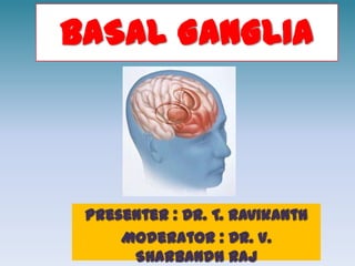
The basal ganglia are a group of interconnected brain structures that include the caudate nucleus, putamen, globus pallidus, subthalamic nucleus, and substantia nigra. They are involved in regulating movement and certain movement disorders. The basal ganglia receive input from the cortex and thalamus and send output to the thalamus and brainstem areas. Dopamine from the substantia nigra helps modulate input and output signaling in the basal ganglia circuits. Disorders of these circuits can result in dyskinesias like tremors, chorea, athetosis, and ballismus or disturbances of muscle tone.
Largest part of hind brain.
Called “ silent area/Little Brain ”
Weight- 150 gms.
Cerebellar cortex is a large folded sheet, each fold is called Folium.
Connected to brain stem by 3 pairs of peduncles- Superior (Brachium conjunctiva), Middle (Brachium Pontis) & Inferior (Restiform body) peduncle.
Largest part of hind brain.
Called “ silent area/Little Brain ”
Weight- 150 gms.
Cerebellar cortex is a large folded sheet, each fold is called Folium.
Connected to brain stem by 3 pairs of peduncles- Superior (Brachium conjunctiva), Middle (Brachium Pontis) & Inferior (Restiform body) peduncle.
Functional Anatomy & physiology of the Basal nucleiRafid Rashid
Provides a good description of the functional anatomy & physiology of the basal nuclei/ basal ganglia for undergraduate medical students. It also describes disorders of the basal ganglia like parkinsonism & chorea.
Factory Supply Best Quality Pmk Oil CAS 28578–16–7 PMK Powder in Stockrebeccabio
Factory Supply Best Quality Pmk Oil CAS 28578–16–7 PMK Powder in Stock
Telegram: bmksupplier
signal: +85264872720
threema: TUD4A6YC
You can contact me on Telegram or Threema
Communicate promptly and reply
Free of customs clearance, Double Clearance 100% pass delivery to USA, Canada, Spain, Germany, Netherland, Poland, Italy, Sweden, UK, Czech Republic, Australia, Mexico, Russia, Ukraine, Kazakhstan.Door to door service
Hot Selling Organic intermediates
Anti ulcer drugs and their Advance pharmacology ||
Anti-ulcer drugs are medications used to prevent and treat ulcers in the stomach and upper part of the small intestine (duodenal ulcers). These ulcers are often caused by an imbalance between stomach acid and the mucosal lining, which protects the stomach lining.
||Scope: Overview of various classes of anti-ulcer drugs, their mechanisms of action, indications, side effects, and clinical considerations.
Prix Galien International 2024 Forum ProgramLevi Shapiro
June 20, 2024, Prix Galien International and Jerusalem Ethics Forum in ROME. Detailed agenda including panels:
- ADVANCES IN CARDIOLOGY: A NEW PARADIGM IS COMING
- WOMEN’S HEALTH: FERTILITY PRESERVATION
- WHAT’S NEW IN THE TREATMENT OF INFECTIOUS,
ONCOLOGICAL AND INFLAMMATORY SKIN DISEASES?
- ARTIFICIAL INTELLIGENCE AND ETHICS
- GENE THERAPY
- BEYOND BORDERS: GLOBAL INITIATIVES FOR DEMOCRATIZING LIFE SCIENCE TECHNOLOGIES AND PROMOTING ACCESS TO HEALTHCARE
- ETHICAL CHALLENGES IN LIFE SCIENCES
- Prix Galien International Awards Ceremony
These simplified slides by Dr. Sidra Arshad present an overview of the non-respiratory functions of the respiratory tract.
Learning objectives:
1. Enlist the non-respiratory functions of the respiratory tract
2. Briefly explain how these functions are carried out
3. Discuss the significance of dead space
4. Differentiate between minute ventilation and alveolar ventilation
5. Describe the cough and sneeze reflexes
Study Resources:
1. Chapter 39, Guyton and Hall Textbook of Medical Physiology, 14th edition
2. Chapter 34, Ganong’s Review of Medical Physiology, 26th edition
3. Chapter 17, Human Physiology by Lauralee Sherwood, 9th edition
4. Non-respiratory functions of the lungs https://academic.oup.com/bjaed/article/13/3/98/278874
Ethanol (CH3CH2OH), or beverage alcohol, is a two-carbon alcohol
that is rapidly distributed in the body and brain. Ethanol alters many
neurochemical systems and has rewarding and addictive properties. It
is the oldest recreational drug and likely contributes to more morbidity,
mortality, and public health costs than all illicit drugs combined. The
5th edition of the Diagnostic and Statistical Manual of Mental Disorders
(DSM-5) integrates alcohol abuse and alcohol dependence into a single
disorder called alcohol use disorder (AUD), with mild, moderate,
and severe subclassifications (American Psychiatric Association, 2013).
In the DSM-5, all types of substance abuse and dependence have been
combined into a single substance use disorder (SUD) on a continuum
from mild to severe. A diagnosis of AUD requires that at least two of
the 11 DSM-5 behaviors be present within a 12-month period (mild
AUD: 2–3 criteria; moderate AUD: 4–5 criteria; severe AUD: 6–11 criteria).
The four main behavioral effects of AUD are impaired control over
drinking, negative social consequences, risky use, and altered physiological
effects (tolerance, withdrawal). This chapter presents an overview
of the prevalence and harmful consequences of AUD in the U.S.,
the systemic nature of the disease, neurocircuitry and stages of AUD,
comorbidities, fetal alcohol spectrum disorders, genetic risk factors, and
pharmacotherapies for AUD.
TEST BANK for Operations Management, 14th Edition by William J. Stevenson, Ve...kevinkariuki227
TEST BANK for Operations Management, 14th Edition by William J. Stevenson, Verified Chapters 1 - 19, Complete Newest Version.pdf
TEST BANK for Operations Management, 14th Edition by William J. Stevenson, Verified Chapters 1 - 19, Complete Newest Version.pdf
These lecture slides, by Dr Sidra Arshad, offer a quick overview of physiological basis of a normal electrocardiogram.
Learning objectives:
1. Define an electrocardiogram (ECG) and electrocardiography
2. Describe how dipoles generated by the heart produce the waveforms of the ECG
3. Describe the components of a normal electrocardiogram of a typical bipolar leads (limb II)
4. Differentiate between intervals and segments
5. Enlist some common indications for obtaining an ECG
Study Resources:
1. Chapter 11, Guyton and Hall Textbook of Medical Physiology, 14th edition
2. Chapter 9, Human Physiology - From Cells to Systems, Lauralee Sherwood, 9th edition
3. Chapter 29, Ganong’s Review of Medical Physiology, 26th edition
4. Electrocardiogram, StatPearls - https://www.ncbi.nlm.nih.gov/books/NBK549803/
5. ECG in Medical Practice by ABM Abdullah, 4th edition
6. ECG Basics, http://www.nataliescasebook.com/tag/e-c-g-basics
2. OUTLINE
• Overview of Basal Ganglia structure
• Basal Ganglia – components and connections
1. The caudate nucleus,
2. The putamen,
3. The globus pallidus (referred to as the paleostriatum or pallidum),
4. The subthalamic nucleus,
5. The substantia nigra
• Neural circuits of the Basal Ganglia
• Modulation of inputs and outputs to the Basal Ganglia
• Summary of extrapyramidal circuitry
• Functional considerations
3. Basal Ganglia System
• The basal ganglia are a collection of nuclei that have been grouped
together on the basis of their interconnections.
• These nuclei play an important role in regulating movement
• Role in certain disorders of movement (dyskinesias), which include
– jerky movements (chorea),
– writhing movements (athetosis),
– rhythmic movements (tremors).
–
• In addition, more recent studies have shown that
certain components of the basal ganglia play an
important role in many cognitive functions.
• Derived from telencephalon and partly diencephalon
4. BASAL GANGLIA
• The basal ganglia are generally considered to include
1. The caudate nucleus,
2. The putamen,
3. The globus pallidus (referred to as the paleostriatum or pallidum),
4. The subthalamic nucleus,
5. The substantia nigra
5. Basal ganglia structures
This cartoon represents a horizontal slice through the brain at the level of the thalamus.
It is a midline view from above, with anterior at the top of the screen and posterior at
the bottom of the screen.
5
9. Major Structures
CORPUS STRIATUM
STRIATUM LENTIFORM NUCLEUS
CAUDATE NUCLEUS PUTAMEN GLOBUS PALLIDUS
10. Caudate nucleus and putamen are continuous rostroventrally, beneath the anterior limb
of internal capsule and dorsal regions where slender grey cellular bridges pass across the
posterior limb of IC.
11. Striatum
• Electron microscope indicate the
striatal neurons fall into 2 categories:
1. Spiny dendrites : mc
– Large nucleus with 7-8 pri. dendrites covered with spiny
processes
• Type I – axons reach GP/S.Nigra ; NT : GABA, Leutenkephalin
• Type II – stubby and less dense spiny processes ; NT - ??Substance P
2. Smooth dendrites
– small varicose and recurring dendrites and short axon , no
spiny processes
– NT : GABA
12. Caudate Nucleus
• The caudate nucleus is a C-shaped structure that
is divided into three general regions.
1. Head
2. Body
3. Tail
• The caudate nucleus is associated with the
contour of the lateral ventricles: the head lies
against the frontal horn of the lateral ventricle,
and the tail lies against the temporal horn.
• The head = continuous with the putamen
• The tail = terminates in the amygdala
13. Putamen
• The putamen lies in the brain
– medial to the insula
– bounded laterally by the
external capsule
– medially by the globus pallidus.
• As noted earlier, the putamen is
continuous with the head of the caudate nucleus.
• Although bridges of neurons between the caudate
nucleus and the putamen show the continuity of
the nuclei, the two structures are separated by fibers of the
anterior limb of the internal capsule.
14.
15. STRIATAL CONNECTIONS
• Afferent connections from
1. Cerebral cortex
2. Amygdala
3. Thalamus
4. Substantia nigra
5. Dorsal nucleus of Raphe
16. Striatal connections (afferent)
1. Cortico striate fibres
i. Primary motor area B/L Putamen
ii. Premotor area I/L CN and Putamen
iii. Prefrontal cortex CN
• NT : Glutamate
2. Amygdalo striate fibres
• Part of limbic sytem = behaviour
17. Striatal connections (afferents)
3. Thalamostriate fibres
– Intra laminar thalamic Nu. to Striatum
4. Nigro striatal fibres
– Pars compacta of S.nigra to striatum
– NT : Dopamine
5. Dorsal Nu. Of Raphe(Mesencephalon)
– Project to striatum ; inhibitory
– NT : 5 HT
18. Striatal connections (efferent)
• Effrent fibres to GP and S.Nigra
1. Striato pallidal fibres:
– CN – IC – GP & SN
– Putamen – medially – GP & SN
– NT : GABA
2. Striato Nigral fibres :
– Project on pars reticulata
– NT : GABA & Enkephelin (spiny 1), substance P(spiny2)
19.
20. Globus Pallidus
• globus pallidus is derived from the
diencephalon.
• lentiform nucleus = forms a cone-like
structure, with its tip directed medially.
• The posterior limb of the internal
capsule
• putamen
• medial medullary lamina
21.
22.
23. PALLIDAL CONNECTIONS
• Pallidal afferent fibres:
– From : Striatum and STN (Sub thalamic Nu.)
– Unlike striatum : not from c.c, thalamus,s.n
1. Striopallidal fibres :
– NT : GABA(M&L) > enkephalin(L)> substance P(M)
– Patients with Huntington’s disease have low levels of NT in
GP
2. Subthalamopallidal fibres :
– NT : GABA
– Inhibitory action on pallidum via interneurons
24. Pallidal connections (efferent)
• Pallidofugal fibres to different brain stem Nu.
• Medial pallidal seg. – Thalamic Nu., mid brain RF & S.Nigra
– Pallidothalamic fibres to ventral anterior and ventro lateral thalamic
nuclei
• Lateral pallidal seg. – Subthalamic Nu & S.Nigra
– Pallido subthalamic projections are inhibitory to STN via GABA.
• Pallido Nigral fibres terminate preferentially upon dopaminergic
neurons in pars compacta (unlike striatonigral fibres on pars
reticulata) via GABA & substance P
25. Subthalamic Nucleus
• The subthalamic nucleus (of Luys) is also derived from the diencephalon.
• The large-celled nucleus lies
– Dorsomedial to the posterior limb of the internal capsule
– Dorsal to the substantia nigra
– Ventral to thalamus
– Lateral and caudal to hypothalamus
Discrete lesions of the
subthalamic nucleus in humans
lead to hemiballism, a syndrome
characterized by violent, forceful
choreiform movements that
occur on the side contralateral to
the lesion and inv. primarily prox.
muscles.
26. Subthalamic connections
• Afferents :
– Motor, premotor and prefrontal cortex
– Thalamus
– Lateral pallidal segment (major)
– Pedunculopontine nucleus
• Efferent projections:
– Both segments of GP (M&L)
– Substantia Nigra
27. Substantia Nigra
• The substantia nigra is present
– in the midbrain
– between the tegmentum and the basis pedunculi
– mesencephalic in origin
– Highest concentration of GABA in CNS
• The substantia nigra consists of two components:
– Pars compacta : dorsal cell–rich portion
• Pigmented(neuromelanin) neurons = contain Dopamine
• Principal source of striatal dopamine
– Pars reticulata : ventral cell–sparse portion
• Inhibitory neurotransmitter GABA.
28. Substantia nigra - connections
• Afferents from :
1. Striatum
2. GP
3. STN
4. Dorsal Nu. Of Raphe
5. Pedunculopontine Nu.
6. Nucleus accumbens
• Efferents fibres from SN broadly classified as :
A. Dopaminergic
A. Pars compacta to striatum and Dorsal nu. of Raphe
B. Non – Dopaminergic
A. pars reticulata to thalamus, tectum, tegmentum
29. Input Output
Substantia nigra Striatum Striatum (from pars
Pallidum compacta – DA)
STN,
PPN,
DNR.
Subthalamic Nucleus Lateral pallidal segment, Globus pallidus
Motor cortex Pars reticulata (S.N)
39. Reciprocal connections with the caudate & putamen allow exitatory inputs from
the substantia nigra to modulate the amount and type of output sent to the
globus pallidus. Dopamine is the neurotransmitter used by these substantia
nigra pathways.
39
40. When the substantia nigra isn’t working properly, input to the basal
ganglia isn’t modulated properly, and the globus pallidus receive
progressvely less information. Without this information, the initiation of
movement (i.e., timing) message is less effective and the person’s
movements progressively become slower (i.e., bradykinesia).
40
41. Parkinson’s disease is related to a deterioration of the substantia nigra
and globus pallidus, and is characterized by resting tremors and
bradykinesia.
41
Basal Ganglia menu
43. Modulation of output from the Basal Ganglia
Output modulation
- part 1
1) The putamen provides
processed information to
the globus pallidus.
44. - part 1
In addition to modulating
input to the basal ganglia,
the substantia nigra also
modulates the output.
45. - part 1
The substantia nigra, in turn, has
many connections.
46. - part 2
2) The subthalamus plays a role
in modulating output from
the basal ganglia
47. - part 2
Deterioration of the
subthalamus results in the
ballisms, or explosive
movements occurring
periodically, that
characterize Huntington’s
disease.
50. Functional considerations
• Over 70 years ago Wilson introduced term
‘extra pyramidal’ motor system in his classic
description of hepatolenticular
degeneration :
– Familial disorder of copper metabolism
– Degeneration of striatum
– Liver cirrhosis
– Flapping tremor
– Rigidity
– K F ring on cornea
• The corpus striatum and related nuclei
exert their inflence on motor activities by
the way of thalamic neurons that project
upon and modulate the motor cortical
areas
51. Functional considerations
• The information from the frontal, prefrontal, and parietal areas of
the cortex passes through the basal ganglia, then returns to the
supplementary motor area via the thalamus.
• The basal ganglia are thus thought to facilitate movement by
channelling information from various regions of the cortex to the
SMA.
• The basal ganglia may also act as a filter, blocking the execution of
movements that are unsuited to the situation.
52. • Dopamine neurons can be more meaningfully organized at a functional
level into dorsal and ventral tiers.
• The DORSAL TIER is formed by a medially–laterally oriented band of neurons that
includes the dopamine-containing cells that are
– (1) located in the medial ventral mesencephalon,
– (2) scattered dorsal to the dense cell clusters in the substantia nigra,
– (3) distributed lateral and caudal to the red nucleus.
• Dorsal tier = low levels of dopamine = input from limbic-related structures = the
pathophysiology of SCHIZOPHRENIA.
• The VENTRAL TIER comprises
1. The dopamine neurons that are densely packed in the substantia nigra
2. The cell columns that penetrate into the substantia nigra pars reticulata.
• Ventral-tier neurons = high levels of dopamine = projections to the sensorimotor
regions of the striatum = the pathology of PARKINSON'S DISEASE
53. Functional considerations
• Clinically 2 types of disturbances are associated with
diseases of corpus striatum :
A. Dyskinesia : various types of abn. Involuntary
movements
1. Tremor
2. Athetosis
3. Chorea
4. Ballism
B. Disturbances of muscle tone
54. Dyskinesia
• Tremor :
– Mc dyskinesia
– Rhythical, alternating, abn involuntary motor activity having relatively regular
frequency and amplitude
– Paralysis agitans (Parkinsonism) reduce with voluntary movement
– Cerebellar lesions : increase with voluntary movements
– Paresis : with weakness
– Emotional excitement :
– Drug induced:
– Disappears during sleep /GA : supporting the role of cortex in the neural
mechanism of dyskinesias
• Athetosis:
– slow, writhing, vermicular involantary movements of esp. extremities
– May involve axial muscles produce severe torsion
55. Dyskinesia
• Chorea
– Brisk , graceful series of sucessice involuntary movements of
considerable complexity which resemble fragments of porpuseful
voluntary movements
– Distal portions of extremities (unlike ballismus), face, tounge and
delutional musculature
– Associated wit hypotonus
– Sydenham’s chorea with RHD – complete recovery
– Hunting ton’s disease – choreiform movements and progressive
dementia
• Ballism
– A voilent , forceful, flinging movement, involves primarily prox.muscle
– Represents most voilent form of dyskinesia
– Almost always associated with discrete lesions in STN
– Associated with marked hypotonus
56. DYSKINESIA - NEURAL MECHANISMS
• Dyskinesia with excessive muscle tone = positive symptoms
• Believed to be result of release phenomena= a lesion in one stucture
removes the controlling and regulating influences which was previously
exerted another neural mechanism.
• This forms the basis of neurosurgical attempts to alleviate or abolish
dyskinesia and rigidity without producing paresis.
• Patients with paralysis agitans exhibit mask like
face, infrequent eye blinking, slowness of
movement, stooped posture, loss of associated
movements = negative symptoms = due to destroyed
neural structures
58. Basal ganglion lesions in Psychiatric Diseases
• MDD :
– One abnormality commonly observed in the depressive disorders is increased
frequency of hyperintensities in subcortical regions such as periventricular
regions, the basal ganglia, and the thalamus.
• TICS :
– Tics are defined as sudden rapid recurrent non-rhythmic stereotyped
movements, gestures, or utterances, which may affect any part of the body,
and typically mimic some aspects or fragments of normal behaviour.
– Tourette's disorder = a diffuse process in the brain involving
corticostriatothalamicortical (CSTC) pathways in the basal ganglia, striatum,
and frontal lobes.
– Several neurotransmitters and neuromodulators have been implicated,
including dopamine, serotonin, and endogenous opioids.
– Volumetric MRI studies = decreased volume of the basal ganglia
– Typical neuroleptic medications block postsynaptic D2 (dopamine) receptors in
the basal ganglia, decreasing dopaminergic input from the substantia nigra
and ventral tegmentum and thus reducing tics.
59. Basal ganglion lesions in Psychiatric Diseases
• ADHD :
– Although the etiology of ADHD yet has to be determined, there is a growing consensus
that the condition involves functional and anatomical dysfunction in the brain's frontal
cortex and basal ganglia segments of the cortico-basal ganglia-thalamo-cortical
circuitry.
– These areas support the regulation of attentional resources, the programming of
complex motor behaviors, and the learning of responses to reinforcement.
• OCD:
– obsessive–compulsive symptoms could be associated with neurological disorders of
motor control, including Tourette's disorder, Huntington's disease, Parkinson's disease,
as well as traumatic or infectious lesions of the basal ganglia
– PET and functional MRI have generally demonstrated metabolic abnormalities in the
circuits involving orbitofrontal/cingulate cortex and the basal ganglia—most particularly
the caudate nuclei—in obsessive–compulsive patients.
– Studies done at rest and during symptom provocation = selective increases in regional
blood flow in the caudate and orbitofrontal cortex, which correlated with symptom
intensity.
60. Reference
• Kaplan and Saddock CTP 9th Ed
• Malcom B Carpenter Neuroanatomy 3rd Ed
• Atlas of the Human Brain and Spinal Cord
(Jones & Bartlett, 2008)
• OTP 2003Ed
• Internet