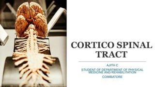
SlideShare a Scribd company logo
CORTICO SPINAL
TRACT
AJITH C
STUDENT OF DEPARTMENT OF PHYSICAL
MEDICINE AND REHABILITATION
COIMBATORE
i. Introduction about descending tracts
ii. Introduction about pyramidal tract
iii. Introduction about Corticospinal tract
iv. Fibres of the Corticospinal tract
v. Origin of the Corticospinal tract
vi. Course of the Corticospinal tract
vii. Lateral Corticospinal tract
viii. Anterior Corticospinal tract
ix. Functions of the Corticospinal tract
x. Clinical significance (or) Applied Anatomy of
the Corticospinal tract
xi. References.
INTRODUCTION ABOUT DESCENDING TRACT :
•Descending pathway are considered with,
SOMATIC &
VISCERAL Motor activities.
• Somatic motor pathway of brain and spinal cord are divided into two
types.
• These tracts are functionally different. Clinically these tracts are
considered together because lesions within the cortex always almost
involve both of them.
• Both these system control the motor activities of body through lower motor
neurons (LMN).
• Have their cells of origin in the cerebral cortex (or) in the brainstem.
• It is otherwise called as motor pathway.
INTRODUCTION ABOUT PYRAMIDAL
TRACT:
• Conventionally, the term pyramidal tract refers specifically to a group of corticospinal
fibres (corticospinal tract) which occupies the pyramid of the Medulla Oblengata,
however clinically. or
(the pyramidal tracts derive their name from the medullary pyramids of the
medulla oblengata, which they pass through).
• This is the longest tract starting from the motor cortex and reaching up to the last
segment of the spinal cord & carry motor impulses from cortex to the spinal cord.
• This is the main Voluntary motor pathway.
• 80% - 90% of the fibres in the pyramidal system are small diameter is 1µm diameter.
• It consists of two neurons, the upper and lower motor neurons. Previously
mentioned that the pyramidal tract are control the motor activities of body
through Lower motor neuron (LMN).
• It is present in the higher animals and man where cerebrum has developed.
• All the pyramidal fibres,
55% end in the Cervical
20% in the Thoracic
25% in the Lumbosacral Region
• Pyramidal tract are considered with,
Corticospinal tract
Corticonuclear tract
Corticospinal tracts - Supplies the musculature of the axial &
Extremity.
 Corticobulbar tracts - Supplies the musculature of the head & neck.
• Corticonuclear fibres otherwise called as Corticobulbar tract.
• That Motor Cranial Nuclei (Particularly 4, 7, 12)
• This is a pathway that begins in the cerebral cortex and ends in the brain stem.
• Bulbar means pertaining to the brainstem where all motor cranial nuclei are
located.
• Throughout the brainstem, the corticobulbar fibres are crossing to reach the
motor cranial nuclei of the opposite side
CORTICOSPINAL
TRACT
• The corticospinal tract are not the sole pathway for serving voluntary movement.
Rather, they form the pathway that confers speed and agility to voluntary
movements and is thus used in performing rapid skilled movements.
• Many of the simple, basic voluntary movements are mediated by other descending
tracts.
• The corticospinal tract are the pathway concerned with voluntary, discrete, skilled
movements, especially those of the distal part of the limbs.
• Corticospinal tract has approximately 1 Million nerve fibres with an avarage
conduction velocity of approximately 60m/s using glutamate as their transmitter
substance.
GROWTH OF CORTICOSPINAL TRACT IN
FETUS:
• 1st corticospinal axons, less than 0.5 microns in diameter.
• MYELINATION:-
The myelination of the pyramidal fibres is incomplete at birth &
gradually progresses in Cranio-caudal (from head to feet)
direction and thereby progressively gaining functionality.
Most of the myelination complete by 2 years of the age (that’s why
under 2 years baby have babinski sign negative).
Myelination commences between postnatal days 10-12.
Myelinate largerly during the 1st & 2nd years after birth.
It progressively slowly in Carnio-caudal direction upto 12the year of
the age.
• The rate of extension of corticospinal axons are not constant.
On the day after birth, labelled corticospinal axons have crossed
in the pyramidal decussating and extended into the dorsal
column of the upper cervical spinal cord level.
Postnatal day-3
Corticospinal tract reach the thoracic segments
Postnatal day-6
It reach Lumbar segments
Postnatal day-9
It reach the sacral segments
FIBERS OF THE CORTICOSPINAL TRACT:
• Are usually 90% between 1 – 4 micro in diameter.
• Are usually 67% myelinated.
• With diameters greater than 20 micra, represent 4% of the tract’s population.
These are the axons of the Giant cells of Betz (these Betz cells are found in
the precentral gyrus and in the anterior paracentral lobule).
• Betz cells are pyramidal cell neurons located within the 5th layer of the
primary motor cortex. They are some of the largest in the central nervous
system, sometimes reaching 100 µm in diameter and send their axons down
the corticospinal tracts to the anterior horn cells of the spinal cord. They are
named after Ukrainian scientist Vladimir Betz, who described them in his
work published in 1874.
(Functions of the Betz cells are see further more in the functions of the
corticospinal tract)*.
• The Archicortex consists of the hippocampus, which is a three-layered
cortex.
• The Neocortex represents the great majority of the cerebral cortex. It has six
layers and contains between 10 and 14 billion neurons.
TYPES OF CORTEX:
Archicortex
Paleocortex
Neocortex
INTRODUCTION ABOUT CORTICOSPINAL
TRACT:
• Corticospinal tract are considered with,
Lateral Corticospinal tract
Anterior Corticospinal tract
Physiologically, the anterior pathways are old, whereas the lateral
pathways are new.
ORIGIN OF CORTICOSPINAL TRACT:
• Fibres of the corticospinal tract arise as axons of pyramidal cells situated in the 5th
layer of the cerebral cortex.
1/3rd of the fibres originate primary motor cortex (Area 4).
1/3rd originate from the Secondary motor cortex (Area 6).
1/3rd originate from the
These fibres do not control motor activity but influence sensory input to
the nervous system.
Primary Somato sensory cortex (postcentral
gyrus)
(Area 3, 1 and 2).
PRIMARY
SOMATOSENSORY
CORTEX
(Post central gyrus)
PRIMARY MOTOR CORTEX
(Precentral gyrus)
SUPPLEMENTAL MOTOR AREA
PRE MOTOR AREA
PRE FRONTAL CORTEX
SECONDARY MOTOR CORTEX
•Electrical stimulation of different part of the
precentral gyrus produces movements of
different parts of the opposite side of the body,
we can represent the parts of the body in the
areas of the cortex, Such a Homunculus.
•Note that the region controlling the face is situated
inferiorly and the region controlling the lower limb is
situated superiorly and on the medial surface of the
hemisphere.
•The Homunculus is a distorted picture of the body,
with the various parts having a size proportional to
the area of the cerebral cortex devoted to their
control. It is intresting to find that the majority of the
corticospinal fibres are myelinated and are relatively
slow-conducting small fibres.
COURSE OF THE
CORTICOSPINAL TRACT:
• These descending fibres converge in the
Corona radiata to reach Internal
capsule. (Located between the
Thalamus and the basal ganglia)
• IN THE INTERNAL CAPSULE:
then pass through the posterior limb of
the Internal capsule.
Where they occupy in the genu and the
anterior 2/3rd of the posterior limb.
INTERNAL CAPSULE INTERNAL CAPSULE
The motor fibres passes through the
posterior limb of the internal capsule
where they are organized in the
sequence of “fibres of UPPER
EXTREMITY, TRUNK, LOWER
EXTREMITY”.
This is clinically important, as the internal
capsule is particularly susceptible to
compression from haemorrhagic
bleeds, known as a ‘capsular
stroke‘. Such an event could cause a
lesion of the descending tracts.
CAUDATE
NUCLEUS PUTAMEN
THALAMUS
GLOBUS
PALLIDUS
IN THE MIDBRAIN:
The tract then continues through the
middle 3/5th of the (Crus cerebri of
Cerebral peduncle) or basis
pedunculi of the midbrain ventral to
the substantia nigra.
The middle fifth carries the pyramidal
tract and medial frontopontine and lateral
temporopontine fibres.
IN THE PONS:
And then passes through the base
(Basilar part) of the pons.
In the pons the corticospinal tracts are
become scattered.
Great Motor
Decussation
IN MEDULLA OBLONGATA:
While coming out of the pons, the
scattered corticospinal fibres are
reunited and enter the medulla as a
thick bundle.
The bundles are become grouped
together in the upper part of medulla
& along the anterior border to form a
swelling known as the pyramid or
Medulla Oblongatary Pyramids
(Cervicomedullary Junction)
(hence the alternative name
pyramidal tract).
Pyramid of the
Medulla Oblangata
Olivary Nuclei
Olivary Nuclei
Pyramid of the
Medulla Oblangata
• The majority of the fibres cross (Decussate) to the opposite side
& enter the lateral white column as the Lateral
Corticospinal Tract of spinal cord.
• The remaining fibres about do not cross in the decussation &
enter the anterior white column as Anterior corticospinal
tract of spinal cord.
LATERAL CORTICOSPINAL
TRACT
In the lower part of medulla(junction between the medulla oblongata
and the spinal cord) the majority of the fibres (75-90%) cross to the
opposite side & descend in the spinal cord occupying the posterior part of
lateral white column as the Lateral Corticospinal Tract of spinal
cord.
It extend throughout the spinal cord.
At each segment some fibres leave the tract, turn inward and end round
the anterior grey horn cells (Motor neurons) either directly or through
interneuron's.
They are also called as “CROSSED CORTICOSPINAL TRACT”.
• The Lateral Corticospinal
Tract (Betz cells fibres)
descend in the Lateral
funiculus of the spinal cord
to terminate mainly in the
lumbosacral region of the
spinal cord.
TERMINATION OF THE
LATERAL CORTICOSPINAL
TRACT:
• It terminates via
Interneurons on ventral horn
motor neurons and sensory
neurons of the dorsal horn
till the lumbosacral region of
the spinal cord.
ANTERIOR CORTICOSPINAL
TRACT
The remaining fibres about (10-25%) do not cross in the decussation &
enter the anterior white column near the median fissure and descend
down as Anterior corticospinal tract of spinal cord.
They are also called as “UNCROSSED CORTICOSPINAL TRACT”.
As a rule, the direct pyramidal tract does not crossed beyond the Lower
cervical (or) Mid thoracic region.
• The Anterior Corticospinal
Tract descend in the Anterior
funiculus of the spinal cord
to terminate mainly in the
anterior horn grey matter of
the cervical and upper thoracic
spinal cord levels.
 Near their termination, fibres of the
anterior corticospinal tract cross the
midline (decussate to the opposite side)
to end round the anterior horn cells of
the opposite side & instead synapse
directly with lower motorneurons.
FUNCTIONS OF THE
CORTICOSPINAL
TRACT:
Thought of the Movement in Prefrontal cortex
(Example: Flexion of Biceps)
ORIGIN: (BETZ cells)
~Primary motor cortex
- pre motor cortex
~Secondary motor cortex - Supplemental Area
~Primary Somatosensory cortex
Basal Ganglia
(Blue print of the
movement)
Special checking mechanism:
~Low/high/perfect
intensity of the movement
Cerebellum
This is the
planned
motor
movements
What type of
movement
can be perform?
Muscle
Receptors
(Proprioception)
Pontine nuclei
(Conveying the information)Spinal cord
1
2
3 4
5
5 5
6
6
~Primary motor cortex
-pre motor cortex
~Secondary motor cortex -pre frontal cortex
-Supplemental Area
~Primary Somatosensory cortex
Internal Capsule
(Special white matter)
Corona radiata
Crus Cerebri of the Cerebro peduncle
Pontine Nuclei
Fibres are scattered
(leg/arm/trunk)
Continue
Scattered fibres are reunited
Lower part of meduul oblengata
(Pyramidal decussation)
Lateral Corticospinal tract Anterior Corticospinal tract
Continue
Lateral Funiculus Anterior Funiculus
Synapse with the same side cell bodies
of Ventral or Anterior grey horn
Synapse (via Anterior commusure) with
the opposite side cell bodies of Venral or
Anterior grey horn
Alpha motor
neuron
Gamma motor
neuron
Stimulate
Extrafeusal fibres
(Maintain the
contractions)
Stimulate the
Muscle Spindles
(Maintain the
length and Tone)
1) The corticospinal tract has many functions which include the,
Control of afferent inputs:–
(These fibres that originate from the sensory cortex (somatosensory cortex)
terminate in the dorsal horn of the spinal cord where they synapse with
interneurns that receive input from somatosensory receptors and are thought
to regulate information pheripheral receptors within the spinal cord).
Spinal reflexes:–
(the 1st order afferent sensory fibres transmitting sensory information from the
muscle spindles also from synapses with the inhibitory interneurons (that
synapse with the Lateral corticospinal tract) to mediate reflex activity.
Motor neuron activity.
MOTOR NEURON ACTIVITY
ANTERIOR CORTICOSPINAL
TRACT
This mediates the execution
of rapid, skilled, voluntary
and Fine movements of the
distal musculature of upper
and lower limbs.
i.e., The intrinsic and
extrinsic muscles of the
hand and foot, especially
the muscles of the hand.
LATERAL CORTICOSPINAL
TRACT
Control of Axial muscles
(Neck, Shoulder, and Trunk)
proximal upper limb (girdle)
musculature.
And they are associated with
the maintenance of upright
posture.
Betz cells are capable of faster nerve impulse transmission to
the spinal cord. The rapid conduction rate is 70m/sec.
CLINICAL
SIGNIFICANSE:
INTRODUCTION:
1) The resulting deficiencies associated with lesions to the respective tracts
will depend on the location of the lesions.
 A lesion proximal to the decussation of the pathway will result in a
contralateral defect.
 In contrast, a lesion distal to the decussation will result in ipsilateral
signs and symptoms.
• Injury to the corticospinal tract caudal to the decussation may present with
varying types of paresis or paralysis of the upper and lower limbs.
Unilateral lesions present with ipsilateral hemiparesis, hemiplegia or
Monoplegia.
While bilateral lesions may result quadriplegia, or bilateral paresis.
2) The lesion types are deviding into two.
Upper Motor Neuron Lesion
Lower Motor Neuron Lesion
1) UPPER MOTOR NEURON LESIONS:
• The pyramidal tracts are susceptible to damage, because they extend almost
the whole length of the central nervous system. As mentioned previously,
they particularly vulnerable as they pass through the internal capsule – a
common site of cerebrovascular accidents (CVA) - (haemorrhagic bleeds,
known as a ‘capsular stroke‘).
(Additionally, lesions of the cortex (cortical lesions) or within the internal
capsule (capsular lesions) may present with both corticospinal and
corticobulbar findings. That is, contralateral muscle weakness in addition to
cranial nerve abnormalities.)
• If there is only a unilateral lesion(upper to the decussation) of the left or right
corticospinal tract, symptoms will appear on the contralateral side of the body.
• (Corticospinal tract syndrome)
• The Cardinal signs of an upper motor neurone lesion are:
1) Hypertonia – an increased muscle tone
2) Hyperreflexia – increased muscle reflexes
3) Clonus – involuntary, rhythmic muscle contractions (an
oscillatory motor response to muscle stretching)
4) Babinski sign – extension of the hallux in response to blunt
stimulation of the sole of the foot.
5) Muscle weakness.
• When the Babinski reflex is present in a child older than 2 years or in an
adult, it is often a sign of a central nervous system disorder. The central
nervous system includes the brain and spinal cord. Disorders may
include:
Amyotrophic lateral sclerosis (Lou Gehrig disease)
Brain tumor or injury
Meningitis (infection of the membranes covering the brain and
spinal cord)
Multiple sclerosis
Spinal cord injury, defect, or tumor
Stroke.
1. THE BABINSKI SIGN:
The normal response in an adult to stroking the sole of the
foot is flexion of the big toe, and often the other toes. Following
damage to descending upper motor neuron pathways,
however, this stimulus elicits extension of the big toe and a
fanning of the other toes.
A similar response occurs in human infants before the
maturation of the corticospinal pathway and presumably
indicates incomplete upper motor neuron control of local
motor neuron circuitry.
2. SAPSTICITY (Hypertonia):
Spasticity is increased muscle tone.
Spasticity is probably caused by the removal of inhibitory influences
exerted by the cortex. It predominates in the Antigravity muscles.
Spasticity is also eliminated by sectioning the dorsal roots.
The form and intensity of spasticity may vary markedly, depending on
the extend and site of the Central Nervous System(CNS) system damage.
It is typically manifested as increased resistance to passive movements.
3. SPINAL REFLEXES (Hyperreflexia):
Spasticity is also eliminated by sectioning the dorsal roots, suggesting
that it represents an abnormal increase in the gain of the spinal
cord reflex due to loss of descending inhibition.
4. HYPOREFLEXIA OF SUPERFICIAL REFLEXES:
 The initial stage of lesion, the superficial reflexes are Areflexia or
Hyporeflexia. That mechanism of diminishment of superficial reflexes
is not well understood.
Further signs are the decreased vigor (and increased threshold) of
superficial reflexes such as the,
Corneal reflex,
Superficial abdominal reflex (tensing of abdominal muscles in
response to stroking the overlying skin), and
The cremasteric reflex in males (elevation of the scrotum in response
to stroking the inner aspect of the thigh).
5. A loss of the ability to perform fine movements:
If the lesion involves the descending pathways that control the lower
motor neurons to the upper limbs, the ability to execute fine movements (such
as independent movements of the fingers) is lost.
6. MUSCLE WEAKNESS:
Weakness may range in severity from mild paresis to total paralysis, depending
on the extent of the lesion.
Types of weakness or paralysis:
1. Hemiplegia/paresis
2. Monoplegia/paresis
3. Paraplegia/paresis
4. Triplegia/paresis
5. Tetraplegia (or) Quadriplegia/paresis
Types of Weakness Definition Common causes
Hemiplegia/Hemiparesis
Paralysis/Weakness of muscles of the arm,
leg &sometimes face on one side of the
body.
Lesion at the Internal capsule,
cerebral hemispheres, pontine
bleed, rarely a high spinal cord
injury .
Monoplegia/Moparesis
Paralysis/Weakness of all the muscles of the
limbs, either Upper extremity & Lower
extremity.
Lesion at the cerebral
hemisphere and spinal cord &
peripheral Neuropathy.
Paraplegia/Paraparesis Paralysis of muscles in both legs Spinal cord lesions &
Peripheral Neuropathy.
Triplegia/Triparesis
Hemiplegia paresis combined with
paralysis of one limb on the opposite side
of the body.
High cervical spinal cord lesion
or multiple lesion.
Tetraplegia/tetraparesis
Paralysis/Weakness of all four
limbs.
Lesion: high cervical spinal cord,
brainstem or Cerebral Hemispheres,
and Acute polyneuropathy,
Radiculopathy, Myopathy
PARTS ARTERIAL SUPPLY
Motor cortex
1) Leg area
2) Face, Trunk & Arm areas
1) Anterior cerebral artery
2) Middle cerebral artery
Internal capsule Branches of middle cerebral artery
Midbrain (Crus cerebri) Posterior cerebral artery
Pons Pontine branches of basilar artery
Medulla Oblangata Anterior spinal branches of vertibral artery
Spinal cord Segmental branches of Anterior & posterior spinal artery
• TRISOMY 18 (or) EDWARDS SYNDROME:
A condition that causes severe developmental delays due to an extra
chromosome 18 .
This is exhibit various developmental abnormalities in the CNS.
Dominant trisomy-18 syndrome is uncrossed pyramidal tract.
The symptoms are,
1. Cleft palate
2. Clenched fists
3. Deformed feet (Rocker-Bottom feet)
4. Feeding problems
5. Small head (microcephaly)
6. Small jaw (micrognathia)
7. Weak cry.
REFERENCE:
1. B.D.Chourasia (Human Anatomy)
2. Sembulingam (Medical Physiology)
3. A.K.Jain (Textbook of Physiology)
4. James D.Fix (NeuroAnatomy)
5. Christoper M.Fredericks (Pathophysiology of the motor
system)
6. Eric R.Kandel (Principles of neural science)
7. Snell’s (Clinical NeuroAnatomy)
8. Vichram Singh (Clinical NeuroAnatomy)
9. Harold Ellis (Clinical Anatomy)
10. Shumway –Cook and Woolcott M.H. (2007) . (Motor control.
Translating research into clinical practice).
11. Crossman, A.R. and Neary, D. (2015) (Neuroanatomy)
12. Bear MF, Connors BW, Paradiso. (Brain and Neuroscience).
13. Stiner CM, Barber PA, Petoe M, Anwar S, (Functional
potential in chronic stroke patients depends on corticospinal
tract integrity).
14. Dale Purves, George J Augustine, David Fitzpatrick, Lawrence
C Katz, Anthony-Samuel LaMantia, James O McNamara, and S
Mark Williams (Neuroscience-2nd edition).
15.Karen J.Jones (Neuro Assessment a Clinical Guide).
16.Pictures from KEN HUB (Licensed Anatomy page on the
internet).
17.Susan B. O’Sullivan (Physical Rehablitation).
18.Gray’s atlas of Human Anatomy.
19.Mlyata H. (Neuro Physiology)
Corticospinal tract (Pyramidal tract)

More Related Content

What's hot

Basal ganglia
Basal gangliaBasal ganglia
Basal ganglia
Dr Nilesh Kate
 
Anatomy of cerebellum
Anatomy of cerebellumAnatomy of cerebellum
Blood supply to the brain
Blood supply to the brainBlood supply to the brain
Blood supply to the brain
Shehzad Hussain Raja
 
Anatomy of cerebellum
Anatomy of cerebellumAnatomy of cerebellum
Anatomy of cerebellumMBBS IMS MSU
 
Corpus callosum
Corpus callosumCorpus callosum
Corpus callosum
Chaitra Chinmayee M K
 
Cerebellum ppt
Cerebellum pptCerebellum ppt
Cerebellum ppt
Dr Nilesh Kate
 
Basal ganglia
Basal gangliaBasal ganglia
Basal ganglia
Enoch R G
 
Anatomy of internal capsule
Anatomy of  internal capsuleAnatomy of  internal capsule
Anatomy of internal capsuleMBBS IMS MSU
 
Anatomy of pons
Anatomy of ponsAnatomy of pons
Anatomy of pons
Dr. Mohammad Mahmoud
 
Descending tracts of sc
Descending tracts of scDescending tracts of sc
Descending tracts of sc
Dr. sana yaseen
 
Cerebellum-Connections and Functions
Cerebellum-Connections and FunctionsCerebellum-Connections and Functions
Cerebellum-Connections and Functions
Ranadhi Das
 
Basal ganglia
Basal gangliaBasal ganglia
Basal ganglia
DrChintansinh Parmar
 
Spinothalamic tract
Spinothalamic tractSpinothalamic tract
Spinothalamic tract
M S
 
Pons Anatomy
Pons AnatomyPons Anatomy
Pons Anatomy
Mehul Tandel
 
motor cortex
motor cortexmotor cortex
motor cortex
Lubna Abu Alrub,DDS
 
Extrapyramidal tract
Extrapyramidal tractExtrapyramidal tract
Extrapyramidal tract
ASNasrullah
 
Anatomy of brainstem
Anatomy of brainstemAnatomy of brainstem
Anatomy of brainstemMBBS IMS MSU
 

What's hot (20)

Basal ganglia
Basal gangliaBasal ganglia
Basal ganglia
 
Anatomy of cerebellum
Anatomy of cerebellumAnatomy of cerebellum
Anatomy of cerebellum
 
Blood supply to the brain
Blood supply to the brainBlood supply to the brain
Blood supply to the brain
 
Anatomy of cerebellum
Anatomy of cerebellumAnatomy of cerebellum
Anatomy of cerebellum
 
Corpus callosum
Corpus callosumCorpus callosum
Corpus callosum
 
ASCENDING TRACTS
ASCENDING TRACTSASCENDING TRACTS
ASCENDING TRACTS
 
Cerebellum ppt
Cerebellum pptCerebellum ppt
Cerebellum ppt
 
Tracts
TractsTracts
Tracts
 
Basal ganglia
Basal gangliaBasal ganglia
Basal ganglia
 
Anatomy of internal capsule
Anatomy of  internal capsuleAnatomy of  internal capsule
Anatomy of internal capsule
 
Anatomy of pons
Anatomy of ponsAnatomy of pons
Anatomy of pons
 
Descending tracts of sc
Descending tracts of scDescending tracts of sc
Descending tracts of sc
 
Cerebellum-Connections and Functions
Cerebellum-Connections and FunctionsCerebellum-Connections and Functions
Cerebellum-Connections and Functions
 
Basal ganglia
Basal gangliaBasal ganglia
Basal ganglia
 
Spinothalamic tract
Spinothalamic tractSpinothalamic tract
Spinothalamic tract
 
Pons Anatomy
Pons AnatomyPons Anatomy
Pons Anatomy
 
motor cortex
motor cortexmotor cortex
motor cortex
 
NEUROANATOMY - I
NEUROANATOMY - INEUROANATOMY - I
NEUROANATOMY - I
 
Extrapyramidal tract
Extrapyramidal tractExtrapyramidal tract
Extrapyramidal tract
 
Anatomy of brainstem
Anatomy of brainstemAnatomy of brainstem
Anatomy of brainstem
 

Similar to Corticospinal tract (Pyramidal tract)

corticospinal tracts.pptx
corticospinal tracts.pptxcorticospinal tracts.pptx
corticospinal tracts.pptx
DunsonNampaso
 
DESCENDING-TRACTS.pptx
DESCENDING-TRACTS.pptxDESCENDING-TRACTS.pptx
DESCENDING-TRACTS.pptx
YubiKim5
 
Cerebellum .pptx
Cerebellum  .pptxCerebellum  .pptx
Cerebellum .pptx
Mr SACHIN
 
Cerebellum
CerebellumCerebellum
Cerebellum
Prakhar Jain
 
Descending tracts of spinal cord.pptx
Descending tracts of spinal cord.pptxDescending tracts of spinal cord.pptx
Descending tracts of spinal cord.pptx
gopal8694
 
TRACTS OF THE SPINAL CORD.pptx
TRACTS OF THE SPINAL CORD.pptxTRACTS OF THE SPINAL CORD.pptx
TRACTS OF THE SPINAL CORD.pptx
OlaniyiEmmanuel5
 
2_Motility-46863 2.pdf
2_Motility-46863 2.pdf2_Motility-46863 2.pdf
2_Motility-46863 2.pdf
nashat3
 
Functional neuroanatomy of spinal cord
Functional neuroanatomy of spinal cordFunctional neuroanatomy of spinal cord
Functional neuroanatomy of spinal cord
NeurologyKota
 
Spinal cord & its tracts II (Descending tracts)
Spinal cord & its tracts II (Descending tracts)Spinal cord & its tracts II (Descending tracts)
Spinal cord & its tracts II (Descending tracts)
ABDUL QADEER MEMON
 
pyramidal, extrapyramidal pathways
pyramidal, extrapyramidal pathways pyramidal, extrapyramidal pathways
pyramidal, extrapyramidal pathways
med_students0
 
embryology_developmentofcentralnervoussystem_100708132545_phpapp02.pdf
embryology_developmentofcentralnervoussystem_100708132545_phpapp02.pdfembryology_developmentofcentralnervoussystem_100708132545_phpapp02.pdf
embryology_developmentofcentralnervoussystem_100708132545_phpapp02.pdf
Joharseid1
 
Embryology developmentofcentralnervoussystem-100708132545-phpapp02
Embryology developmentofcentralnervoussystem-100708132545-phpapp02Embryology developmentofcentralnervoussystem-100708132545-phpapp02
Embryology developmentofcentralnervoussystem-100708132545-phpapp02PTCHANDAN221
 
Motor system. Extrapyramydal system. (1,2,3 lectures).pdf
Motor system. Extrapyramydal system. (1,2,3 lectures).pdfMotor system. Extrapyramydal system. (1,2,3 lectures).pdf
Motor system. Extrapyramydal system. (1,2,3 lectures).pdf
Saicharitha15
 
white fibres of cns modified
white fibres of cns modifiedwhite fibres of cns modified
white fibres of cns modified
Med Study
 
Spinal cord
Spinal cordSpinal cord
SPINAL CORD.2.pptx
SPINAL CORD.2.pptxSPINAL CORD.2.pptx
SPINAL CORD.2.pptx
Deepak Khedekar
 
Medulla oblongata
Medulla oblongata Medulla oblongata
Medulla oblongata
Prabhakar Yadav
 
Central nervous system
Central nervous system Central nervous system
Central nervous system
Sunil Pahari
 
CEREBELLUM- with clinical and radiology
CEREBELLUM- with clinical and radiologyCEREBELLUM- with clinical and radiology
CEREBELLUM- with clinical and radiology
lalit yadav
 
Anatomy of inner ear
Anatomy of inner earAnatomy of inner ear
Anatomy of inner ear
Mohammed Nishad N
 

Similar to Corticospinal tract (Pyramidal tract) (20)

corticospinal tracts.pptx
corticospinal tracts.pptxcorticospinal tracts.pptx
corticospinal tracts.pptx
 
DESCENDING-TRACTS.pptx
DESCENDING-TRACTS.pptxDESCENDING-TRACTS.pptx
DESCENDING-TRACTS.pptx
 
Cerebellum .pptx
Cerebellum  .pptxCerebellum  .pptx
Cerebellum .pptx
 
Cerebellum
CerebellumCerebellum
Cerebellum
 
Descending tracts of spinal cord.pptx
Descending tracts of spinal cord.pptxDescending tracts of spinal cord.pptx
Descending tracts of spinal cord.pptx
 
TRACTS OF THE SPINAL CORD.pptx
TRACTS OF THE SPINAL CORD.pptxTRACTS OF THE SPINAL CORD.pptx
TRACTS OF THE SPINAL CORD.pptx
 
2_Motility-46863 2.pdf
2_Motility-46863 2.pdf2_Motility-46863 2.pdf
2_Motility-46863 2.pdf
 
Functional neuroanatomy of spinal cord
Functional neuroanatomy of spinal cordFunctional neuroanatomy of spinal cord
Functional neuroanatomy of spinal cord
 
Spinal cord & its tracts II (Descending tracts)
Spinal cord & its tracts II (Descending tracts)Spinal cord & its tracts II (Descending tracts)
Spinal cord & its tracts II (Descending tracts)
 
pyramidal, extrapyramidal pathways
pyramidal, extrapyramidal pathways pyramidal, extrapyramidal pathways
pyramidal, extrapyramidal pathways
 
embryology_developmentofcentralnervoussystem_100708132545_phpapp02.pdf
embryology_developmentofcentralnervoussystem_100708132545_phpapp02.pdfembryology_developmentofcentralnervoussystem_100708132545_phpapp02.pdf
embryology_developmentofcentralnervoussystem_100708132545_phpapp02.pdf
 
Embryology developmentofcentralnervoussystem-100708132545-phpapp02
Embryology developmentofcentralnervoussystem-100708132545-phpapp02Embryology developmentofcentralnervoussystem-100708132545-phpapp02
Embryology developmentofcentralnervoussystem-100708132545-phpapp02
 
Motor system. Extrapyramydal system. (1,2,3 lectures).pdf
Motor system. Extrapyramydal system. (1,2,3 lectures).pdfMotor system. Extrapyramydal system. (1,2,3 lectures).pdf
Motor system. Extrapyramydal system. (1,2,3 lectures).pdf
 
white fibres of cns modified
white fibres of cns modifiedwhite fibres of cns modified
white fibres of cns modified
 
Spinal cord
Spinal cordSpinal cord
Spinal cord
 
SPINAL CORD.2.pptx
SPINAL CORD.2.pptxSPINAL CORD.2.pptx
SPINAL CORD.2.pptx
 
Medulla oblongata
Medulla oblongata Medulla oblongata
Medulla oblongata
 
Central nervous system
Central nervous system Central nervous system
Central nervous system
 
CEREBELLUM- with clinical and radiology
CEREBELLUM- with clinical and radiologyCEREBELLUM- with clinical and radiology
CEREBELLUM- with clinical and radiology
 
Anatomy of inner ear
Anatomy of inner earAnatomy of inner ear
Anatomy of inner ear
 

More from Ajith lolita

Manual muscle test (MMT)
Manual muscle test (MMT)Manual muscle test (MMT)
Manual muscle test (MMT)
Ajith lolita
 
Deep layer of (Iliotibial) IT band
Deep layer of (Iliotibial) IT bandDeep layer of (Iliotibial) IT band
Deep layer of (Iliotibial) IT band
Ajith lolita
 
Ligaments of ankle joint (Ankle complex)
Ligaments of ankle joint (Ankle complex)Ligaments of ankle joint (Ankle complex)
Ligaments of ankle joint (Ankle complex)
Ajith lolita
 
Structural adaptations to weight bearing of Hip
Structural adaptations to weight bearing of HipStructural adaptations to weight bearing of Hip
Structural adaptations to weight bearing of Hip
Ajith lolita
 
Elbow complex (Biomechanics)
Elbow complex (Biomechanics)Elbow complex (Biomechanics)
Elbow complex (Biomechanics)
Ajith lolita
 
Anatomy of Femoral nerve
Anatomy of Femoral nerve Anatomy of Femoral nerve
Anatomy of Femoral nerve
Ajith lolita
 
Immobility (Bed Rest Complications)
Immobility (Bed Rest Complications)Immobility (Bed Rest Complications)
Immobility (Bed Rest Complications)
Ajith lolita
 

More from Ajith lolita (7)

Manual muscle test (MMT)
Manual muscle test (MMT)Manual muscle test (MMT)
Manual muscle test (MMT)
 
Deep layer of (Iliotibial) IT band
Deep layer of (Iliotibial) IT bandDeep layer of (Iliotibial) IT band
Deep layer of (Iliotibial) IT band
 
Ligaments of ankle joint (Ankle complex)
Ligaments of ankle joint (Ankle complex)Ligaments of ankle joint (Ankle complex)
Ligaments of ankle joint (Ankle complex)
 
Structural adaptations to weight bearing of Hip
Structural adaptations to weight bearing of HipStructural adaptations to weight bearing of Hip
Structural adaptations to weight bearing of Hip
 
Elbow complex (Biomechanics)
Elbow complex (Biomechanics)Elbow complex (Biomechanics)
Elbow complex (Biomechanics)
 
Anatomy of Femoral nerve
Anatomy of Femoral nerve Anatomy of Femoral nerve
Anatomy of Femoral nerve
 
Immobility (Bed Rest Complications)
Immobility (Bed Rest Complications)Immobility (Bed Rest Complications)
Immobility (Bed Rest Complications)
 

Recently uploaded

Biological Screening of Herbal Drugs in detailed.
Biological Screening of Herbal Drugs in detailed.Biological Screening of Herbal Drugs in detailed.
Biological Screening of Herbal Drugs in detailed.
Ashokrao Mane college of Pharmacy Peth-Vadgaon
 
A Strategic Approach: GenAI in Education
A Strategic Approach: GenAI in EducationA Strategic Approach: GenAI in Education
A Strategic Approach: GenAI in Education
Peter Windle
 
How to Make a Field invisible in Odoo 17
How to Make a Field invisible in Odoo 17How to Make a Field invisible in Odoo 17
How to Make a Field invisible in Odoo 17
Celine George
 
Unit 2- Research Aptitude (UGC NET Paper I).pdf
Unit 2- Research Aptitude (UGC NET Paper I).pdfUnit 2- Research Aptitude (UGC NET Paper I).pdf
Unit 2- Research Aptitude (UGC NET Paper I).pdf
Thiyagu K
 
The French Revolution Class 9 Study Material pdf free download
The French Revolution Class 9 Study Material pdf free downloadThe French Revolution Class 9 Study Material pdf free download
The French Revolution Class 9 Study Material pdf free download
Vivekanand Anglo Vedic Academy
 
CACJapan - GROUP Presentation 1- Wk 4.pdf
CACJapan - GROUP Presentation 1- Wk 4.pdfCACJapan - GROUP Presentation 1- Wk 4.pdf
CACJapan - GROUP Presentation 1- Wk 4.pdf
camakaiclarkmusic
 
Phrasal Verbs.XXXXXXXXXXXXXXXXXXXXXXXXXX
Phrasal Verbs.XXXXXXXXXXXXXXXXXXXXXXXXXXPhrasal Verbs.XXXXXXXXXXXXXXXXXXXXXXXXXX
Phrasal Verbs.XXXXXXXXXXXXXXXXXXXXXXXXXX
MIRIAMSALINAS13
 
Polish students' mobility in the Czech Republic
Polish students' mobility in the Czech RepublicPolish students' mobility in the Czech Republic
Polish students' mobility in the Czech Republic
Anna Sz.
 
Model Attribute Check Company Auto Property
Model Attribute  Check Company Auto PropertyModel Attribute  Check Company Auto Property
Model Attribute Check Company Auto Property
Celine George
 
Supporting (UKRI) OA monographs at Salford.pptx
Supporting (UKRI) OA monographs at Salford.pptxSupporting (UKRI) OA monographs at Salford.pptx
Supporting (UKRI) OA monographs at Salford.pptx
Jisc
 
CLASS 11 CBSE B.St Project AIDS TO TRADE - INSURANCE
CLASS 11 CBSE B.St Project AIDS TO TRADE - INSURANCECLASS 11 CBSE B.St Project AIDS TO TRADE - INSURANCE
CLASS 11 CBSE B.St Project AIDS TO TRADE - INSURANCE
BhavyaRajput3
 
678020731-Sumas-y-Restas-Para-Colorear.pdf
678020731-Sumas-y-Restas-Para-Colorear.pdf678020731-Sumas-y-Restas-Para-Colorear.pdf
678020731-Sumas-y-Restas-Para-Colorear.pdf
CarlosHernanMontoyab2
 
Chapter 3 - Islamic Banking Products and Services.pptx
Chapter 3 - Islamic Banking Products and Services.pptxChapter 3 - Islamic Banking Products and Services.pptx
Chapter 3 - Islamic Banking Products and Services.pptx
Mohd Adib Abd Muin, Senior Lecturer at Universiti Utara Malaysia
 
Introduction to AI for Nonprofits with Tapp Network
Introduction to AI for Nonprofits with Tapp NetworkIntroduction to AI for Nonprofits with Tapp Network
Introduction to AI for Nonprofits with Tapp Network
TechSoup
 
Guidance_and_Counselling.pdf B.Ed. 4th Semester
Guidance_and_Counselling.pdf B.Ed. 4th SemesterGuidance_and_Counselling.pdf B.Ed. 4th Semester
Guidance_and_Counselling.pdf B.Ed. 4th Semester
Atul Kumar Singh
 
Palestine last event orientationfvgnh .pptx
Palestine last event orientationfvgnh .pptxPalestine last event orientationfvgnh .pptx
Palestine last event orientationfvgnh .pptx
RaedMohamed3
 
aaaaaaaaaaaaaaaaaaaaaaaaaaaaaaaaaaaaaaaaa
aaaaaaaaaaaaaaaaaaaaaaaaaaaaaaaaaaaaaaaaaaaaaaaaaaaaaaaaaaaaaaaaaaaaaaaaaaaaaaaaaa
aaaaaaaaaaaaaaaaaaaaaaaaaaaaaaaaaaaaaaaaa
siemaillard
 
The geography of Taylor Swift - some ideas
The geography of Taylor Swift - some ideasThe geography of Taylor Swift - some ideas
The geography of Taylor Swift - some ideas
GeoBlogs
 
TESDA TM1 REVIEWER FOR NATIONAL ASSESSMENT WRITTEN AND ORAL QUESTIONS WITH A...
TESDA TM1 REVIEWER  FOR NATIONAL ASSESSMENT WRITTEN AND ORAL QUESTIONS WITH A...TESDA TM1 REVIEWER  FOR NATIONAL ASSESSMENT WRITTEN AND ORAL QUESTIONS WITH A...
TESDA TM1 REVIEWER FOR NATIONAL ASSESSMENT WRITTEN AND ORAL QUESTIONS WITH A...
EugeneSaldivar
 
1.4 modern child centered education - mahatma gandhi-2.pptx
1.4 modern child centered education - mahatma gandhi-2.pptx1.4 modern child centered education - mahatma gandhi-2.pptx
1.4 modern child centered education - mahatma gandhi-2.pptx
JosvitaDsouza2
 

Recently uploaded (20)

Biological Screening of Herbal Drugs in detailed.
Biological Screening of Herbal Drugs in detailed.Biological Screening of Herbal Drugs in detailed.
Biological Screening of Herbal Drugs in detailed.
 
A Strategic Approach: GenAI in Education
A Strategic Approach: GenAI in EducationA Strategic Approach: GenAI in Education
A Strategic Approach: GenAI in Education
 
How to Make a Field invisible in Odoo 17
How to Make a Field invisible in Odoo 17How to Make a Field invisible in Odoo 17
How to Make a Field invisible in Odoo 17
 
Unit 2- Research Aptitude (UGC NET Paper I).pdf
Unit 2- Research Aptitude (UGC NET Paper I).pdfUnit 2- Research Aptitude (UGC NET Paper I).pdf
Unit 2- Research Aptitude (UGC NET Paper I).pdf
 
The French Revolution Class 9 Study Material pdf free download
The French Revolution Class 9 Study Material pdf free downloadThe French Revolution Class 9 Study Material pdf free download
The French Revolution Class 9 Study Material pdf free download
 
CACJapan - GROUP Presentation 1- Wk 4.pdf
CACJapan - GROUP Presentation 1- Wk 4.pdfCACJapan - GROUP Presentation 1- Wk 4.pdf
CACJapan - GROUP Presentation 1- Wk 4.pdf
 
Phrasal Verbs.XXXXXXXXXXXXXXXXXXXXXXXXXX
Phrasal Verbs.XXXXXXXXXXXXXXXXXXXXXXXXXXPhrasal Verbs.XXXXXXXXXXXXXXXXXXXXXXXXXX
Phrasal Verbs.XXXXXXXXXXXXXXXXXXXXXXXXXX
 
Polish students' mobility in the Czech Republic
Polish students' mobility in the Czech RepublicPolish students' mobility in the Czech Republic
Polish students' mobility in the Czech Republic
 
Model Attribute Check Company Auto Property
Model Attribute  Check Company Auto PropertyModel Attribute  Check Company Auto Property
Model Attribute Check Company Auto Property
 
Supporting (UKRI) OA monographs at Salford.pptx
Supporting (UKRI) OA monographs at Salford.pptxSupporting (UKRI) OA monographs at Salford.pptx
Supporting (UKRI) OA monographs at Salford.pptx
 
CLASS 11 CBSE B.St Project AIDS TO TRADE - INSURANCE
CLASS 11 CBSE B.St Project AIDS TO TRADE - INSURANCECLASS 11 CBSE B.St Project AIDS TO TRADE - INSURANCE
CLASS 11 CBSE B.St Project AIDS TO TRADE - INSURANCE
 
678020731-Sumas-y-Restas-Para-Colorear.pdf
678020731-Sumas-y-Restas-Para-Colorear.pdf678020731-Sumas-y-Restas-Para-Colorear.pdf
678020731-Sumas-y-Restas-Para-Colorear.pdf
 
Chapter 3 - Islamic Banking Products and Services.pptx
Chapter 3 - Islamic Banking Products and Services.pptxChapter 3 - Islamic Banking Products and Services.pptx
Chapter 3 - Islamic Banking Products and Services.pptx
 
Introduction to AI for Nonprofits with Tapp Network
Introduction to AI for Nonprofits with Tapp NetworkIntroduction to AI for Nonprofits with Tapp Network
Introduction to AI for Nonprofits with Tapp Network
 
Guidance_and_Counselling.pdf B.Ed. 4th Semester
Guidance_and_Counselling.pdf B.Ed. 4th SemesterGuidance_and_Counselling.pdf B.Ed. 4th Semester
Guidance_and_Counselling.pdf B.Ed. 4th Semester
 
Palestine last event orientationfvgnh .pptx
Palestine last event orientationfvgnh .pptxPalestine last event orientationfvgnh .pptx
Palestine last event orientationfvgnh .pptx
 
aaaaaaaaaaaaaaaaaaaaaaaaaaaaaaaaaaaaaaaaa
aaaaaaaaaaaaaaaaaaaaaaaaaaaaaaaaaaaaaaaaaaaaaaaaaaaaaaaaaaaaaaaaaaaaaaaaaaaaaaaaaa
aaaaaaaaaaaaaaaaaaaaaaaaaaaaaaaaaaaaaaaaa
 
The geography of Taylor Swift - some ideas
The geography of Taylor Swift - some ideasThe geography of Taylor Swift - some ideas
The geography of Taylor Swift - some ideas
 
TESDA TM1 REVIEWER FOR NATIONAL ASSESSMENT WRITTEN AND ORAL QUESTIONS WITH A...
TESDA TM1 REVIEWER  FOR NATIONAL ASSESSMENT WRITTEN AND ORAL QUESTIONS WITH A...TESDA TM1 REVIEWER  FOR NATIONAL ASSESSMENT WRITTEN AND ORAL QUESTIONS WITH A...
TESDA TM1 REVIEWER FOR NATIONAL ASSESSMENT WRITTEN AND ORAL QUESTIONS WITH A...
 
1.4 modern child centered education - mahatma gandhi-2.pptx
1.4 modern child centered education - mahatma gandhi-2.pptx1.4 modern child centered education - mahatma gandhi-2.pptx
1.4 modern child centered education - mahatma gandhi-2.pptx
 

Corticospinal tract (Pyramidal tract)

  • 1. CORTICO SPINAL TRACT AJITH C STUDENT OF DEPARTMENT OF PHYSICAL MEDICINE AND REHABILITATION COIMBATORE
  • 2.
  • 3. i. Introduction about descending tracts ii. Introduction about pyramidal tract iii. Introduction about Corticospinal tract iv. Fibres of the Corticospinal tract v. Origin of the Corticospinal tract vi. Course of the Corticospinal tract vii. Lateral Corticospinal tract viii. Anterior Corticospinal tract ix. Functions of the Corticospinal tract x. Clinical significance (or) Applied Anatomy of the Corticospinal tract xi. References.
  • 4. INTRODUCTION ABOUT DESCENDING TRACT : •Descending pathway are considered with, SOMATIC & VISCERAL Motor activities.
  • 5. • Somatic motor pathway of brain and spinal cord are divided into two types. • These tracts are functionally different. Clinically these tracts are considered together because lesions within the cortex always almost involve both of them.
  • 6. • Both these system control the motor activities of body through lower motor neurons (LMN). • Have their cells of origin in the cerebral cortex (or) in the brainstem. • It is otherwise called as motor pathway.
  • 7. INTRODUCTION ABOUT PYRAMIDAL TRACT: • Conventionally, the term pyramidal tract refers specifically to a group of corticospinal fibres (corticospinal tract) which occupies the pyramid of the Medulla Oblengata, however clinically. or (the pyramidal tracts derive their name from the medullary pyramids of the medulla oblengata, which they pass through). • This is the longest tract starting from the motor cortex and reaching up to the last segment of the spinal cord & carry motor impulses from cortex to the spinal cord. • This is the main Voluntary motor pathway. • 80% - 90% of the fibres in the pyramidal system are small diameter is 1µm diameter.
  • 8. • It consists of two neurons, the upper and lower motor neurons. Previously mentioned that the pyramidal tract are control the motor activities of body through Lower motor neuron (LMN). • It is present in the higher animals and man where cerebrum has developed. • All the pyramidal fibres, 55% end in the Cervical 20% in the Thoracic 25% in the Lumbosacral Region
  • 9. • Pyramidal tract are considered with, Corticospinal tract Corticonuclear tract Corticospinal tracts - Supplies the musculature of the axial & Extremity.  Corticobulbar tracts - Supplies the musculature of the head & neck.
  • 10.
  • 11. • Corticonuclear fibres otherwise called as Corticobulbar tract. • That Motor Cranial Nuclei (Particularly 4, 7, 12) • This is a pathway that begins in the cerebral cortex and ends in the brain stem. • Bulbar means pertaining to the brainstem where all motor cranial nuclei are located. • Throughout the brainstem, the corticobulbar fibres are crossing to reach the motor cranial nuclei of the opposite side
  • 13. • The corticospinal tract are not the sole pathway for serving voluntary movement. Rather, they form the pathway that confers speed and agility to voluntary movements and is thus used in performing rapid skilled movements. • Many of the simple, basic voluntary movements are mediated by other descending tracts. • The corticospinal tract are the pathway concerned with voluntary, discrete, skilled movements, especially those of the distal part of the limbs. • Corticospinal tract has approximately 1 Million nerve fibres with an avarage conduction velocity of approximately 60m/s using glutamate as their transmitter substance.
  • 14.
  • 15. GROWTH OF CORTICOSPINAL TRACT IN FETUS: • 1st corticospinal axons, less than 0.5 microns in diameter. • MYELINATION:- The myelination of the pyramidal fibres is incomplete at birth & gradually progresses in Cranio-caudal (from head to feet) direction and thereby progressively gaining functionality. Most of the myelination complete by 2 years of the age (that’s why under 2 years baby have babinski sign negative). Myelination commences between postnatal days 10-12. Myelinate largerly during the 1st & 2nd years after birth. It progressively slowly in Carnio-caudal direction upto 12the year of the age. • The rate of extension of corticospinal axons are not constant.
  • 16. On the day after birth, labelled corticospinal axons have crossed in the pyramidal decussating and extended into the dorsal column of the upper cervical spinal cord level. Postnatal day-3 Corticospinal tract reach the thoracic segments Postnatal day-6 It reach Lumbar segments Postnatal day-9 It reach the sacral segments
  • 17. FIBERS OF THE CORTICOSPINAL TRACT: • Are usually 90% between 1 – 4 micro in diameter. • Are usually 67% myelinated. • With diameters greater than 20 micra, represent 4% of the tract’s population. These are the axons of the Giant cells of Betz (these Betz cells are found in the precentral gyrus and in the anterior paracentral lobule). • Betz cells are pyramidal cell neurons located within the 5th layer of the primary motor cortex. They are some of the largest in the central nervous system, sometimes reaching 100 µm in diameter and send their axons down the corticospinal tracts to the anterior horn cells of the spinal cord. They are named after Ukrainian scientist Vladimir Betz, who described them in his work published in 1874. (Functions of the Betz cells are see further more in the functions of the corticospinal tract)*.
  • 18. • The Archicortex consists of the hippocampus, which is a three-layered cortex. • The Neocortex represents the great majority of the cerebral cortex. It has six layers and contains between 10 and 14 billion neurons. TYPES OF CORTEX: Archicortex Paleocortex Neocortex
  • 19.
  • 20.
  • 21. INTRODUCTION ABOUT CORTICOSPINAL TRACT: • Corticospinal tract are considered with, Lateral Corticospinal tract Anterior Corticospinal tract Physiologically, the anterior pathways are old, whereas the lateral pathways are new.
  • 22. ORIGIN OF CORTICOSPINAL TRACT: • Fibres of the corticospinal tract arise as axons of pyramidal cells situated in the 5th layer of the cerebral cortex. 1/3rd of the fibres originate primary motor cortex (Area 4). 1/3rd originate from the Secondary motor cortex (Area 6). 1/3rd originate from the These fibres do not control motor activity but influence sensory input to the nervous system. Primary Somato sensory cortex (postcentral gyrus) (Area 3, 1 and 2).
  • 24. SUPPLEMENTAL MOTOR AREA PRE MOTOR AREA PRE FRONTAL CORTEX SECONDARY MOTOR CORTEX
  • 25.
  • 26. •Electrical stimulation of different part of the precentral gyrus produces movements of different parts of the opposite side of the body, we can represent the parts of the body in the areas of the cortex, Such a Homunculus. •Note that the region controlling the face is situated inferiorly and the region controlling the lower limb is situated superiorly and on the medial surface of the hemisphere. •The Homunculus is a distorted picture of the body, with the various parts having a size proportional to the area of the cerebral cortex devoted to their control. It is intresting to find that the majority of the corticospinal fibres are myelinated and are relatively slow-conducting small fibres.
  • 27. COURSE OF THE CORTICOSPINAL TRACT: • These descending fibres converge in the Corona radiata to reach Internal capsule. (Located between the Thalamus and the basal ganglia) • IN THE INTERNAL CAPSULE: then pass through the posterior limb of the Internal capsule. Where they occupy in the genu and the anterior 2/3rd of the posterior limb.
  • 29. The motor fibres passes through the posterior limb of the internal capsule where they are organized in the sequence of “fibres of UPPER EXTREMITY, TRUNK, LOWER EXTREMITY”. This is clinically important, as the internal capsule is particularly susceptible to compression from haemorrhagic bleeds, known as a ‘capsular stroke‘. Such an event could cause a lesion of the descending tracts.
  • 32.
  • 33. IN THE MIDBRAIN: The tract then continues through the middle 3/5th of the (Crus cerebri of Cerebral peduncle) or basis pedunculi of the midbrain ventral to the substantia nigra. The middle fifth carries the pyramidal tract and medial frontopontine and lateral temporopontine fibres.
  • 34. IN THE PONS: And then passes through the base (Basilar part) of the pons. In the pons the corticospinal tracts are become scattered.
  • 35. Great Motor Decussation IN MEDULLA OBLONGATA: While coming out of the pons, the scattered corticospinal fibres are reunited and enter the medulla as a thick bundle. The bundles are become grouped together in the upper part of medulla & along the anterior border to form a swelling known as the pyramid or Medulla Oblongatary Pyramids (Cervicomedullary Junction) (hence the alternative name pyramidal tract).
  • 36. Pyramid of the Medulla Oblangata Olivary Nuclei
  • 37. Olivary Nuclei Pyramid of the Medulla Oblangata
  • 38. • The majority of the fibres cross (Decussate) to the opposite side & enter the lateral white column as the Lateral Corticospinal Tract of spinal cord. • The remaining fibres about do not cross in the decussation & enter the anterior white column as Anterior corticospinal tract of spinal cord.
  • 40.
  • 41. In the lower part of medulla(junction between the medulla oblongata and the spinal cord) the majority of the fibres (75-90%) cross to the opposite side & descend in the spinal cord occupying the posterior part of lateral white column as the Lateral Corticospinal Tract of spinal cord. It extend throughout the spinal cord. At each segment some fibres leave the tract, turn inward and end round the anterior grey horn cells (Motor neurons) either directly or through interneuron's. They are also called as “CROSSED CORTICOSPINAL TRACT”.
  • 42. • The Lateral Corticospinal Tract (Betz cells fibres) descend in the Lateral funiculus of the spinal cord to terminate mainly in the lumbosacral region of the spinal cord.
  • 43.
  • 44.
  • 45. TERMINATION OF THE LATERAL CORTICOSPINAL TRACT: • It terminates via Interneurons on ventral horn motor neurons and sensory neurons of the dorsal horn till the lumbosacral region of the spinal cord.
  • 47.
  • 48. The remaining fibres about (10-25%) do not cross in the decussation & enter the anterior white column near the median fissure and descend down as Anterior corticospinal tract of spinal cord. They are also called as “UNCROSSED CORTICOSPINAL TRACT”. As a rule, the direct pyramidal tract does not crossed beyond the Lower cervical (or) Mid thoracic region.
  • 49. • The Anterior Corticospinal Tract descend in the Anterior funiculus of the spinal cord to terminate mainly in the anterior horn grey matter of the cervical and upper thoracic spinal cord levels.
  • 50.  Near their termination, fibres of the anterior corticospinal tract cross the midline (decussate to the opposite side) to end round the anterior horn cells of the opposite side & instead synapse directly with lower motorneurons.
  • 52. Thought of the Movement in Prefrontal cortex (Example: Flexion of Biceps) ORIGIN: (BETZ cells) ~Primary motor cortex - pre motor cortex ~Secondary motor cortex - Supplemental Area ~Primary Somatosensory cortex Basal Ganglia (Blue print of the movement) Special checking mechanism: ~Low/high/perfect intensity of the movement Cerebellum This is the planned motor movements What type of movement can be perform? Muscle Receptors (Proprioception) Pontine nuclei (Conveying the information)Spinal cord 1 2 3 4 5 5 5 6 6
  • 53. ~Primary motor cortex -pre motor cortex ~Secondary motor cortex -pre frontal cortex -Supplemental Area ~Primary Somatosensory cortex Internal Capsule (Special white matter) Corona radiata Crus Cerebri of the Cerebro peduncle Pontine Nuclei Fibres are scattered (leg/arm/trunk) Continue
  • 54. Scattered fibres are reunited Lower part of meduul oblengata (Pyramidal decussation) Lateral Corticospinal tract Anterior Corticospinal tract Continue Lateral Funiculus Anterior Funiculus Synapse with the same side cell bodies of Ventral or Anterior grey horn Synapse (via Anterior commusure) with the opposite side cell bodies of Venral or Anterior grey horn Alpha motor neuron Gamma motor neuron Stimulate Extrafeusal fibres (Maintain the contractions) Stimulate the Muscle Spindles (Maintain the length and Tone)
  • 55. 1) The corticospinal tract has many functions which include the, Control of afferent inputs:– (These fibres that originate from the sensory cortex (somatosensory cortex) terminate in the dorsal horn of the spinal cord where they synapse with interneurns that receive input from somatosensory receptors and are thought to regulate information pheripheral receptors within the spinal cord). Spinal reflexes:– (the 1st order afferent sensory fibres transmitting sensory information from the muscle spindles also from synapses with the inhibitory interneurons (that synapse with the Lateral corticospinal tract) to mediate reflex activity. Motor neuron activity.
  • 56. MOTOR NEURON ACTIVITY ANTERIOR CORTICOSPINAL TRACT This mediates the execution of rapid, skilled, voluntary and Fine movements of the distal musculature of upper and lower limbs. i.e., The intrinsic and extrinsic muscles of the hand and foot, especially the muscles of the hand. LATERAL CORTICOSPINAL TRACT Control of Axial muscles (Neck, Shoulder, and Trunk) proximal upper limb (girdle) musculature. And they are associated with the maintenance of upright posture.
  • 57. Betz cells are capable of faster nerve impulse transmission to the spinal cord. The rapid conduction rate is 70m/sec.
  • 59. INTRODUCTION: 1) The resulting deficiencies associated with lesions to the respective tracts will depend on the location of the lesions.  A lesion proximal to the decussation of the pathway will result in a contralateral defect.  In contrast, a lesion distal to the decussation will result in ipsilateral signs and symptoms. • Injury to the corticospinal tract caudal to the decussation may present with varying types of paresis or paralysis of the upper and lower limbs. Unilateral lesions present with ipsilateral hemiparesis, hemiplegia or Monoplegia. While bilateral lesions may result quadriplegia, or bilateral paresis.
  • 60. 2) The lesion types are deviding into two. Upper Motor Neuron Lesion Lower Motor Neuron Lesion
  • 61. 1) UPPER MOTOR NEURON LESIONS: • The pyramidal tracts are susceptible to damage, because they extend almost the whole length of the central nervous system. As mentioned previously, they particularly vulnerable as they pass through the internal capsule – a common site of cerebrovascular accidents (CVA) - (haemorrhagic bleeds, known as a ‘capsular stroke‘). (Additionally, lesions of the cortex (cortical lesions) or within the internal capsule (capsular lesions) may present with both corticospinal and corticobulbar findings. That is, contralateral muscle weakness in addition to cranial nerve abnormalities.)
  • 62. • If there is only a unilateral lesion(upper to the decussation) of the left or right corticospinal tract, symptoms will appear on the contralateral side of the body. • (Corticospinal tract syndrome) • The Cardinal signs of an upper motor neurone lesion are: 1) Hypertonia – an increased muscle tone 2) Hyperreflexia – increased muscle reflexes 3) Clonus – involuntary, rhythmic muscle contractions (an oscillatory motor response to muscle stretching) 4) Babinski sign – extension of the hallux in response to blunt stimulation of the sole of the foot. 5) Muscle weakness.
  • 63. • When the Babinski reflex is present in a child older than 2 years or in an adult, it is often a sign of a central nervous system disorder. The central nervous system includes the brain and spinal cord. Disorders may include: Amyotrophic lateral sclerosis (Lou Gehrig disease) Brain tumor or injury Meningitis (infection of the membranes covering the brain and spinal cord) Multiple sclerosis Spinal cord injury, defect, or tumor Stroke.
  • 64. 1. THE BABINSKI SIGN: The normal response in an adult to stroking the sole of the foot is flexion of the big toe, and often the other toes. Following damage to descending upper motor neuron pathways, however, this stimulus elicits extension of the big toe and a fanning of the other toes. A similar response occurs in human infants before the maturation of the corticospinal pathway and presumably indicates incomplete upper motor neuron control of local motor neuron circuitry.
  • 65. 2. SAPSTICITY (Hypertonia): Spasticity is increased muscle tone. Spasticity is probably caused by the removal of inhibitory influences exerted by the cortex. It predominates in the Antigravity muscles. Spasticity is also eliminated by sectioning the dorsal roots. The form and intensity of spasticity may vary markedly, depending on the extend and site of the Central Nervous System(CNS) system damage. It is typically manifested as increased resistance to passive movements. 3. SPINAL REFLEXES (Hyperreflexia): Spasticity is also eliminated by sectioning the dorsal roots, suggesting that it represents an abnormal increase in the gain of the spinal cord reflex due to loss of descending inhibition.
  • 66. 4. HYPOREFLEXIA OF SUPERFICIAL REFLEXES:  The initial stage of lesion, the superficial reflexes are Areflexia or Hyporeflexia. That mechanism of diminishment of superficial reflexes is not well understood. Further signs are the decreased vigor (and increased threshold) of superficial reflexes such as the, Corneal reflex, Superficial abdominal reflex (tensing of abdominal muscles in response to stroking the overlying skin), and The cremasteric reflex in males (elevation of the scrotum in response to stroking the inner aspect of the thigh).
  • 67. 5. A loss of the ability to perform fine movements: If the lesion involves the descending pathways that control the lower motor neurons to the upper limbs, the ability to execute fine movements (such as independent movements of the fingers) is lost. 6. MUSCLE WEAKNESS: Weakness may range in severity from mild paresis to total paralysis, depending on the extent of the lesion. Types of weakness or paralysis: 1. Hemiplegia/paresis 2. Monoplegia/paresis 3. Paraplegia/paresis 4. Triplegia/paresis 5. Tetraplegia (or) Quadriplegia/paresis
  • 68. Types of Weakness Definition Common causes Hemiplegia/Hemiparesis Paralysis/Weakness of muscles of the arm, leg &sometimes face on one side of the body. Lesion at the Internal capsule, cerebral hemispheres, pontine bleed, rarely a high spinal cord injury . Monoplegia/Moparesis Paralysis/Weakness of all the muscles of the limbs, either Upper extremity & Lower extremity. Lesion at the cerebral hemisphere and spinal cord & peripheral Neuropathy. Paraplegia/Paraparesis Paralysis of muscles in both legs Spinal cord lesions & Peripheral Neuropathy. Triplegia/Triparesis Hemiplegia paresis combined with paralysis of one limb on the opposite side of the body. High cervical spinal cord lesion or multiple lesion. Tetraplegia/tetraparesis Paralysis/Weakness of all four limbs. Lesion: high cervical spinal cord, brainstem or Cerebral Hemispheres, and Acute polyneuropathy, Radiculopathy, Myopathy
  • 69. PARTS ARTERIAL SUPPLY Motor cortex 1) Leg area 2) Face, Trunk & Arm areas 1) Anterior cerebral artery 2) Middle cerebral artery Internal capsule Branches of middle cerebral artery Midbrain (Crus cerebri) Posterior cerebral artery Pons Pontine branches of basilar artery Medulla Oblangata Anterior spinal branches of vertibral artery Spinal cord Segmental branches of Anterior & posterior spinal artery
  • 70. • TRISOMY 18 (or) EDWARDS SYNDROME: A condition that causes severe developmental delays due to an extra chromosome 18 . This is exhibit various developmental abnormalities in the CNS. Dominant trisomy-18 syndrome is uncrossed pyramidal tract. The symptoms are, 1. Cleft palate 2. Clenched fists 3. Deformed feet (Rocker-Bottom feet) 4. Feeding problems 5. Small head (microcephaly) 6. Small jaw (micrognathia) 7. Weak cry.
  • 71.
  • 72. REFERENCE: 1. B.D.Chourasia (Human Anatomy) 2. Sembulingam (Medical Physiology) 3. A.K.Jain (Textbook of Physiology) 4. James D.Fix (NeuroAnatomy) 5. Christoper M.Fredericks (Pathophysiology of the motor system) 6. Eric R.Kandel (Principles of neural science) 7. Snell’s (Clinical NeuroAnatomy) 8. Vichram Singh (Clinical NeuroAnatomy) 9. Harold Ellis (Clinical Anatomy)
  • 73. 10. Shumway –Cook and Woolcott M.H. (2007) . (Motor control. Translating research into clinical practice). 11. Crossman, A.R. and Neary, D. (2015) (Neuroanatomy) 12. Bear MF, Connors BW, Paradiso. (Brain and Neuroscience). 13. Stiner CM, Barber PA, Petoe M, Anwar S, (Functional potential in chronic stroke patients depends on corticospinal tract integrity). 14. Dale Purves, George J Augustine, David Fitzpatrick, Lawrence C Katz, Anthony-Samuel LaMantia, James O McNamara, and S Mark Williams (Neuroscience-2nd edition). 15.Karen J.Jones (Neuro Assessment a Clinical Guide). 16.Pictures from KEN HUB (Licensed Anatomy page on the internet). 17.Susan B. O’Sullivan (Physical Rehablitation). 18.Gray’s atlas of Human Anatomy. 19.Mlyata H. (Neuro Physiology)