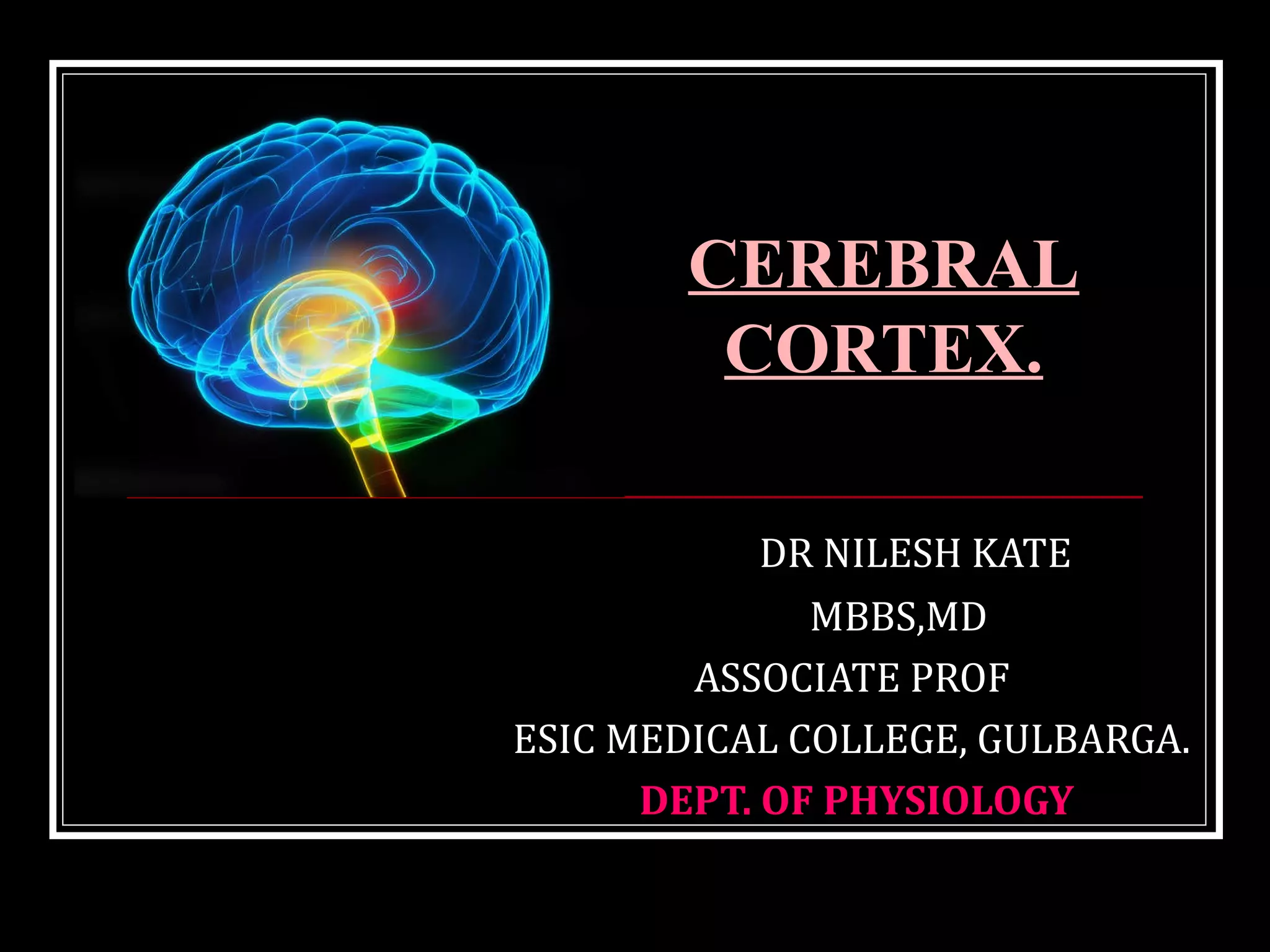
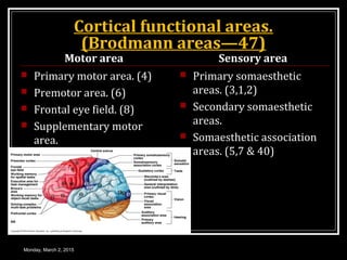
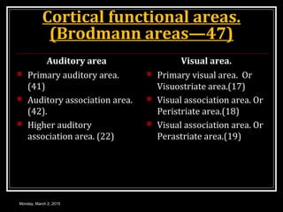
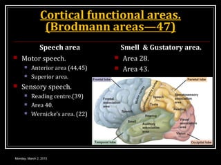
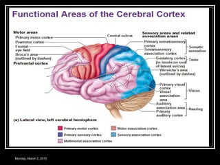
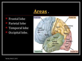
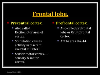
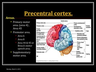
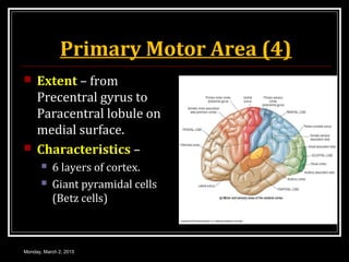
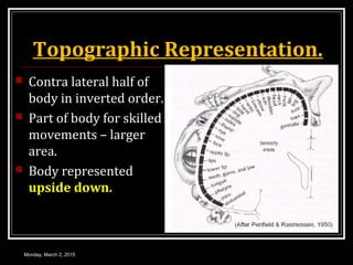
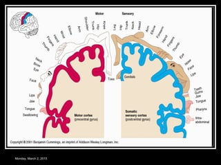
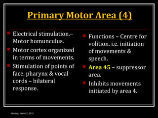
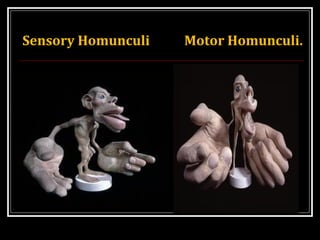
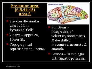
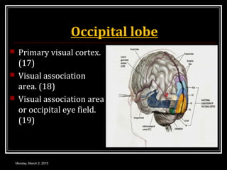
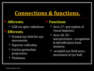
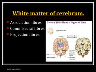
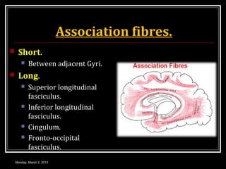
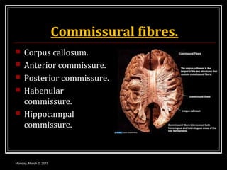
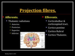
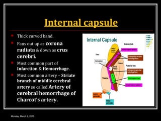
This document provides an overview of the cerebral cortex, including its external features, functional areas, lobes, connections, and histological structure. It discusses the different areas of the cortex such as the frontal lobe (including the precentral, premotor, and prefrontal cortex), parietal lobe (primary and secondary sensory areas), temporal lobe (primary auditory and association areas), occipital lobe, and their functions. It also covers applied aspects like frontal lobe syndrome and temporal lobe syndrome.
14. Laminae.
Outer pyramidal layer.
Mainly pyramidal cells,
stellate cells & basket
cells.
Inner granular layer.
Densely packed stellate
cells.
Inner zone prominent
transverse fibres.”
External Band of
Baillarger.
Monday, March 2, 2015
15. Laminae.
Inner pyramidal layer.
(Ganglionic)
Large pyramidal cells.
Developed in motor
cortex.
Inner Band of
Baillarger.
Polymorphous or
multiform layer.
Modified pyramidal cells.
Many fusiform cells.
Monday, March 2, 2015
16. Types of Neocortex.
Type I or agranular
cortex.
Granule cells & stellate
cells absent.
Pyramidal cells – large,
Betz cells.
Seen in Motor cortex
(4) & Broca’s area. (44)
Type II or frontal
cortex.
Triangular granular
cells
Seen in frontal lobe.
Monday, March 2, 2015
17. Types of Neocortex.
Type III or Parietal
cortex.
Depth & density of layer
II & III increased.
Granule cells – round.
Seen in parietal lobe &
junction of parietal,
temporal & occipital
lobe.
Type IV or Polar Type.
Cortex narrow
All layers reduced depth.
Seen in frontal &
occipital pole.
Type V or granular
cortex.
Excess granule cells
Seen in sensory cortex &
calcarine region.
Monday, March 2, 2015
18. Areas .
Frontal lobe
Parietal lobe
Temporal lobe
Occipital lobe.
Monday, March 2, 2015
19. Frontal lobe.
Precentral cortex.
Also called
Excitomotor area of
cortex.
Stimulation causes
activity in discrete
skeletal muscles
Sensorimotor cortex.—
sensory & motor
cortex.
Prefrontal cortex.
Also called prefrontal
lobe or Orbitofrontal
cortex.
Ant to area 8 & 44.
Monday, March 2, 2015
20. Precentral cortex.
Areas.
Primary motor
area. (area 4) ,
Area 45
Premotor area.
Area 6
Area 8
Area 44 & 45 or
Broca’s motor
speech area.
Supplementary
motor area.
Monday, March 2, 2015
21. Primary Motor Area (4)
Extent – from
Precentral gyrus to
Paracentral lobule on
medial surface.
Characteristics –
6 layers of cortex.
Giant pyramidal cells
(Betz cells)
Monday, March 2, 2015
22. Topographic Representation.
Contra lateral half of
body in inverted order.
Part of body for skilled
movements – larger
area.
Body represented
upside down.
Monday, March 2, 2015
24. Primary Motor Area (4)
Electrical stimulation.–
Motor homunculus.
Motor cortex organized
in terms of movements.
Stimulation of points of
face, pharynx & vocal
cords – bilateral
response.
Functions – Centre for
volition. i.e. initiation
of movements &
speech.
Area 45 – suppressor
area.
Inhibits movements
initiated by area 4.
Monday, March 2, 2015
26. Premotor area.
(6,8,44,45)
area 6
Structurally similar
except Giant
Pyramidal Cells.
2 parts – Upper 2a,
Lower 2b.
Topographical
representation – same.
Functions –
Integration of
voluntary movements.
Make skilled
movements accurate &
smooth.
Lesions – Hemiplegia
with Spastic paralysis.
Monday, March 2, 2015
27. Area 8
Frontal Eye field.
Location – ant to area 6.
Function – control eye
movements.
Lesions – turn eye to
opposite side.
Conjugate movements --
lost
Afferent – occipital
lobe & Dorsomedial N.
of Thalamus.
Efferents – N. of III, IV
& VI Cranial nerves.
Monday, March 2, 2015
28. Area 44 & 45.
Broca’s motor
speech area.
Area 44 – pars
Triangularis.
Area 45 – pars
Opercularis.
Functions – in
Dominant
Hemisphere
concerned with
movements
responsible for
production of voice &
articulation of speech.
Monday, March 2, 2015
29. Supplementary Motor Area.
Locations – medial
surface of frontal lobe
behind primary motor
area.
Topographical
representation.–
upper body located
dorsal to lower body.
Functions – with
Premotor area provide
attitudinal
movements., fixational
movements &
positional movements
of head & eye.
Monday, March 2, 2015
30. Connections of pre central cortex.
Afferents Efferents.
Monday, March 2, 2015
32. Prefrontal cortex.
Afferents
Dorsomedial nucleus of
thalamus.
Anterior nuclei of thalamus.
Form Papez circuit– close
circuit connection with
thalamus.
Responsible for resting
EEG & genesis of
Emotions.
Efferents
Thalamus. (V &M)
Tegmental reticular
formation.
Pontine nuclei
Caudate nucleus.
Mammilary bodies.
Monday, March 2, 2015
33. Functions.
Centre of planned
actions
Centre for higher
functions.
Emotions, learning,
memory & social
behaviour.
Causes autonomic
changes during emotional
conditions
Seat of intelligence.
Register short term
memory
Called An organ of
mind.
Control of intellectual
activities.
Monday, March 2, 2015
34. Control of Intellectual Activities.
To prognosticate.
Plan future.
Concentrate – Central
theme of thought.
Delay actions in
response to sensory
signals.
Consequences of
motor action before it.
Solution of
complicated
mathematical &
philosophical
problems.
Controls one’s activity
according to moral
laws.
Monday, March 2, 2015
35. Applied aspects.
Frontal lobe syndrome.
Cause – ablation of
prefrontal cortex.
Features. (FEEL –
FILL)
Flight of ideas.
Emotional instability.
Euphoria.
Loss of moral & social
sense.
Functional abnormal—
Hyperphagia.
Sphincters control loss.
Disorientation & tremors.
Impairment of memory.
Lack of attention &
concentration.
Lack of initiative & decreased
intellectual activity.
Monday, March 2, 2015
37. Primary sensory area. (3,1,& 2)
Location – Post
central Gyrus & Post
wall of Central sulcus.
Histo – dense stellate
cells , few pyramidal
cells.
Topographical
representations. –
Sensory Homunculus.
Lesion – without
thalamus only
discriminative
functions lost
With thalamus – all
sensation from opp
half of body.
Monday, March 2, 2015
38. Secondary sensory area.
Location – post central
Gyrus below 1st
somatic
sensory area.
Topographical
representation – body
represented twice in
area I & area II.
Lesion – discriminative
power lost.
Monday, March 2, 2015
39. Sensory Association Areas. (5, 7)
Post to area S1.
Point to point
representation.
Lesion – Tactile
Agnosia.
(Asteriognosis &
Tactile Aphasia)
Monday, March 2, 2015
40. Connections of Parietal Lobe
Afferents.
SI – Thalamus
(VPM,VPL)
Exteroceptive &
Proprioceptive from opp
side & both sides of face.
SII – from SI & thalamus
Sensory Association
area. – SI & SII
Efferents.
Corticospinal
Corticobulbar
Corticonuclear tracts.
Monday, March 2, 2015
41. Connections of Parietal Lobe
Association fibres.
To other cortical
areas.
Involve in somatic
sensation.
Commissural fibres.
Mostly pyramidal cells
connect to opp
hemisphere.
Opp area SI & SII
Monday, March 2, 2015
42. Functions of Parietal Lobe
First somatic sensory area. (SI)
Localize, analyze & Discriminate Cutaneous &
Proprioceptive senses.
Area 3 – touch, pressure, position & vibrations.
Area 1 – Cutaneous & joint sense.
Area 2 – deep sense from muscles & joints.
Monday, March 2, 2015
43. Parietal Lobe
Second somatic sensory area. (SII)
From S1 & thalamus directly.
Perception of sensation.
Sensory association area. (5,7)
Discriminate stimuli related to intensity.
Warm, warmer, cold, colder
Monday, March 2, 2015
44. Temporal lobe.
Primary auditory area.
41, 42 Audio sensory Area.
HEARING CENTRE.
Middle of superior
temporal gyrus.
From – MGB, Thalamus.
To – MGB, Superior
colliculus.
Perceives – loudness,
pitch, source & direction of
sound.
Auditory association area
22,21 & 20
22- Wernicke's area, also
called sensory speech area
in dominant hemisphere.
Interpretation of meaning,
comprehension of spoken
language & formation of
ideas.
21,20 – interpretation &
integration of auditory
impulses.
Monday, March 2, 2015
45. Applied Aspects
Unilateral removal
of temporal lobe
Causes no deafness
As ear bilaterally
represented in
auditory pathway &
project equally to 2
cerebral hemispheres.
Temporal lobe
syndrome. (Kluver-
Bucy syndrome)
Bilateral removal of
temporal lobe with
amygdala & Uncus.
Monday, March 2, 2015
53. Internal capsule
Thick curved band.
Fans out up as corona
radiata & down as crus
cerebri.
Most common part of
Infarction & Hemorrhage.
Most common artery – Striate
branch of middle cerebral
artery so called Artery of
cerebral hemorrhage of
Charcot’s artery.
Monday, March 2, 2015