Downloaded 217 times
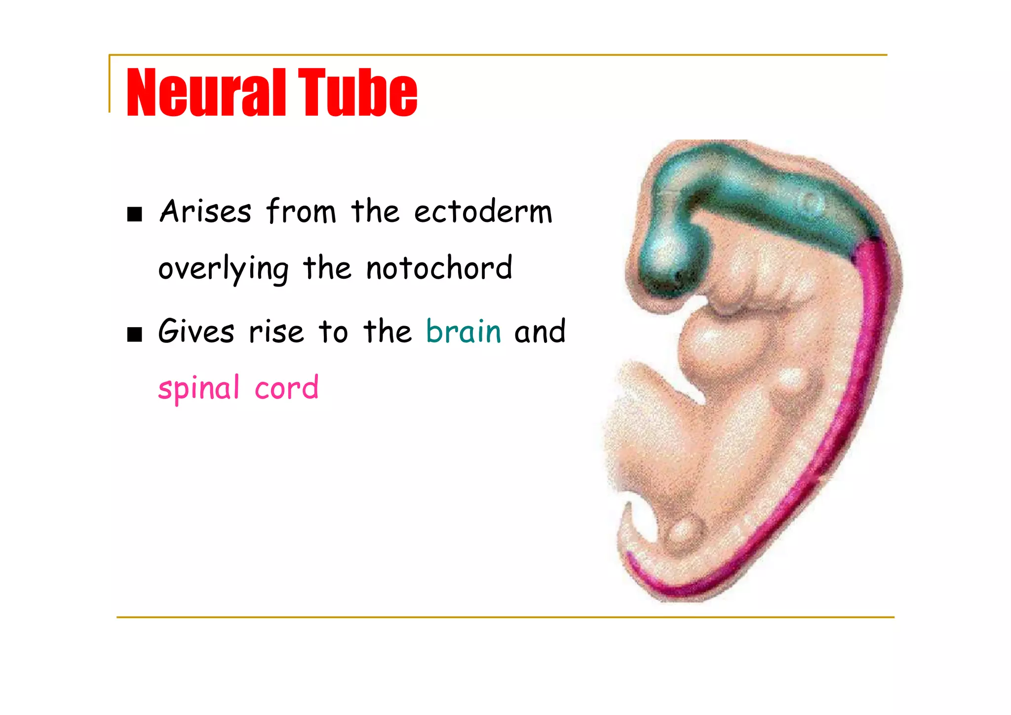
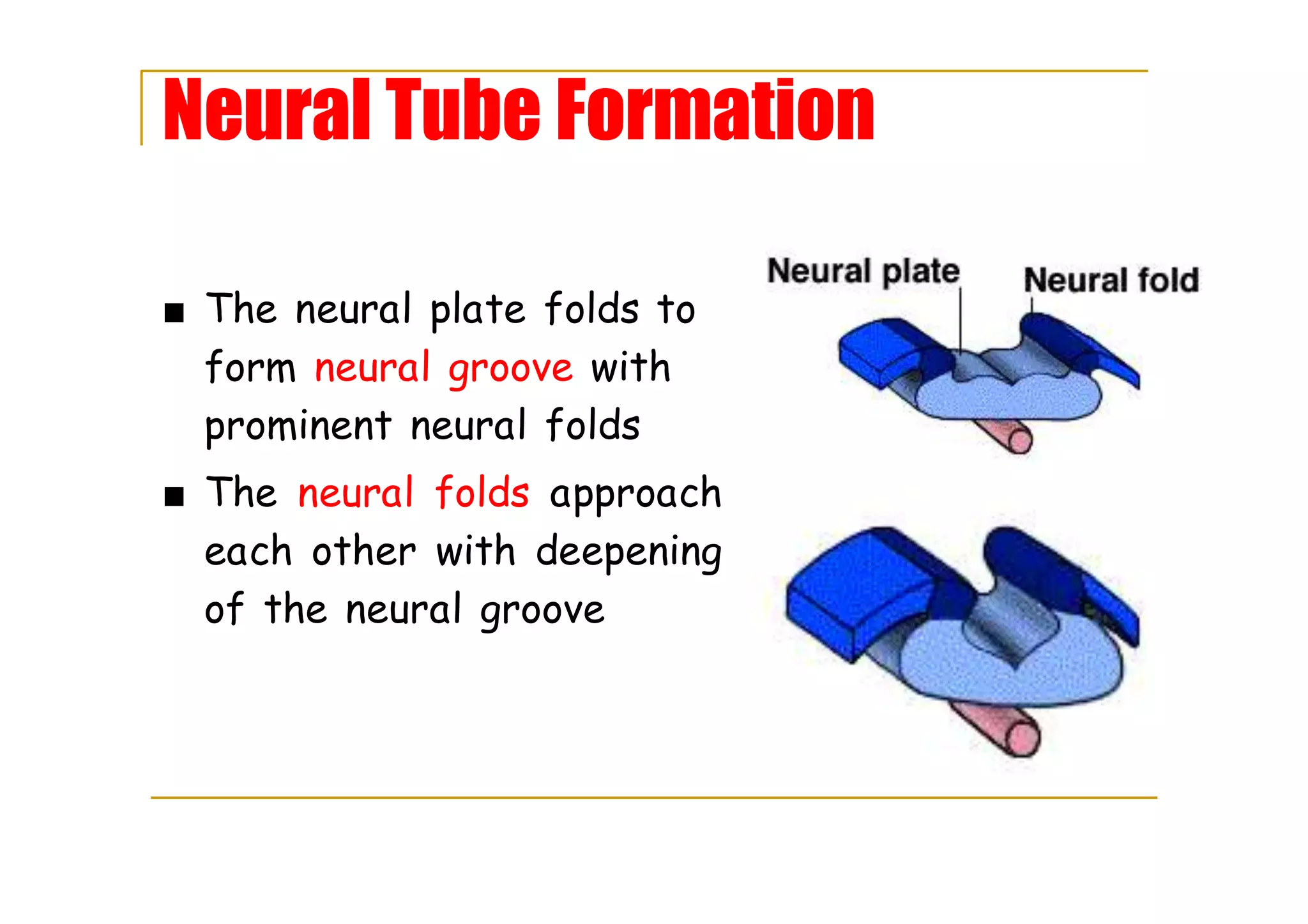
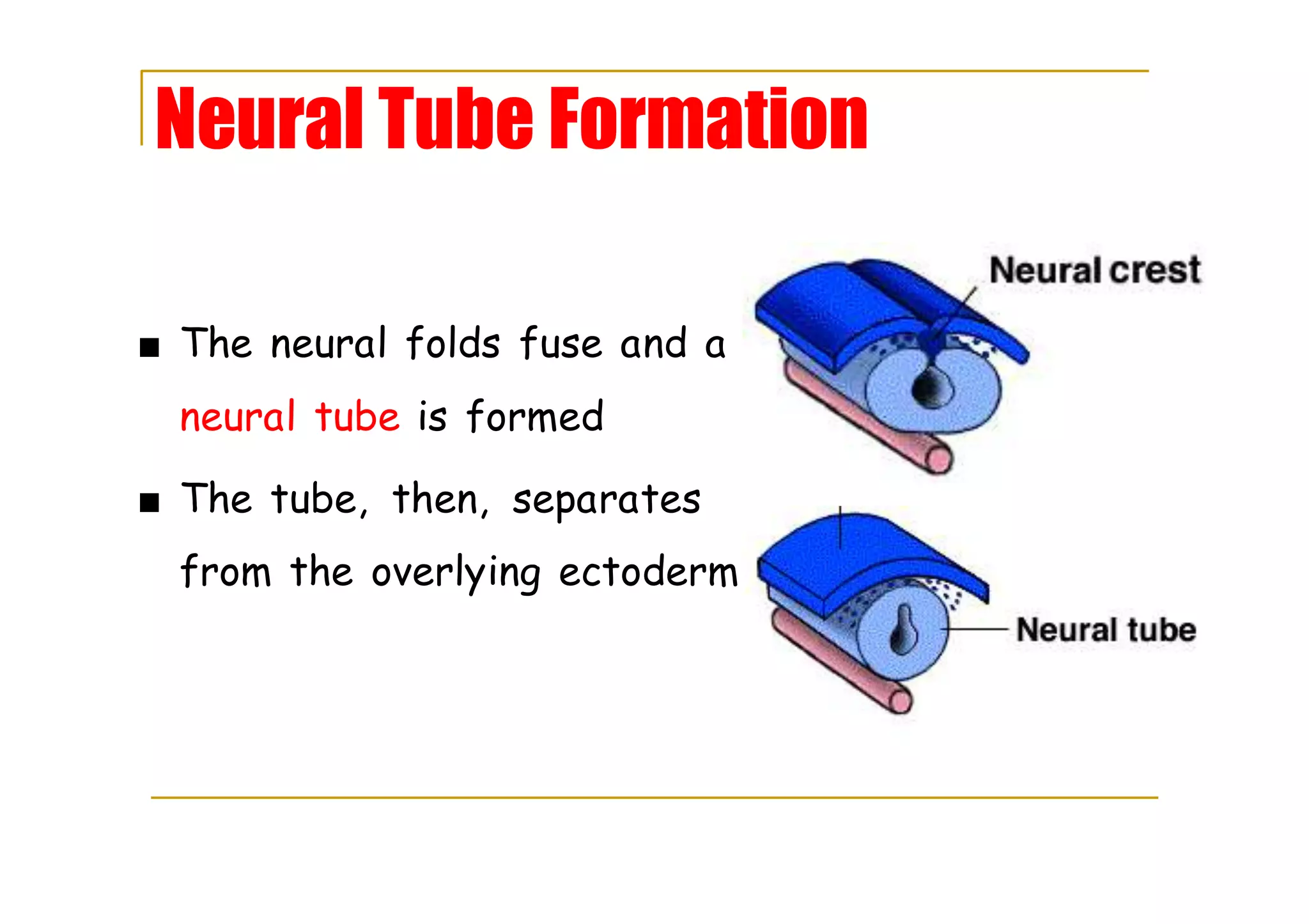
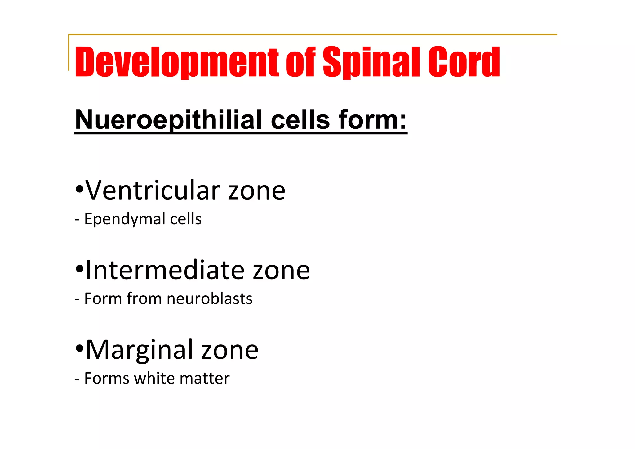
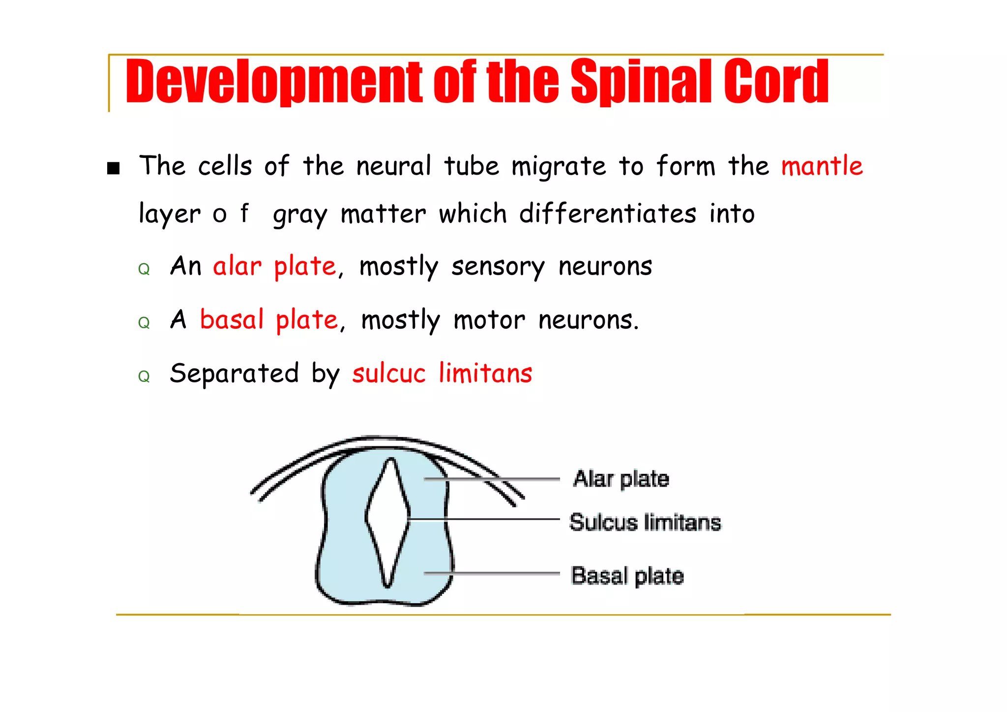
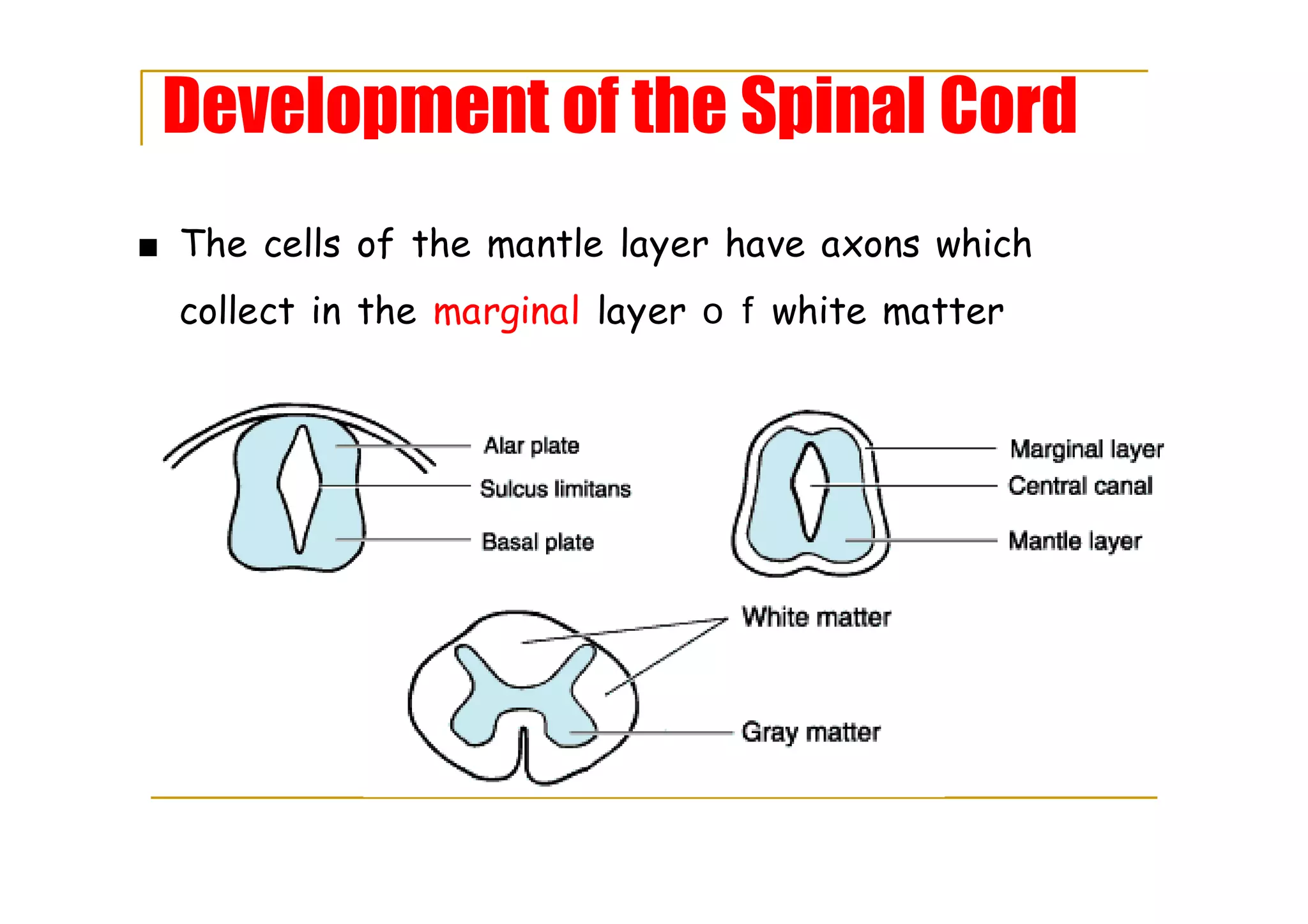
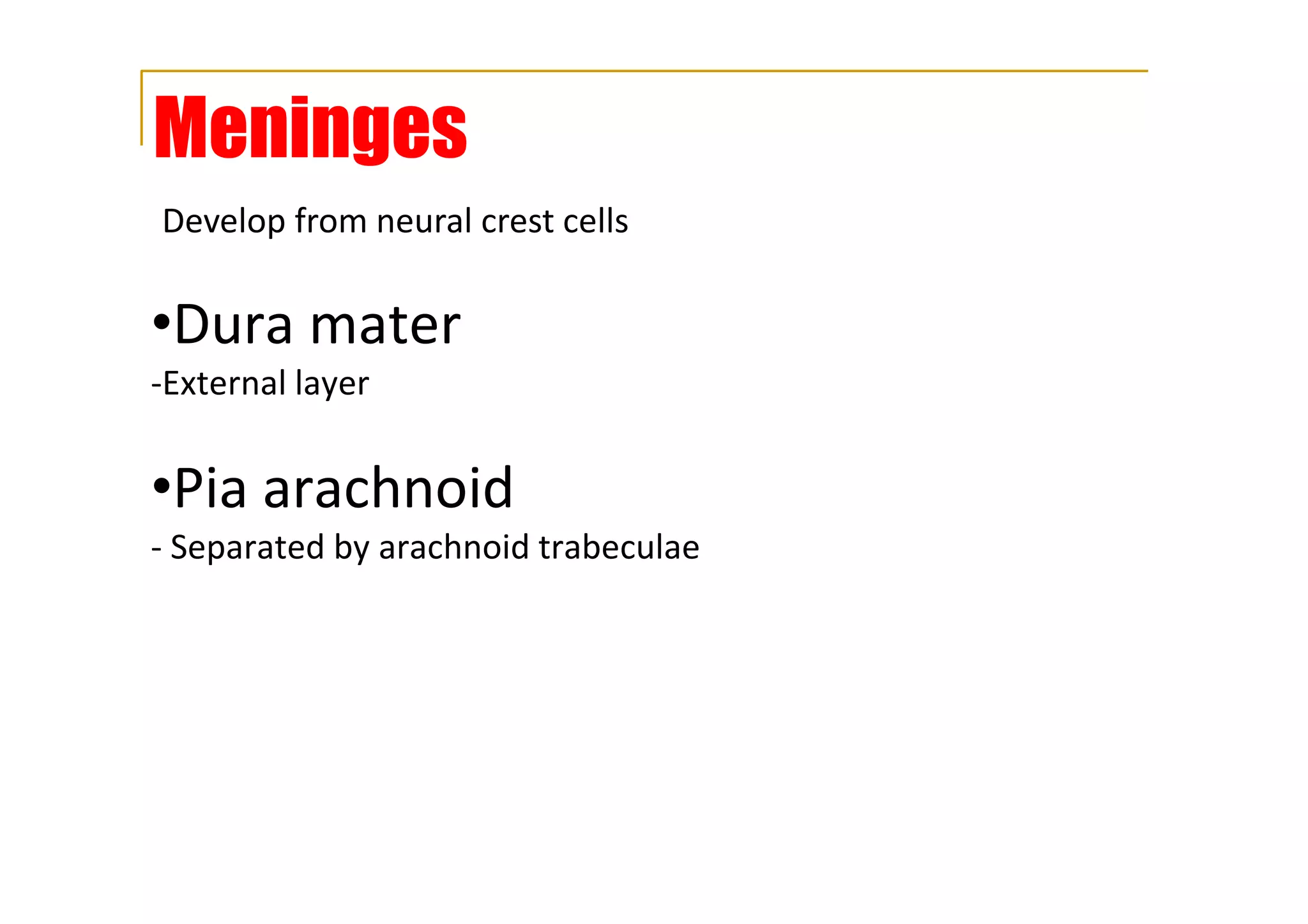
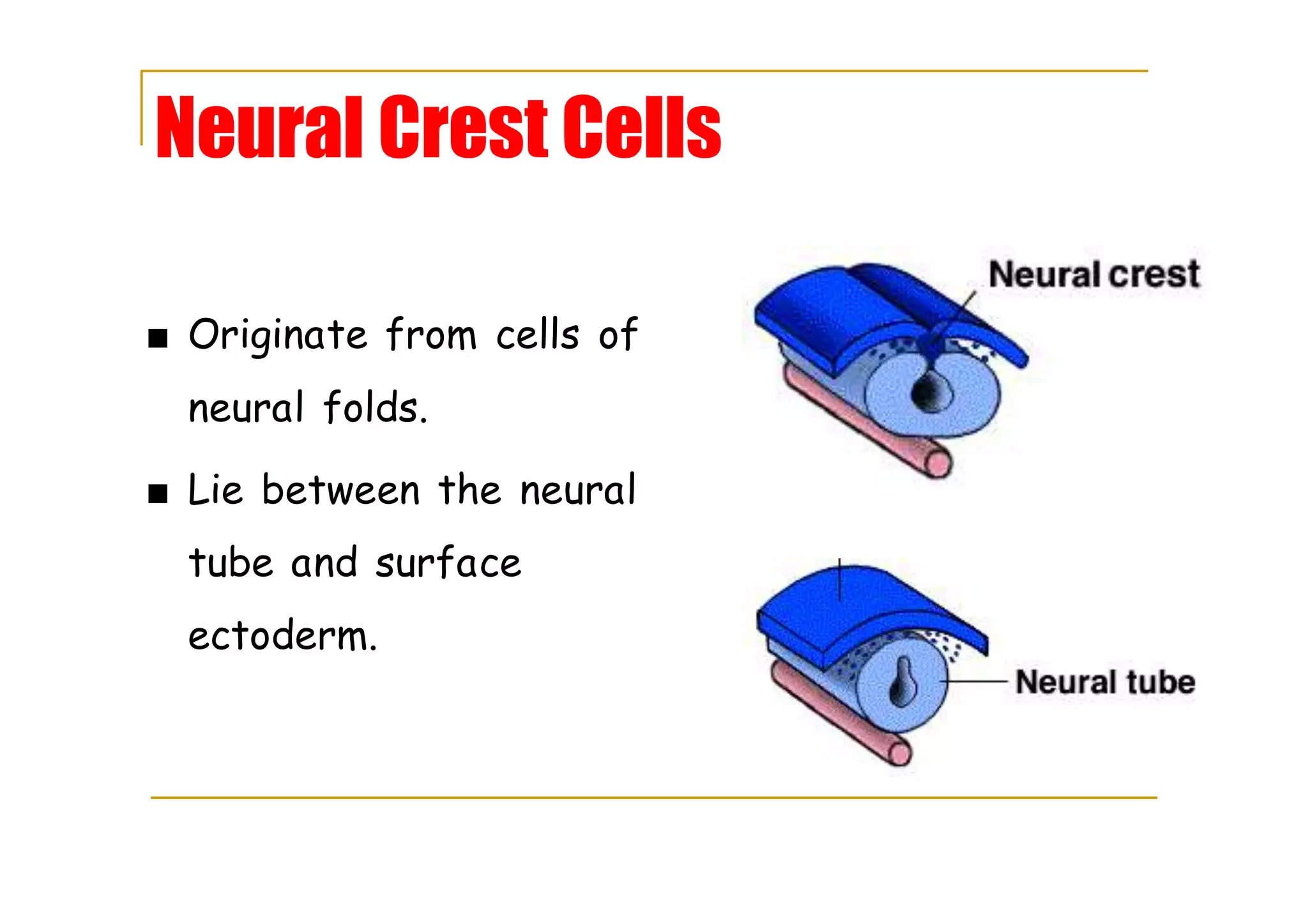
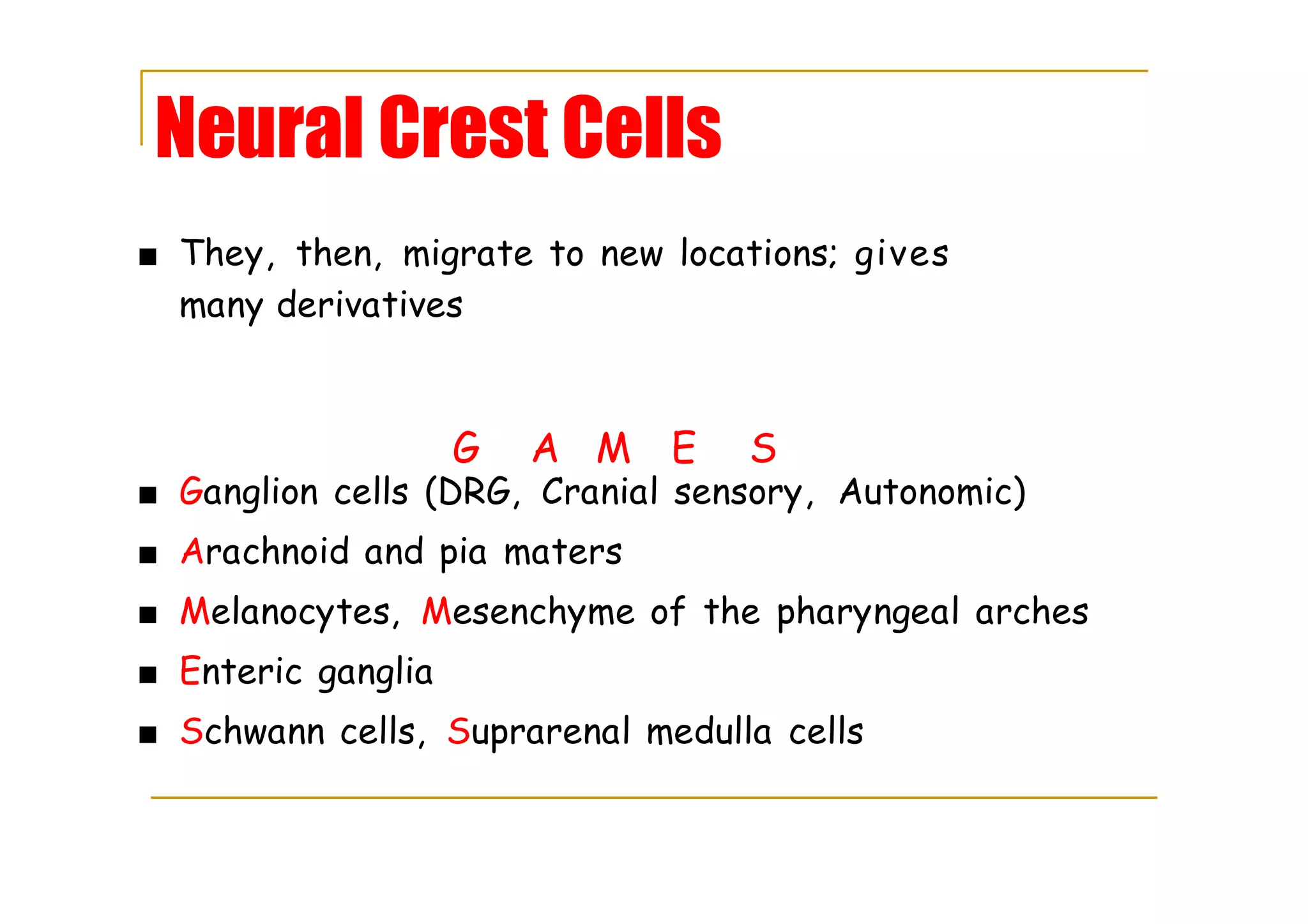


The spinal cord arises from the neural tube which forms when the neural plate folds and fuses. The neural tube is made of neuroepithelial cells that migrate and differentiate to form the gray matter of the spinal cord, consisting of the alar plate and basal plate. Axons from cells in the mantle layer collect in the marginal layer to form the white matter. Neural crest cells originate between the neural tube and ectoderm, migrate, and give rise to many structures including ganglion cells, meninges, melanocytes, and Schwann cells.


![Development of the Muscular System [Human Embryology]](https://cdn.slidesharecdn.com/ss_thumbnails/developmentofthemuscularsystemhumanembryology-190416145251-thumbnail.jpg?width=640&height=640&fit=bounds)


