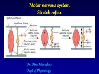
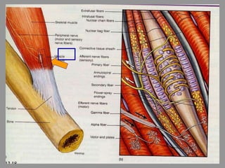
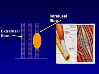
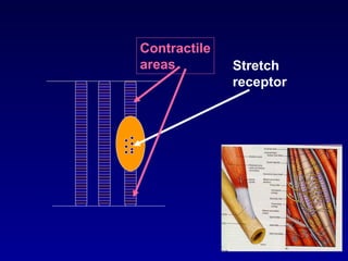
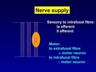
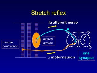
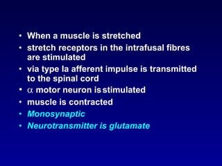
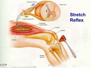
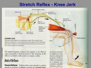
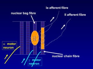
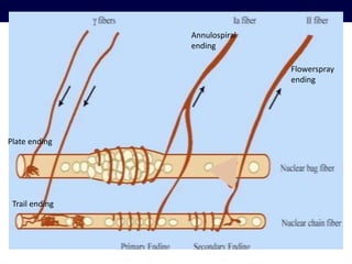
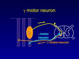
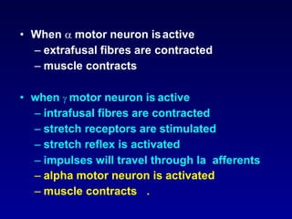
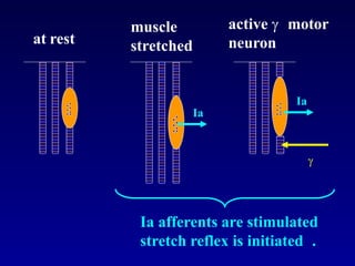
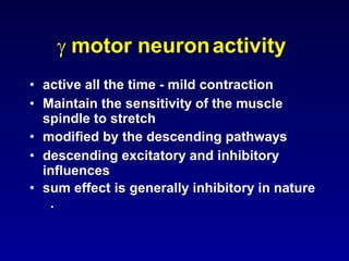
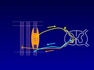
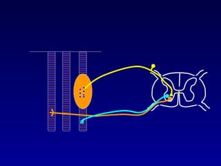
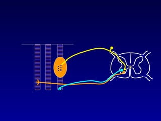
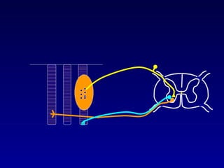
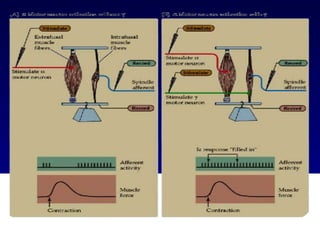
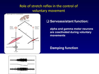
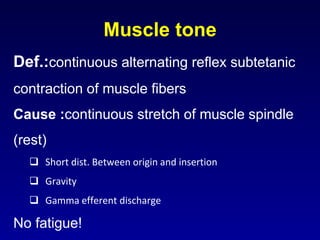
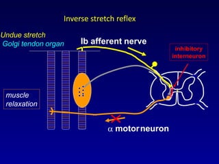
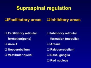
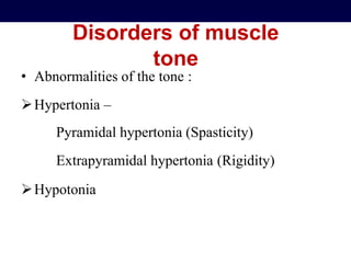
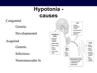
Dr: Dina Merzeban
Dept ofPhysiology
Motor nervous system
Stretch reflex
What is a reflex?
• Response to a stimulus
• Stimulus Response
Task:
Write down 3 reflexes .
What is a reflex?
Afferent nerve
Central
connections
Efferent nerve
Receptor
Stimulus
Response
Effector organ
Stretch reflex
• This is a basic reflex present in the
spinal cord
• Stimulus: muscle stretch
• Response: contraction of the muscle
• Receptors: stretch receptors located
in the muscle spindle .
skeletal muscle
• two types of muscle fibres
– extrafusal
• normally contracting fibres
– intrafusal
• non contractile fibres present inside the
muscle spindle
• lie parallel to extrafusal fibres
• contains stretch receptors .
Extrafusal
fibre
Intrafusal
fibre
Contractile
areas Stretch
receptor
Nerve supply
Sensory to intrafusal fibre:
Ia afferent
II afferent
Motor:
to extrafusal fibre
 motor neuron
to intrafusal fibre
 motor neuron .
Ia afferent nerve
 motorneuron
one
synapse
muscle
stretchmuscle
contraction
Stretch reflex
• When a muscle is stretched
• stretch receptors in the intrafusal fibres
are stimulated
• via type Ia afferent impulse is transmitted
to the spinal cord
•  motor neuron isstimulated
• muscle is contracted
• Monosynaptic
• Neurotransmitter is glutamate
Stretch
Reflex
Stretch Reflex - Knee Jerk
– nuclear bag fibre
• primary (Ia) afferent
– supplies annulospiral ending in the centre
– nuclear chain fibre
• primary (Ia) and secondary (II) afferent
– supplies flower spray ending .
two types of intrafusal fibres
Ia afferent fibre
II afferent fibre
nuclear bag fibre
 motor
neuron
nuclear chain fibre
 motor
neuron
Plate ending
Trail ending
Annulospiral
ending
Flowerspray
ending
Importance of stretch reflex
• detects muscle length and changes
in muscle length .
 motorneuron
• cell body is located in the anterior
horn
• motor neuron travels through the
motor nerve
• supplies the intrafusal fibres
(contractile elements at either end) .
 motor
neuron
 motorneuron
 motor neuron
• When  motor neuron isactive
– extrafusal fibres are contracted
– muscle contracts
• when  motor neuron is active
– intrafusal fibres are contracted
– stretch receptors are stimulated
– stretch reflex is activated
– impulses will travel through Ia afferents
– alpha motor neuron is activated
– muscle contracts .
at rest
muscle
stretched
active  motor
neuron
Ia
Ia

Ia afferents are stimulated
stretch reflex is initiated .
 motor neuronactivity
• active all the time - mild contraction
• Maintain the sensitivity of the muscle
spindle to stretch
• modified by the descending pathways
• descending excitatory and inhibitory
influences
• sum effect is generally inhibitory in nature
.
Alpha gamma co-activation
• gamma motoneurons are activated in
parallel with alpha motoneurons to
maintain the firing of spindle afferents
when the extrafusal muscles shorten
• Prevent unloading
Stretch reflex 2 types
-Response that is transmitted:
Dynemic:
-when there is change in the length of the spindle receptor
(stretching of the sensory receptor area of the muscle spindle
by stretching of the muscle spindle or the whole muscle).
Detect Change in length.
-transmitted by the primary fiber Aα type
Static
continuous information about the length of the muscle
(not the change in length).
transmitted by both the primary Aα and secondary (Aβ
and Aγ)
STATIC AND DYNAMIC RESPONSE
OF MUSCLE
 SPINDLE AFFRENTS
 Static response is the discharge at any constant length of the
muscle. The greater the muscle length greater is the stretch in the
spindle and the higher is the static response of the spindle
affrents. Both the primary (Iα) and secondary II spindle affrents
gives static or length sensitive responses.
 The dynamic response of a spindle affrents refer to the discharge
during stretch of the muscle. If the spindle affrents gives greater
response during a fast stretch than it dose during a slow stretch
(velocity different but distance of stretch same) it is said to poses
a dynamic response component. Only the primary spindle affrents
gives a dynamic or velocity sensitive response.
STATIC AND DYNAMIC FUSIMOTOR
NEURONS
 Dynamic fusimotor fiber increase the dynamic response
of the primary spindle affrents (Iα) and have little or no
effect on secondary.
 Static fusimotor fibers increases the static response of
both the primary and secondary spindle affrents.
However the effect of static fusimotor fiber on primary
spindle affrents is less marked than their effect on the
secondary.
 Static fusimotor fiber terminate as trail endings (mostly
present in nuclear chain fibers).
 Experiment using depletion of muscle glycogen as an
index of muscle fiber activity have shown that repetitive
stimulation of the static fusimotor fiber result primarily in
chain fiber glycogen depletion.
 Dynamic fusimotor stimulation produces mostly bag
fiber glycogen depletion.
Significance of stretch
reflex
 Maintain muscle length
 Control of voluntary movement
 Muscle tone
 Servoassistant function:
alpha and gamma motor neurons
are coactivated during voluntary
movements
Damping function
Role of stretch reflex in the control of
voluntary movement
Muscle tone
Def.:continuous alternating reflex subtetanic
contraction of muscle fibers
Cause :continuous stretch of muscle spindle
(rest)
 Short dist. Between origin and insertion
 Gravity
 Gamma efferent discharge
No fatigue!
Function:
 Posture against gravity
 Background for voluntary movement
 Regulation of body temp.
 Venous and lymph return
Inverse stretch reflex
• When the muscle is strongly
stretched
• Golgi tendon organs are stimulated
• Via type Ib afferents impulse is
transmitted to the spinal cord
• inhibitory interneuron is stimulated
•  motor neuron is inhibited
• muscle is relaxed .
GOLGI TENDON ORGAN
Inverse Stretch Reflex
 motorneuron
Undue stretch
Golgi tendon organ
muscle
relaxation
Ib afferent nerve
inhibitory
interneuron
Inverse stretch reflex
Importance of inverse
stretch reflex
• detects muscle tension .
Supraspinal regulation
Facilitatory areas
 Facilitatory reticular
formation(pons)
 Area 4
 Neocerebellum
 Vestibular nuclei
Inhibitory areas
 Inhibitory reticular
formation (medulla)
 Area4s
 Paleocerebellum
 Basal ganglia
 Red nucleus
Disorders of muscle
tone
• Abnormalities of the tone :
Hypertonia –
Pyramidal hypertonia (Spasticity)
Extrapyramidal hypertonia (Rigidity)
Hypotonia
Pyramidal hypertonia (Spasticity)
• Spasticity – a motor disorder characterized by
velocity- dependent increase in muscle tone with
exaggerated tendon jerks, resulting from
hyperexcitability of the stretch reflex.
• Pyramidal hypertonia is most pronounced in the
muscle groups most used in voluntary
movements.
CLASP KNIFE REFLEX
 Seen in decerebrate rigidity
 On stretching the muscle beyond a point causes Ib
affrent inhibitory discharge from GTO which reflexly
inhibits homonymous stretched muscle
Spasticity
• Physiologic evidence suggests that interruption of
reticulospinal projections is important in the genesis of
spasticity.
• In spinal cord lesions, bilateral damage to the
pyramidal and reticulospinal pathways can produce
severe spasticity and flexor spasms, reflecting increased
tone in flexor muscle groups and weakness of extensor
muscles.
Clonus
• Clonus is the phenomenon of involuntary rhythmic contractions
in response to sudden sustained stretch.
• A sudden stretch activates muscle spindles, resulting inthe
stretch reflex.
• Tension produced by the muscle contraction activates the Golgi
tendon organs, which in turn activate an „inverse stretch reflex‟,
relaxing the muscle.
• If the stretch is sustained, the muscle spindles are again activated,
causing a cycle of alternating contractions and relaxations.
Cerebellum and muscle
tone
• The cerebellum does not seem to have a direct effect on
muscle tone determining spinal reflex pathways as
there is no direct descending cerebellospinal tract.
• The cerebellum mainly influences muscle tone through
its connections with the vestibular and brain stem
reticular nuclei.
• Pure cerebellar lesions classically produce hypotonia.
Extrapyramidal hypertonia
(Rigidity)
• Rigidity is characterized by an increase in muscle tone causing
resistance to externally imposed joint movements.
• It does not depend on imposed speed and can be elicited at
very low speeds of passive movement.
• It is felt in both agonist and antagonist muscles and in
movements in both directions.
Extrapyramidal hypertonia
(Rigidity)
• 'Cogwheel' rigidity and 'leadpipe' rigidity are two types.
• 'Leadpipe' rigidity results when an increase in muscle tone causes
a sustained resistance to passive movement throughout the
whole range of motion, with no fluctuations.
• 'Cogwheel' rigidity occus in association with tremor which
presents as a jerky resistance to passive movement as muscles
tense and relax.
• Basal ganglia structures are clearly implicated in pathophysiology
of rigidity.
Hypotonia
• Hypotonia may affect muscleresistance to passive
movement and/or its extensibility.
• Aetiological types of hypotonia :
1. Nerve trunk and root lesion
2. A lesion of anterior horn
3. Cerebellar lesions
4. Cerebral lesions
Hypotonia -
causes
Congenital
Genetic
Developmental
Acquired
Genetic
Infectious
Neuromuscular Jn
Clinical
Examination
 Tone is difficult to assess.
 The determination of tone is subjective and prone to interexaminer
variability.
 The most important part of the examination of tone is determination
of the resistance of relaxed muscles to passive manipulation as well
as the extensibility, flexibility, and range of motion.
 The examination of tone needs a relaxed & cooperative patient
Methods
• Inspection : Attitude of the limb atrest.
• Palpation : Feel of the muscle – normal, firm orflabby.
• Range of movement at the joints.
• Passive movement - first slowly and through complete range ofmotion
and then at varying speeds.
• Shake the distal part of the limb.
• Brace a limb and suddenly remove support.
• Bilateral examination of homologous parts helps comparefor
differences in tone on the two sides of the body.

motor nervous system : Stretch reflex

  • 1.
    Dr: Dina Merzeban DeptofPhysiology Motor nervous system Stretch reflex
  • 2.
    What is areflex? • Response to a stimulus • Stimulus Response Task: Write down 3 reflexes .
  • 3.
    What is areflex? Afferent nerve Central connections Efferent nerve Receptor Stimulus Response Effector organ
  • 4.
    Stretch reflex • Thisis a basic reflex present in the spinal cord • Stimulus: muscle stretch • Response: contraction of the muscle • Receptors: stretch receptors located in the muscle spindle .
  • 6.
    skeletal muscle • twotypes of muscle fibres – extrafusal • normally contracting fibres – intrafusal • non contractile fibres present inside the muscle spindle • lie parallel to extrafusal fibres • contains stretch receptors .
  • 7.
  • 8.
  • 9.
    Nerve supply Sensory tointrafusal fibre: Ia afferent II afferent Motor: to extrafusal fibre  motor neuron to intrafusal fibre  motor neuron .
  • 10.
    Ia afferent nerve motorneuron one synapse muscle stretchmuscle contraction Stretch reflex
  • 11.
    • When amuscle is stretched • stretch receptors in the intrafusal fibres are stimulated • via type Ia afferent impulse is transmitted to the spinal cord •  motor neuron isstimulated • muscle is contracted • Monosynaptic • Neurotransmitter is glutamate
  • 12.
  • 13.
  • 14.
    – nuclear bagfibre • primary (Ia) afferent – supplies annulospiral ending in the centre – nuclear chain fibre • primary (Ia) and secondary (II) afferent – supplies flower spray ending . two types of intrafusal fibres
  • 15.
    Ia afferent fibre IIafferent fibre nuclear bag fibre  motor neuron nuclear chain fibre  motor neuron
  • 16.
  • 17.
    Importance of stretchreflex • detects muscle length and changes in muscle length .
  • 18.
     motorneuron • cellbody is located in the anterior horn • motor neuron travels through the motor nerve • supplies the intrafusal fibres (contractile elements at either end) .
  • 19.
  • 20.
    • When motor neuron isactive – extrafusal fibres are contracted – muscle contracts • when  motor neuron is active – intrafusal fibres are contracted – stretch receptors are stimulated – stretch reflex is activated – impulses will travel through Ia afferents – alpha motor neuron is activated – muscle contracts .
  • 21.
    at rest muscle stretched active motor neuron Ia Ia  Ia afferents are stimulated stretch reflex is initiated .
  • 22.
     motor neuronactivity •active all the time - mild contraction • Maintain the sensitivity of the muscle spindle to stretch • modified by the descending pathways • descending excitatory and inhibitory influences • sum effect is generally inhibitory in nature .
  • 24.
    Alpha gamma co-activation •gamma motoneurons are activated in parallel with alpha motoneurons to maintain the firing of spindle afferents when the extrafusal muscles shorten • Prevent unloading
  • 29.
    Stretch reflex 2types -Response that is transmitted: Dynemic: -when there is change in the length of the spindle receptor (stretching of the sensory receptor area of the muscle spindle by stretching of the muscle spindle or the whole muscle). Detect Change in length. -transmitted by the primary fiber Aα type Static continuous information about the length of the muscle (not the change in length). transmitted by both the primary Aα and secondary (Aβ and Aγ)
  • 30.
    STATIC AND DYNAMICRESPONSE OF MUSCLE  SPINDLE AFFRENTS  Static response is the discharge at any constant length of the muscle. The greater the muscle length greater is the stretch in the spindle and the higher is the static response of the spindle affrents. Both the primary (Iα) and secondary II spindle affrents gives static or length sensitive responses.  The dynamic response of a spindle affrents refer to the discharge during stretch of the muscle. If the spindle affrents gives greater response during a fast stretch than it dose during a slow stretch (velocity different but distance of stretch same) it is said to poses a dynamic response component. Only the primary spindle affrents gives a dynamic or velocity sensitive response.
  • 31.
    STATIC AND DYNAMICFUSIMOTOR NEURONS  Dynamic fusimotor fiber increase the dynamic response of the primary spindle affrents (Iα) and have little or no effect on secondary.  Static fusimotor fibers increases the static response of both the primary and secondary spindle affrents. However the effect of static fusimotor fiber on primary spindle affrents is less marked than their effect on the secondary.  Static fusimotor fiber terminate as trail endings (mostly present in nuclear chain fibers).  Experiment using depletion of muscle glycogen as an index of muscle fiber activity have shown that repetitive stimulation of the static fusimotor fiber result primarily in chain fiber glycogen depletion.  Dynamic fusimotor stimulation produces mostly bag fiber glycogen depletion.
  • 32.
    Significance of stretch reflex Maintain muscle length  Control of voluntary movement  Muscle tone
  • 33.
     Servoassistant function: alphaand gamma motor neurons are coactivated during voluntary movements Damping function Role of stretch reflex in the control of voluntary movement
  • 34.
    Muscle tone Def.:continuous alternatingreflex subtetanic contraction of muscle fibers Cause :continuous stretch of muscle spindle (rest)  Short dist. Between origin and insertion  Gravity  Gamma efferent discharge No fatigue!
  • 35.
    Function:  Posture againstgravity  Background for voluntary movement  Regulation of body temp.  Venous and lymph return
  • 36.
    Inverse stretch reflex •When the muscle is strongly stretched • Golgi tendon organs are stimulated • Via type Ib afferents impulse is transmitted to the spinal cord • inhibitory interneuron is stimulated •  motor neuron is inhibited • muscle is relaxed .
  • 37.
  • 38.
  • 39.
     motorneuron Undue stretch Golgitendon organ muscle relaxation Ib afferent nerve inhibitory interneuron Inverse stretch reflex
  • 40.
    Importance of inverse stretchreflex • detects muscle tension .
  • 41.
    Supraspinal regulation Facilitatory areas Facilitatory reticular formation(pons)  Area 4  Neocerebellum  Vestibular nuclei Inhibitory areas  Inhibitory reticular formation (medulla)  Area4s  Paleocerebellum  Basal ganglia  Red nucleus
  • 43.
    Disorders of muscle tone •Abnormalities of the tone : Hypertonia – Pyramidal hypertonia (Spasticity) Extrapyramidal hypertonia (Rigidity) Hypotonia
  • 44.
    Pyramidal hypertonia (Spasticity) •Spasticity – a motor disorder characterized by velocity- dependent increase in muscle tone with exaggerated tendon jerks, resulting from hyperexcitability of the stretch reflex. • Pyramidal hypertonia is most pronounced in the muscle groups most used in voluntary movements.
  • 45.
    CLASP KNIFE REFLEX Seen in decerebrate rigidity  On stretching the muscle beyond a point causes Ib affrent inhibitory discharge from GTO which reflexly inhibits homonymous stretched muscle
  • 46.
    Spasticity • Physiologic evidencesuggests that interruption of reticulospinal projections is important in the genesis of spasticity. • In spinal cord lesions, bilateral damage to the pyramidal and reticulospinal pathways can produce severe spasticity and flexor spasms, reflecting increased tone in flexor muscle groups and weakness of extensor muscles.
  • 47.
    Clonus • Clonus isthe phenomenon of involuntary rhythmic contractions in response to sudden sustained stretch. • A sudden stretch activates muscle spindles, resulting inthe stretch reflex. • Tension produced by the muscle contraction activates the Golgi tendon organs, which in turn activate an „inverse stretch reflex‟, relaxing the muscle. • If the stretch is sustained, the muscle spindles are again activated, causing a cycle of alternating contractions and relaxations.
  • 48.
    Cerebellum and muscle tone •The cerebellum does not seem to have a direct effect on muscle tone determining spinal reflex pathways as there is no direct descending cerebellospinal tract. • The cerebellum mainly influences muscle tone through its connections with the vestibular and brain stem reticular nuclei. • Pure cerebellar lesions classically produce hypotonia.
  • 49.
    Extrapyramidal hypertonia (Rigidity) • Rigidityis characterized by an increase in muscle tone causing resistance to externally imposed joint movements. • It does not depend on imposed speed and can be elicited at very low speeds of passive movement. • It is felt in both agonist and antagonist muscles and in movements in both directions.
  • 50.
    Extrapyramidal hypertonia (Rigidity) • 'Cogwheel'rigidity and 'leadpipe' rigidity are two types. • 'Leadpipe' rigidity results when an increase in muscle tone causes a sustained resistance to passive movement throughout the whole range of motion, with no fluctuations. • 'Cogwheel' rigidity occus in association with tremor which presents as a jerky resistance to passive movement as muscles tense and relax. • Basal ganglia structures are clearly implicated in pathophysiology of rigidity.
  • 51.
    Hypotonia • Hypotonia mayaffect muscleresistance to passive movement and/or its extensibility. • Aetiological types of hypotonia : 1. Nerve trunk and root lesion 2. A lesion of anterior horn 3. Cerebellar lesions 4. Cerebral lesions
  • 52.
  • 53.
    Clinical Examination  Tone isdifficult to assess.  The determination of tone is subjective and prone to interexaminer variability.  The most important part of the examination of tone is determination of the resistance of relaxed muscles to passive manipulation as well as the extensibility, flexibility, and range of motion.  The examination of tone needs a relaxed & cooperative patient
  • 54.
    Methods • Inspection :Attitude of the limb atrest. • Palpation : Feel of the muscle – normal, firm orflabby. • Range of movement at the joints. • Passive movement - first slowly and through complete range ofmotion and then at varying speeds. • Shake the distal part of the limb. • Brace a limb and suddenly remove support. • Bilateral examination of homologous parts helps comparefor differences in tone on the two sides of the body.