Downloaded 298 times
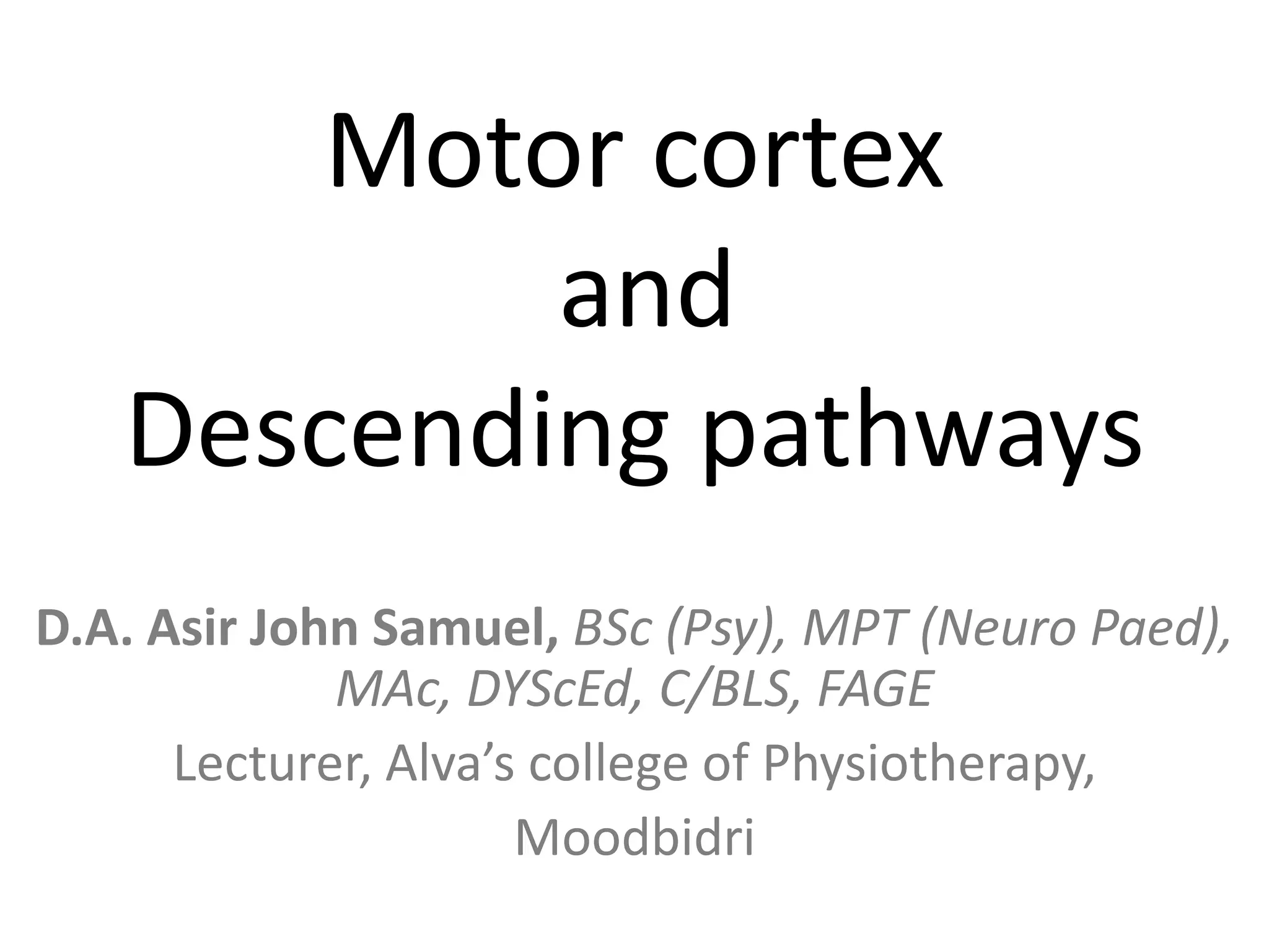
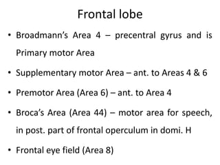
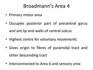
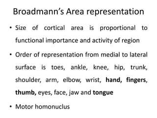
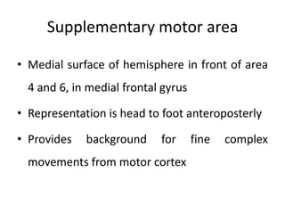
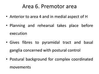
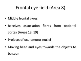

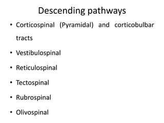
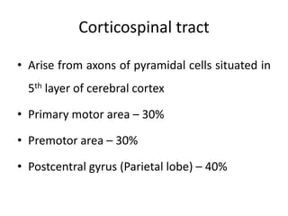
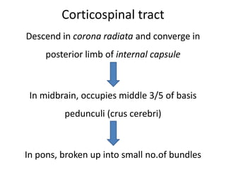
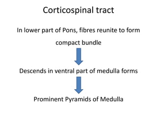
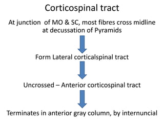
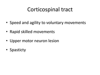
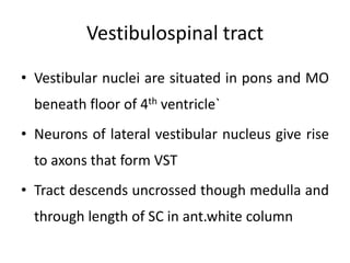
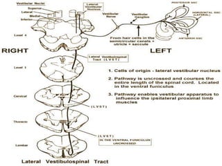
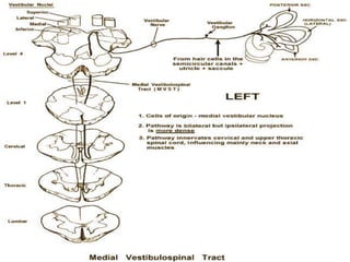
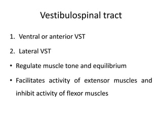
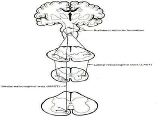
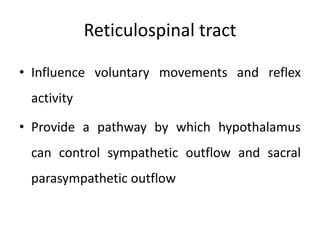
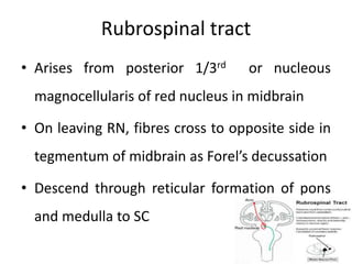
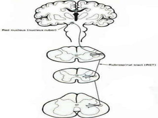
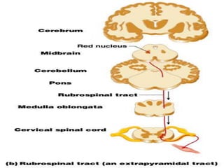
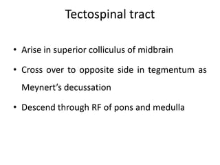
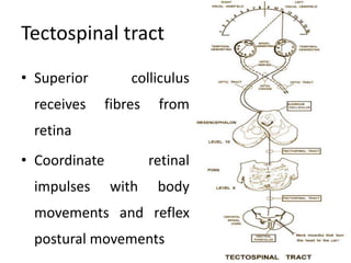
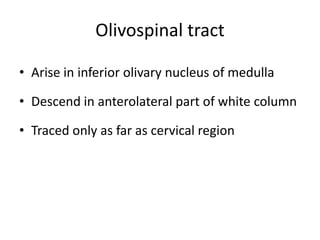

The document discusses the structure and function of the motor cortex and its descending pathways, including the roles of different brain regions like the frontal and parietal lobes in motor control. It details specific areas within the motor cortex, such as the primary motor area and supplementary motor area, along with various descending pathways like corticospinal and vestibulospinal tracts that regulate muscle tone and voluntary movements. Additional pathways mentioned include reticulospinal, rubrospinal, tectospinal, and olivospinal tracts, highlighting their respective contributions to motor function and coordination.


![ONFH[AVN HIP] -TRIPLE REGIME -A NOVAL SURGICAL CONCEPT .pptx](https://cdn.slidesharecdn.com/ss_thumbnails/onfhavnhip2026koaconcalicutdrgokuldevdrmashraf-260210064517-213ec005-thumbnail.jpg?width=640&height=640&fit=bounds)
![PERI-PROSTHETIC FRACTURE NAIL-PLATE CONSTRUCT [NPC].pptx](https://cdn.slidesharecdn.com/ss_thumbnails/drarunkumardrmohamedashrafperiprostheticfrasturenail-plateconstructnpc-260209164459-7e9d15a1-thumbnail.jpg?width=640&height=640&fit=bounds)


