Downloaded 12 times


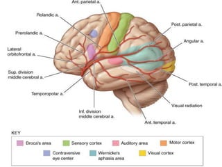


![*Destruction of brodmann area 3, 1, and 2 results in contralateral hemihypesthesia and
astereognosis.
*It could also reduce nociception, thermoception, and crude touch, but, since
information from the spinothalamic tract is interpreted mainly by other areas of the brain
(see insular cortex and cingulate gyrus), it is not as relevant as the other
symptoms[citation needed].](https://image.slidesharecdn.com/cns-160413160930/85/Cns-24-320.jpg)


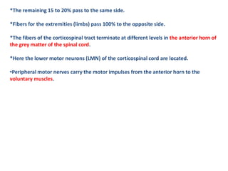
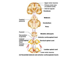


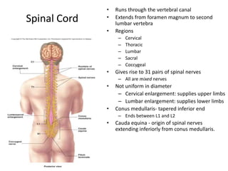

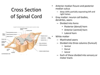


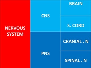
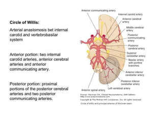
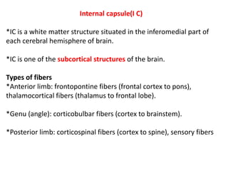
The document provides information about the nervous system including the central nervous system (CNS), peripheral nervous system (PNS), and their components. The CNS is made up of the brain and spinal cord. The brain is divided into the cerebrum, brainstem, cerebellum, and ventricles. The PNS includes the cranial and spinal nerves. The document also discusses the arterial blood supply of the brain, internal capsule, sensory and motor systems, and structure and components of the spinal cord.


![Introduction to the nervous system and nerve tissue[1]](https://cdn.slidesharecdn.com/ss_thumbnails/may2013introductiontothenervoussystemandnervetissue1-150530193624-lva1-app6891-thumbnail.jpg?width=640&height=640&fit=bounds)


