Neuroanatomy seminar with special reference to trigeminal,facial and glossopharyngeal nerve
•Download as PPTX, PDF•
13 likes•334 views

This presentation includes course of trigeminal nerve,facial nerve and glossopharyngeal nerve and its pedodontic implications

Recommended
More Related Content
What's hot
What's hot (20)
Similar to Neuroanatomy seminar with special reference to trigeminal,facial and glossopharyngeal nerve
Similar to Neuroanatomy seminar with special reference to trigeminal,facial and glossopharyngeal nerve (20)
More from Karishma Sirimulla
More from Karishma Sirimulla (20)
Recently uploaded
Recently uploaded (20)
Neuroanatomy seminar with special reference to trigeminal,facial and glossopharyngeal nerve
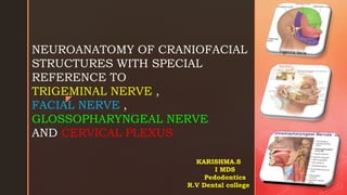
- 1. z NEUROANATOMY OF CRANIOFACIAL STRUCTURES WITH SPECIAL REFERENCE TO TRIGEMINAL NERVE , FACIAL NERVE , GLOSSOPHARYNGEAL NERVE AND CERVICAL PLEXUS KARISHMA.S I MDS Pedodontics R.V Dental college
- 2. CONTENTS Terminologies Functional components Parts of nervous system Cranial nerves Trigeminal nerve -course and branches Facial nerve - course and branches Glossopharyngeal nerve - course and branches Cervical plexus Conclusion References
- 3. TERMINOLOGIES NEURON : Structural & Functional unit of Nervous System. NUCLEUS: Applies to an aggregate of nerve cell bodies located within the CNS. GANGLION: is a collection of nerve cell bodies outside the brain and spinal cord(CNS) e.g: a) sensory ganglia of cranial nerves semilunar , geniculate b) parasympathetic ganglia ciliary, submandibular
- 4. TRACT: defined as a group of nerve cell processes within the CNS. NERVE: is a bundle of neuronal processes outside the CNS. Sensory, Motor, Mixed. PLEXUS: site regrouping of peripheral nerve fibers deriving from diverse origins.
- 5. FUNCTIONAL COMPONENTS: A cranial nerve consists of motor fibers or sensory fibers or both motor and sensory fibers TYPES: MOTOR: 1. GSE: They supply the striated muscles which develop from somites.These fibers convey motor impulses to skeletal muscles. 2. GVE: They supply the glands,smooth muscles of viscera and vessels.They are preganglionic parasympathetic fibers. 3. SVE: They supply the muscles which develop from mesoderm (special) of pharyngeal arches.
- 6. SENSORY: 1.GSA: They carry general sensations of pain, touch and temperature from skin and proprioceptive sensation of vibration and muscle and joint sense. 2.GVA: They carry general sensation of distention and ischemic pain from viscera. 3.SVA: They carry special sensation of taste from tongue. (special) 4.SSA: They carry special sensation of smell,hearing and (special somatic) balance.
- 7. PARTS OF THE NERVOUS SYSTEM
- 8. CENTRAL NERVOUS SYSTEM Brain- control center of nervous system receives sensory input from spinal cord as well its own nerves. Ex: olfactory, optic It consists of six major parts Cerebrum Diencephalon Midbrain Pons Medulla oblongata Cerebrllum Spinal cord- Conducts sensory information from PNS to brain.Conducts motor information from brain to various effectors.
- 9. PERIPHERAL NERVOUS SYSTEM It consists of 12 pairs of cranial nerves 31 pairs of spinal nerves SPINAL NERVES : 8 pairs of cervical nerves (C1 – C8) 12 pairs of thorasic nerves (T1-T12) 5 pairs of lumbar nerves (L1-L5) 5 pairs of sacral nerves (S1-S5) And one coccygeal pair of nerves
- 10. CRANIAL NERVES: Ⅰ Olfactory nerve Ⅱ Optic nerve Ⅲ Oculomotor nerve Ⅳ Trochlear nerve Ⅴ Trigeminal nerve Ⅵ Abducent nerve Ⅶ Facial nerve Ⅷ Vestibulocochlear nerve Ⅸ Glossopharyngeal nerve Ⅹ Vagus nerve Ⅺ Spinal Accessory nerve Ⅻ Hypoglossal nerve
- 11. The peripheral nervous system is further divided into a) Somatic b) Visceral The somatic part innervates structures derived from somites in the embryo,and is mainly involved in receiving and responding to information from external environment. The visceral part innervates organs and other visceral elements such as smooth muscles and glands, in peripheral regions of the body.It is concerned with detecting and responding to information from the internal environment. The visceral motor component is commonly referred as the autonomic division of peripheral nervous system. The visceral component is further divide into a)sympathetic b)Parasympathetic
- 12. DIVISIONS OF ANS SYMPATHETIC NERVOUS SYSTEM: contains chiefly adrenergic fibers and tends to depress secretions, decrease the tone and contractility, and increase heart rate. E.g : increase in heart rate, slowing the movement of intestines. PARA SYMPATHETIC NERVOUS SYSTEM: the part that contains chiefly cholinergic fibers, that tends to induce secretion, to increase the tone and contractility , and to slow heart rate E.g. : slowing heart rate, speeding up movement of intestines
- 13. The cranial nerves carry efferent fibres that innervate the musculature derived from the pharyngeal arches First arch – Trigeminal nerve Second arch – Facial nerve Third arch – Glossopharyngeal Fourth arch – Superior laryngeal branch of vagus nerve Sixth arch – Reccurent laryngeal branch of vagus nerve
- 14. TRIGEMINAL NERVE
- 15. Largest cranial nerve Nerve of 1st brachial arch It carries general somatic afferent(GSA) and brachial efferent fibres(BE) The trigeminal ganglion has a small motor and larger sensory root. The trigeminal ganglion resides in the middle cranial fossa. It is situated in a fold of dura mater that forms an invagination around the posterior two-thirds of the ganglion. This region is referred to as Meckel's cavity. The motor root of trigeminal is connected with the motor nucleus in the pons.
- 16. The sensory root is connected with the three nuclei Mesenchephalic – proprioception from muscles of mastication from mandibular nerve Central pontine – fine touch fibers from face,and mucosal areas from nose and palate Spinal nucleus – pain and temperature fibres. The ganglion divides to form 3 divisions: OPTHALMIC – V1 MAXILLARY – V2 MANDIBULAR – V3
- 17. OPHTHALMIC DIVISION [V1] Exclusively Sensory & Smallest (trunk 2.5cm). Enters Cavernous Sinus and comes out through Superior Orbital Fissure. 3 main branches before leaving cavernous sinus: i. Lacrimal (lateral most) ii. Frontal iii. Nasociliary (medially) Lacrimal & Frontal enter outside the Common Tendinous Ring, where as the nasocilliary branch enters inside the common tendinous ring between the superios and inferior branches of oculomotor nerve
- 18. The Lacrimal Nerve • Smallest Branch of the 3 branches • In orbit it passes along the upper border of the lateral rectus • Receives branch from zygomatico temporal nerve(inf.orbital fissure), which carries Parasympathetic and sympathetic post ganglionic fibers to lacrimal gland. • Supplies the lacrimal gland, conjunctiva & lateral Part of the upper eyelid.
- 19. The Frontal Nerve • Largest branch of the 3 branches • Receives sensory input from areas outside the orbit • From superior orbital fissure it passes forward between levator palpebrae superioris and roof of orbit About midway it divides into a) supra trochlear b) supra orbital
- 20. • Supra trochlear exits medial to supra orbital foramen and supplies Conjuctiva, skin of upper eyelid and skin on lower medial part of forehead. • Supra orbital exits through supra orbital notch and ascends across the forehead, scalp, eyelids,conjunctiva,as posteriorly as middle of scalp
- 21. The NasoCiliary Nerve • First branch of opthalmic division. • Placed between superior and inferior branch of oculomotor nerve • In the orbit it first branches with the ciliary ganglion and then Continues forward to give:
- 22. Long Ciliary Nerve : sensory to eyeball which are 2 or 3 in number supply the Iris & Cornea(pupillary dilation) Post. Ethmoidal : supplies Ethmoidal & Sphenoidal sinuses. Infra Trochlear : supplies medial part of upper and lower eyelid, Lacrimal Sac & skin of upper half of nose. Ant. Ethmoidal : supplies anterior cranial fossa,nasal cavity,skin of lower half of nose.
- 23. Ciliary ganglion : Located about 1cm anterior to the medial end of superior orbital fissure.it is lateral to optic nerve and medial to lateral rectus. Functionally related to oculomotor nerve and anatomically to nasociliary nerve. Only parasympathetic branch rely in the ganglion where as sensory and sympathetic only pass through the ganglion Sympathetic root is dervied from internal carotid artery which contains postganglionic fibers of superior cervical ganglion.
- 24. Only parasympathetic rely in the ganglion where as sensory and sympathetic only pass through the ganglion. The parasympathetic fibers supply the ciliaris muscle of eye ball and constrictor pupili. Sensory nerves to the ciliary ganglion are through the maxillary nerve which continues as nasociliary nerve. Long ciliary nerve carries the general somatic sensations from cornea iris and ciliary body Some general somatic fibers can also enters the short ciliary nerve
- 25. Maxillary division [V2] Purely sensory Origin: middle of trigeminal ganglion Exit from skull: through lateral wall of cavernous sinus exits through foramen rotundum (located in sphenoid) supplying skin of face, lower eyelid, nose & upper lip. Leaves middle cranial fossa through rotundum into Pterygopalatine Fossa and enters orbit through Inf. Orbital fissure & becomes Infraorbital nerve Branches in 4 regions: - within the cranium - in the pterygopalatine fossa - in the infraorbital canal - on the face.
- 26. Within the cranium Small branch of the middle meningeal nerve , which travels with middle meningeal artery to provide sensory innervations to duramater. In the Pterygopalatine fossa Branches given off are : A) Zygomatic Nerve (Z.facial,Z.temporal,communicating) B) Pterygopalatine Nerves C) Post. Superior Alveolar Nerve
- 27. In InfraOrbital Canal Within this , it gives off 2 branches : A) Middle Superior Alveolar nerve (infra orbital fissure) B) Anterior Superior Alveolar nerve (infra orbital canal) The superior dental plexus is formed by posterior, middle and anterior alveolar nerves (PSA+MSA+ASA)
- 28. On Face It emerges through the infraorbital foramen into it’s terminal branches : 1. Inferior Palpebral-lower eyelid,conjuctiva 2. External Nasal-lateral part of nose 3. Superior Labial-upper lip
- 29. Pterygopalatine ganglion or sphenopalatine ganglion Largest parasympathetic ganglion.it is a rely station of secretomotor fibers for lacrimal glands,mucosal glands of nose,paranasal sinuses,palate and pharynx. Anatomically related to maxillary nerve but functionally related to • Internal carotid-through deep petrosal • Facial nerve- through greater petrosal Lies in pterygopalatine fossa in front of pterygopalatine canal. Sensory root – from maxillary nerve Sympathetic root – internal carotid plexus(vasomotor supply) Parasympathetic root – superior salivatory nucleus(supplies secretomotor nerves)
- 30. Post ganglionic fibers(secretomotor) from the pterygopalatine ganglion exit through the 1. inferior orbital canal to reach the orbit to supply orbital mucosa. 2. Sphenopalatine foramen to give medial and lateral branches. nasopalatine branch i.e one of the medial nerve continues downward to exit through the incisive canal. 3. Greater palatine canal and gives greater palatine and lesser palatine nerves 4. Pharyngeal branch passes through the palatinovaginal canal to supply part of nasopharynx
- 31. Mandibular division [V3] Unlike Ophthalmic[V1] and Maxillary[V2] which are purely sensory,the mandibular nerve is both motor and sensory. All braches of the mandibular nerve orginate in the infra temporal fossa SENSORY – Drops vertically through foramen ovale and enters infra temporal fossa between tensor veli palatine and lateral pterygoid. MOTOR – Passes medial to the trigeminal ganglion in cranial cavity and passes through foramen ovale and joins the sensory nerve.
- 32. Branches of mandibular division Branches from undivided nerve: - Nerve to median pterygoid muscle - Also sends motor branches to tensor veli palatini tensor tympani Branches from divided nerve: Anterior division - Nerve to lateral pterygoid - To masseter muscle - To temporal muscles - Long buccal nerve* - Meningeal Posterior division: -Auriculotemporal -Lingual -Inferior alveolar -Nerve to mylohyoid*
- 33. BRANCHES INNERVATIONS MENINGEAL Sensory for duramater,middle cranial fossa,middle ear LATERAL PTERYGOID Branch of anterior trunk of V3 ,passes directly into deep lateralpterygoid TO MASSETER MUSCLE passes over lateral pterygoid to supply masseter muscle DEEP TEMPORAL MUSCLE two in number, supplies temporalis muscles from inside LONG BUCCAL sensory nerves to skin,oral mucosa ,buccal gingivae of lower molars AURICULO-TEMPORAL Sensory innervations to external ear and auditory meatus,tympanic membrane and TMJ LINGUAL Gen sensation frm ant. 2/3rd,oral mucosa and lingual gingivae of lower teeth INFERIOR ALVEOLAR Supplies to the mandibular molars and second premolar,then divides to supply anteriors MYLOHYOID Motor supply to mylohyoid muscle and anterior belly of digastric POSTERIOR ANTERIOR
- 34. Branches from the Anterior Division Significantly smaller than posterior division. Provides motor innervation to the muscles of mastication & sensory innervation to mucous membrane of cheek & mandibular molars. Runs forward between the upper border of Lateral Pterygoid & gives off Buccal Nerve. Gives off several branches : 1. Deep Temporal Nerve to Temporal Muscle 2. Massetric Nerve to Masseter. 3. Nerve to Lateral Pterygoid. Sensory fibers are distributed over the skin of cheek & other fibres run into the retromolar triangle.
- 35. Branches from the Posterior Division Primarily sensory with a small motor component Descends downward & medially to lateral pterygoid at which it branches : 1. Auriculotemporal Nerve : traverses upper part of the parotid & gives off sensory , secretomotor & vasomotor fibres to parotid. 2. Lingual nerve : passes downward parallel to Inf. Alveolar nerve passes along the hyoglossus and genioglossus to reach the tongue It is sensory to the anterior 2/3rd of tongue & gives sensory innervation to the floor of the mouth and to gingiva.
- 36. 3.Inferior alveolar and Nerve to mylohyoid: It originates deep to the lateral pterygoid muscle from posterior trunk of mandibular nerve in association with lingual nerve. • It descends between sphenomandibular ligament and ramus of mandible and enters the mandibular canal through mandibular foramen. • Just before entering it gives origin to nerve of the mylohyoid which lies in mylohyoid groove to innervate mylohyoid muscle and anterior belly of digastric muscle. • It supplies to the three molars and second premolar and then divides into • Incisive nerves • Mental nerves
- 37. Applied anatomy/ 5th nerve TRIGEMINAL NEURALGIA : or tic douloureux It is a complex sensory disorder of sensory root of trigeminal nerve, characterized by extremely severe shock like or lancinating pain limited to one or more branches of trigeminal nerve. Etiology: damage to myelin sheath,blood vessel compression,injury during surgery or trauma C/F: persons > 50 yrs women > men right side > left Pain searing, stabbing or lancinating Initiated by touching trigger zone Spasmodic contractions of facial muscles Types: 1.typical/classic 2.atypical
- 38. 1st line of treatment is medication like Carbamezapine , Gaba pentin , Phenytoin. Tricyclic antidepressants like amitriptyline,nortriptyline. Later other procedures can be considered like Rhizotomy/Rhizolysis(nerve fibers are damaged to block the pain) 1. Ballon compression 2. Glycerol injection 3. Stereotactic radiosurgery 4. Microvascular decompression 5. Neurectomy 6. Compensatary approaches(botulin toxin injection)
- 39. FREY’S SYNDROME It is the damage to auriculotemporal nerve & subsequent reinnervation of sweat glands. Etiology: caused due to either sequelae of parotidectomy or any other surgical or traumatic injuries C/F: Flushing & sweating on ipsilateral facial skin during mastication Treatment: - By topical anticonvulsant medications - Glycopyrolate roll-on lotion - Intra cutaneous injection of botulinum toxin
- 40. Refactory Trigeminal Neuralgia successfully treated by combination therapy (Pregabalin plus Lamotrigine).Unit of Rehabilitation and Functional Recovery, IRCCS Istituti Clinici Scientifici Maugeri, Lissone (MB), Italy. Electronic address: giorgio.ferriero@icsmaugeri.it. Trigeminal Neuralgia (TN) that is probably the most widely recognized neurophatic pain syndrome with a prevalence range from 1.9% to 6.3%.TN treatment primarily consists of antiepileptic medications acting on voltage-dependent sodium channels, such as Carbamazepine (CBZ) and Lamotrigine (LTG). CBZ is reported to be not well tolerated by Multiple sclerosis patients for its side effects that mimic an Multiple sclerosis exacerbation. In order to reduce side effects occurrence both CBZ and LTG were successfully used in association with Gabapentin suggesting combination therapy as a useful therapeutic strategy. Pregabalin (PGB) is an antiepileptic drug (AED), selective ligand for α2δ subunit dependent on voltage calcium channels. Potential binding at this site reduces calcium influx at hyper-excited nerve terminals and the release of several neurotransmitters, including glutamate, noradrenaline and substance.PGB was successfully used in treating paroxysmal symptoms in Multiple sclerosis patients.
- 41. FACIAL NERVE
- 42. -Nerve of 2nd Brachial Arch. -Motor nerve to the muscles of Facial Expression -Has both Sensory & Motor Functions Functional components SVE – muscles of facial expressions GVE – secretomotor to submandibular,lingual and lacrimal glands,glands of nose,palate and pharynx SVA – carry taste sensation from anterior 2/3rd of tongue. GSA – these fibers innervate the ear Exit from skull: in a bony canal at stylomastoid foramen FACIAL NERVE
- 43. Facial nerve Nuclei Arise from 4 nuclei situated in lower pons: i. Motor nucleus : Branchiomotor nucleus ii. ParaSympathetic: Superior salivatory and lacrimatory iii. Nucleus of tractus solitarius : is Gustatory Ganglia associated : i. Geniculate ganglia ii. Submandibular ganglia & iii. Pterygopalatine ganglia.
- 44. Intracranial Course of Facial Nerve Attached to the brainstem by 1 sensory & 1 motor root. The 2 roots run laterally with the 8th nerve to reach Int. Auditory Meatus, & at the bottom fuse to form a single trunk. Within the canal , the course can be divided into 3 parts by 2 bends : 1. 1st part directed laterally over vestibule. 2. 2nd part runs backward above Promontory. 3. 3rd part directed vertically downward behind Promontory. The 1st bend is sharp & is known as GENU as Geniculate Ganglion is present. 1 2 3
- 45. Branches of distribution of Facial nerve I. Within the facial canal : a) Greater Petrosal Nerve b) Nerve to Stapedius c) The Chorda Tympani II. At its exit from stylomastoid foramen : - Nerve to Stylohyoid - Nerve to Digastric - Posterior Auricular Nerve Terminal branches within parotid : -Temporal -Zygomatic -Buccal -Marginal Mandibular -Cervical
- 46. CHORDA TYMPANI: Carries taste from anterior 2/3rd of the tongue and parasympathetic innervations to all salivary glands below the level of oral tissues. Originates from facial nerve Enters lateral aspect of middle ear Leaves middle ear Enters infratemporal fossa Descends to join lingual nerve after 2cm below the skull
- 47. Extracranial course/ 7th nerve Emerges from the base of the skull at stylomastoid foramen and gives off nerve as posterior auricular branch(back of ear) and supplies auricular muscle and fronto-occipital muscle. Posteriorly another branch to Anterior Belly of Digastric & Stylohyoid. Nerve then enters the parotid & within the gland branches into Temporofacial & Cervicofacial branches. The trunks branch further to form a Parotid Plexus. 5 terminal branches arise from the plexus , diverge & supply the muscles of facial expression. -Temporal -Zygomatic -Buccal -Marginal mandibular -Cervival
- 48. Ganglia associated with facial nerve 1) Geniculate ganglion: sensory ganglion. not a para symphathetic ganglion as it has cell bodies of sensory neurons. After reaching here ,Salivatory fibres into middle ear followed by ending up in submandibular ganglion , whereas the Lacrimatory fibres go to middle cranial fossa & enter foramen lacerum to enter Pterygopalatine Fossa
- 49. 2) Submandibular ganglion: -parasympathetic -relay of secretomotor fibers to submandibular and sublingual glands 3) Pterygopalatine ganglion: -parasympathetic -secretomotor fibers are relayed to lacrimal gland.
- 50. Within Facial Canal & Stylomastoid Foramen Nerve to stapedius : supplies the stapedius muscle. Chorda Tympani : enters the middle ear and joins the lingual nerve Posterior auricular : arises below the stylomastoid foramen & supplies the a) orbicularis posterior b) occipitalis c) intrinsic muscles back of auricle. Digastric Branch : supplies the Post. Belly of Digastric. Stylohyoid Branch : arises with the digastric & supplies the stylohyoid.
- 51. THE TERMINAL BRANCHES Temporalis branch : crosses the zygomatic arch & supplies the Frontalis , Orbicularis Oculi , Corrugator Supercilii , Auricularis Sup. & Anterior Zygomatic : runs across the zygomatic bone & Supplies the Orbicularis Oculi Buccal Branch : above & below the Parotid supplying the Buccinator. Marginal Mandibular : runs deep to the platysma & supplies muscles of lower lip & chin. Cervical : emerges from the apex of parotid & supplies the platysma.
- 52. Applied anatomy of facial nerve Infranuclear lesions: ex- Bells palsy (at stylomastoid foramen),upper and lower quarters of face on same side gets paralysed. Supranuclear lesions: only the lower quarter of opposite side of face is paralysed Crocodile tears : in proximal part of nervous intermedius some regenerating salivary fibers find their way into greater petrosal nerve
- 53. Acute crocodile tear syndrome without antecedent facial nerve palsy Chi Yun Doreen Ho MBBS ,Thomas G Hardy FRANZCO First published: 14 February 2018 The case of a 47-year-old male with an unremarkable past medical and ocular history who sustained a motor vehicle accident in 1995 resulting in partial amputation of his left pinna that required partial pinnectomy in 2000. In October 2013, he underwent residual left pinnectomy and implantation of three osseointegrated implants for prosthetic ear reconstruction. These were uncovered in a second- stage procedure in July 2014. Three weeks after the second-stage procedure, he described new left- sided symptoms of otalgia, hearing loss and lacrimation stimulated only when eating. At no stage was there any history or clinical evidence of facial weakness. (a) Crocodile tear syndrome (CTS) with antecedent facial nerve palsy. Misdirection of regenerating salivary fibres destined for the salivary glands become secretory fibres to the lacrimal gland thus establishing a new reflex arc, causing ipsilateral tearing while the patient is eating. If the lesion is in the facial nerve proximal to or involving the geniculate ganglion (explaining its association with an antecedent facial nerve palsy) where the lacrimal and salivary fibres run together, abnormal regeneration of gustatory fibres occurs through the greater superficial petrosal nerve to reach the lacrimal gland. (b) CTS without antecedent facial nerve palsy. Misdirection of regenerating salivary fibres of glossopharyngeal nerve where tympanic branch divides into tympanic plexus to aberrantly reinnervate the sphenopalatine ganglion and thus stimulate the lacrimal gland.
- 54. BELLS PALSY & FACIAL PARALYSIS A syndrome that consisted of ipsilateral facial paralysis with intact facial sensation that occurred after the transection of facial nerve. Etiology: Ischemia, edema and compression of the nerve. - Central lesion - Infra nuclear lesion - Lesion around geniculate ganglion - Lesion at or around stylomastoid foramen
- 55. Treatment : Proper care of the eye -eyedrops/artificial tears - ointment at bed time -goggles for dust protection Steroids Antiviral agents Alcohol injections Physiotherapy Clinical Features : Lesion unilateral Unable to laugh/ smile Cannot blink his eyes Unable to raise eyebrows Absence of wrinkles on forehead Corner of mouth droops Infection of eye
- 57. GLOSSOPHARYNGEAL NERVE Nerve of the 3rd Brachial arch. It is the 9th cranial nerve. Mixed nerve Exit from skull: jugular foramen. functions: Secretomotor to parotid gland Gustatory to post. 1/3rd of tongue Motor to stylopharyngeus Sensory to pharynx, tonsil, post. 1/3rd of tongue. NUCLEI PRESENT : 1. Nucleus Ambigius : Brachiomotor 2. Inf. Salivatory : Parasymphathetic 3. Nucleus of Tractus Solitarius : Gustatory Functional components; SVE : supply stylopharyngeus GVE : supply parotid GVA : supply the oro & laryngo pharynx SVA : from post. 1/3rd of tongue.
- 58. Course of 9th nerve Intracranial: -Arises from the upper part of the lateral aspect of medulla by 3-4 rootlets -Filaments unite to form a single trunk , passes lateraly to leave cranial cavity through jugular foramen -superior and inferior sensory ganglia are located on the nerve as it passes through jugular foramen -inferior ganglion contains most of the sensory fibers.
- 59. Extra cranial: -Descends between Internal Jugular Vein & Internal Carotid Artery -Pass toward the lateral aspect of stylopharyngeous -Pass between external & internal carotid arteries reaches side of pharynx -Then curves along the lateral aspect of stylopharyngeus muscle deep to stylohyoid ligament and gives terminal branches
- 60. Ganglion associated with Glossopharyngeal nerve & it’s Connections ‘OTIC’ • Peripheral parasympathetic • Relays secretomotor fibers to parotid gland • Situated in infratemporal fossa • Motor root formed by lesser petrosal nerve • Sympathetic root by plexus around middle meningeal artery • Sensory root from auriculotemporal nerve
- 61. 1.Tymphanic Nerve : takes part in the formation of tympanic plexus 1 branch of PLEXUS is called Lesser Petrosal Nerve. 2. Carotid Branch : supplies the Carotid Sinus & the Carotid Body. 3. Pharyngeal Branch : takes part in forming the pharyngeal plexus. 4. Muscular Branch : Supplies the Stylopharyngeus. 5 Tosillar Branches : supplies the tonsils , joins the plexus with lesser palatine. 6. Lingual Branch : Carry Taste & General sensation from the post. 1/3rd of the tongue. Branches of 9th nerve
- 62. Applied anatomy/ 9th nerve Glossopharyngeal neuralgia: Rare condition – paroxysmal pain i.e similar to trigeminal neuralgia.pain in ear,throat and neck Cause: oropharyngeal tumors,stylohyoid ligament ossifications,multiple sclerosis. Types: 1.classic/episodic pain 2.symptomatic/continuos pain Trigger zones: pharynx, post. 1/3rd of tongue, ear & infra auricular areas. Treatment: similar to trigeminal neuralgia -anticonvulsant drugs -NSAIDS -Surgical procedures(rhizotomy)
- 63. Orofacial neuralgia associated with a middle cerebral artery aneurysm Raoul Julio Mascarenhas ,Narada Dhitimantha Hapangama ,Peter James Mews ,Arjun Burlakoti,Sarbin Ranjitkar First published: 07 December 2018 Case report A 62-year-old female patient presented for a general dental examination with a chief concern of mild pain around the right mandible. Dental history revealed nocturnal bruxism, for which the patient had been wearing an occlusal splint for 20 years.The masseter and temporalis muscles as well as the lateral pole of the right mandibular condyle were tender to palpation. There was no clicking or significant deviation or limitation to mandibular opening, closing and lateral movements. Furthermore, multiple carious teeth and failing restorations were identified, for which a management plan was formulated. The patient was counselled with regards to the role of bruxism in the aetiology of facial pain, and her symptoms were monitored over the course of restorative care. Three months after the initial appointment, the pattern and severity of the pain changed. The pain became moderately intense and stabbing in nature. It was precipitated by swallowing, and radiated through the right throat, posterior border of the mandible, ear and temporomandibular joint (TMJ)Generally, differential diagnoses of chronic facial pain (in decreasing order of occurrence) include odontogenic pain, temporomandibular joint disorders, temporal arteritis, cranial nerve neuropathies, salivary gland pathology, and Eagle’s Syndrome. subsequently an MRI scan of the head was obtained, which revealed a 7 mm × 6 mm aneurysm in the M2 segment of the Middle cranial artery (MCA). Differentiating between TMD and other causes of facial pain can be a diagnostic challenge. It is the first case of orofacial pain that was relieved by surgical clipping of an Middle cranial artery aneurysm.
- 64. CERVICAL PLEXUS
- 65. THE CERVICAL PLEXUS Formed by the ventral rami of the upper 4 cervical nerves C1 – C4 with contribution Of C5. The plexus is related posteriorly to the muscles that arise from the posterior tubercle Of the transverse process i.e. the Levator Scapulae and scalenus medius Anteriorly, to the prevertebral fascia, the Internal jugular vein and the sternocleidomastoid. The four roots forming the plexus connect to each other to form three loops. E.g. C1-C2, C2-C3, C3-C4.
- 66. Branches 1. Communicating branches: a. Grey rami pass from superior cervical ganglion to the roots of C1-4 nerves. b. Branch from C1 that joins the hypoglossal nerve c. Branch from C2 to sternocleidomastoid and d. Branches from C3 and 4 to the spinal accessory nerve which supplies the trapezius muscle
- 67. 2. Superficial (sensory) branches, which supply cutaneous fibers to the neck. a. Ascending branches; Lesser occipital (C2) and Greater auricular (C2,3) b. Transverse branch; Transverse (anterior) cutaneous nerve of the neck (C2,3) c. Descending branches; Supraclavicular (C3,4) 3. Deep branches, to the neck muscles. 4.The phrenic nerve, which is the motor nerve of the diaphragm. 5.Motor branches
- 68. CONCLUSION
- 69. REFERENCES GRAY’S anatomy for students (third edition) Anatomy of head neck and brain by VISHRAM SINGH Human anatomy-head and neck by B.D CHAURASIA Internet sources