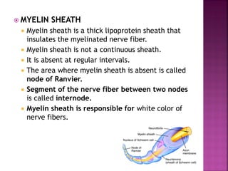
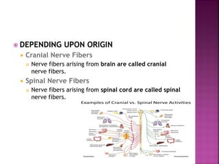
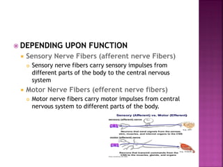
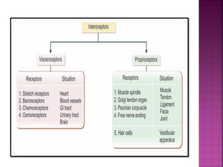
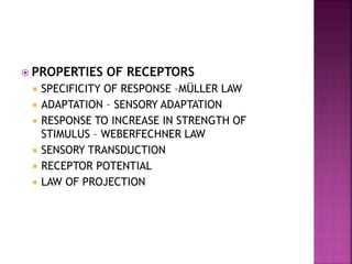
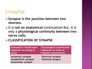
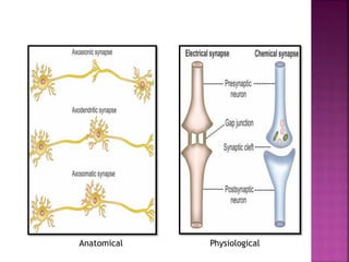
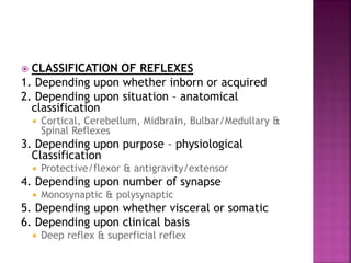
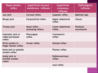
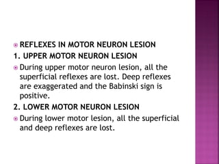
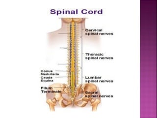
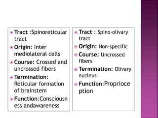
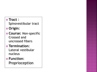
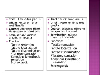
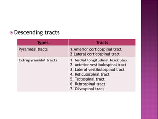
understanding spinal cord, its bransches, lesions, functions and anatomy.
hope to give you better knowledge of spinal cord by the end of it.
plese review ans comment for my future updates and corrections that iw ill be needing in this.
Neuroanatomy | 1. Introduction to NeuroanatomyAhmed Eljack
This is the first lecture in neuroanatomy presented and taught by Ahmed Eljack to second level medical students at Alneelain University.
This lecture discussed the basics of neuroanatomy regarding anatomical terms, planes of section, anatomical divisions of the nervous system, and cells of the nervous system and their major functions.
Describes the overview of the skeletal muscles, its description, functons, and properties. It also inccludes the gross organization of the skeletal system.
1. Locomotion
2. Vasoconstriction and vasodilatation- constriction and
dilation of blood vessel Walls are the results of smooth muscle
contraction.
3. Peristalsis – wavelike motion along the digestive tract is
produced by the Smooth muscle.
4. Cardiac motion
5. Posture maintenance- contraction of skeletal muscles
maintains body posture and muscle tone.
6. Heat generation – about 75% of ATP energy used in
muscle contraction is released as heat. 1. Contracts for a longer time than skeletal muscle
because transverse tubules supply extra Ca+2 ions .
2. intercalated disc connects the ends of adjacent
muscles and hold cells together as a unit (syncytium) .
3. Fibers contracts as a unit .
4. Muscle fibers are self – exiting , rhythmic , and
remain refractory until a contraction is completed.
Indian Dental Academy: will be one of the most relevant and exciting training center with best faculty and flexible training programs for dental professionals who wish to advance in their dental practice,Offers certified courses in Dental implants,Orthodontics,Endodontics,Cosmetic Dentistry, Prosthetic Dentistry, Periodontics and General Dentistry.
CNS Introduction, Neurons, Type of Neurons and functions, Neuroglia and types, Receptors and their types, Synapse, Neurotransmitters and their functions
basic nervous system-CNS-PNS -cell bodie- axon-dendron-grye matter- white mat...shailesh sangle
The nervous system is a complex network of cells, tissues, and organs that coordinates and regulates the body's responses to internal and external stimuli. It is responsible for the control and coordination of all the body's functions, including movement, sensation, thought, and behavior.
The nervous system can be divided into two main parts: the central nervous system (CNS) and the peripheral nervous system (PNS). The CNS consists of the brain and spinal cord, while the PNS consists of all the nerves that extend from the CNS to the rest of the body.
The nervous system is made up of different types of cells, including neurons and glial cells. Neurons are specialized cells that transmit signals through the body in the form of electrical impulses. Glial cells, on the other hand, support and protect the neurons and help maintain the proper functioning of the nervous system.
The nervous system is responsible for many vital functions, including:
Sensory processing: The nervous system receives sensory information from the environment and the body's internal organs, and processes and interprets this information to generate appropriate responses.
Motor control: The nervous system controls the muscles and other organs of the body to produce movement and other responses.
Cognitive functions: The nervous system is responsible for the processes of learning, memory, language, and other complex mental activities.
Autonomic functions: The nervous system regulates the body's automatic functions, such as breathing, heart rate, digestion, and other bodily processes that are not under conscious control.
Overall, the nervous system is a complex and intricate system that plays a critical role in maintaining the body's homeostasis and overall well-being.
IF YOU ARE INTRESTED TO SHOP MAMA EARTH'S PRODUCTES ....
Mamaearth is an Indian personal care brand that offers natural, toxin-free, and sustainable products for all age groups. Their product range includes skincare, haircare, baby care, and men's grooming products. The brand is focused on promoting a healthy and sustainable lifestyle, and all their products are cruelty-free and environmentally friendly.
COPY THAT LINK AND PASTE ON SEARCH BAR OF GOOGLE
https://ekaro.in/enkr20230328s23089700
CHOOSE YOUR AND ORDER NOW.......
thank you....
The central nervous system (CNS) is made up of the brain and spinal cord. The brain controls most body functions, including awareness, movements, sensations, thoughts, speech and memory. The spinal cord is connected to the brain at the brain stem and is covered by the vertebrae of the spine.
In biology, the classical doctrine of the nervous system determines that it is a highly complex part of an animal that coordinates its actions and sensory information by transmitting signals to and from different parts of its body. nervous system is your body's command center. Originating from your brain, it controls your movements, thoughts and automatic responses to the world around you. It also controls other body systems and processes, such as digestion, breathing and sexual development (puberty).
SOUND, TYPES OF SOUND, INTERFERENCE OF SOUND, CALCULATION OF VELOCITY OF SOUND IN AIR, NEWTON'S FORMULA, LAPLACE'S FORMULA, DOPPLER EFFECT, ECHO, RESONANCE, MAGNETO STRICTION & PIEZO ELECTRIC PRODUCTION OF SOUND, APPLICATION OF SOUND
Growth- stages of growth, growth of different tissues & parameters of growth measurements
Developments - Gross motor, fine motor, social & Language development, low birth weight, microcephaly & macrocephaly
Deep Leg Vein Thrombosis (DVT): Meaning, Causes, Symptoms, Treatment, and Mor...The Lifesciences Magazine
Deep Leg Vein Thrombosis occurs when a blood clot forms in one or more of the deep veins in the legs. These clots can impede blood flow, leading to severe complications.
Explore our infographic on 'Essential Metrics for Palliative Care Management' which highlights key performance indicators crucial for enhancing the quality and efficiency of palliative care services.
This visual guide breaks down important metrics across four categories: Patient-Centered Metrics, Care Efficiency Metrics, Quality of Life Metrics, and Staff Metrics. Each section is designed to help healthcare professionals monitor and improve care delivery for patients facing serious illnesses. Understand how to implement these metrics in your palliative care practices for better outcomes and higher satisfaction levels.
ICH Guidelines for Pharmacovigilance.pdfNEHA GUPTA
The "ICH Guidelines for Pharmacovigilance" PDF provides a comprehensive overview of the International Council for Harmonisation of Technical Requirements for Pharmaceuticals for Human Use (ICH) guidelines related to pharmacovigilance. These guidelines aim to ensure that drugs are safe and effective for patients by monitoring and assessing adverse effects, ensuring proper reporting systems, and improving risk management practices. The document is essential for professionals in the pharmaceutical industry, regulatory authorities, and healthcare providers, offering detailed procedures and standards for pharmacovigilance activities to enhance drug safety and protect public health.
R3 Stem Cells and Kidney Repair A New Horizon in Nephrology.pptxR3 Stem Cell
R3 Stem Cells and Kidney Repair: A New Horizon in Nephrology" explores groundbreaking advancements in the use of R3 stem cells for kidney disease treatment. This insightful piece delves into the potential of these cells to regenerate damaged kidney tissue, offering new hope for patients and reshaping the future of nephrology.
One of the most developed cities of India, the city of Chennai is the capital of Tamilnadu and many people from different parts of India come here to earn their bread and butter. Being a metropolitan, the city is filled with towering building and beaches but the sad part as with almost every Indian city
CRISPR-Cas9, a revolutionary gene-editing tool, holds immense potential to reshape medicine, agriculture, and our understanding of life. But like any powerful tool, it comes with ethical considerations.
Unveiling CRISPR: This naturally occurring bacterial defense system (crRNA & Cas9 protein) fights viruses. Scientists repurposed it for precise gene editing (correction, deletion, insertion) by targeting specific DNA sequences.
The Promise: CRISPR offers exciting possibilities:
Gene Therapy: Correcting genetic diseases like cystic fibrosis.
Agriculture: Engineering crops resistant to pests and harsh environments.
Research: Studying gene function to unlock new knowledge.
The Peril: Ethical concerns demand attention:
Off-target Effects: Unintended DNA edits can have unforeseen consequences.
Eugenics: Misusing CRISPR for designer babies raises social and ethical questions.
Equity: High costs could limit access to this potentially life-saving technology.
The Path Forward: Responsible development is crucial:
International Collaboration: Clear guidelines are needed for research and human trials.
Public Education: Open discussions ensure informed decisions about CRISPR.
Prioritize Safety and Ethics: Safety and ethical principles must be paramount.
CRISPR offers a powerful tool for a better future, but responsible development and addressing ethical concerns are essential. By prioritizing safety, fostering open dialogue, and ensuring equitable access, we can harness CRISPR's power for the benefit of all. (2998 characters)
The Importance of Community Nursing Care.pdfAD Healthcare
NDIS and Community 24/7 Nursing Care is a specific type of support that may be provided under the NDIS for individuals with complex medical needs who require ongoing nursing care in a community setting, such as their home or a supported accommodation facility.
How many patients does case series should have In comparison to case reports.pdfpubrica101
Pubrica’s team of researchers and writers create scientific and medical research articles, which may be important resources for authors and practitioners. Pubrica medical writers assist you in creating and revising the introduction by alerting the reader to gaps in the chosen study subject. Our professionals understand the order in which the hypothesis topic is followed by the broad subject, the issue, and the backdrop.
https://pubrica.com/academy/case-study-or-series/how-many-patients-does-case-series-should-have-in-comparison-to-case-reports/
Antibiotic Stewardship by Anushri Srivastava.pptxAnushriSrivastav
Stewardship is the act of taking good care of something.
Antimicrobial stewardship is a coordinated program that promotes the appropriate use of antimicrobials (including antibiotics), improves patient outcomes, reduces microbial resistance, and decreases the spread of infections caused by multidrug-resistant organisms.
WHO launched the Global Antimicrobial Resistance and Use Surveillance System (GLASS) in 2015 to fill knowledge gaps and inform strategies at all levels.
ACCORDING TO apic.org,
Antimicrobial stewardship is a coordinated program that promotes the appropriate use of antimicrobials (including antibiotics), improves patient outcomes, reduces microbial resistance, and decreases the spread of infections caused by multidrug-resistant organisms.
ACCORDING TO pewtrusts.org,
Antibiotic stewardship refers to efforts in doctors’ offices, hospitals, long term care facilities, and other health care settings to ensure that antibiotics are used only when necessary and appropriate
According to WHO,
Antimicrobial stewardship is a systematic approach to educate and support health care professionals to follow evidence-based guidelines for prescribing and administering antimicrobials
In 1996, John McGowan and Dale Gerding first applied the term antimicrobial stewardship, where they suggested a causal association between antimicrobial agent use and resistance. They also focused on the urgency of large-scale controlled trials of antimicrobial-use regulation employing sophisticated epidemiologic methods, molecular typing, and precise resistance mechanism analysis.
Antimicrobial Stewardship(AMS) refers to the optimal selection, dosing, and duration of antimicrobial treatment resulting in the best clinical outcome with minimal side effects to the patients and minimal impact on subsequent resistance.
According to the 2019 report, in the US, more than 2.8 million antibiotic-resistant infections occur each year, and more than 35000 people die. In addition to this, it also mentioned that 223,900 cases of Clostridoides difficile occurred in 2017, of which 12800 people died. The report did not include viruses or parasites
VISION
Being proactive
Supporting optimal animal and human health
Exploring ways to reduce overall use of antimicrobials
Using the drugs that prevent and treat disease by killing microscopic organisms in a responsible way
GOAL
to prevent the generation and spread of antimicrobial resistance (AMR). Doing so will preserve the effectiveness of these drugs in animals and humans for years to come.
being to preserve human and animal health and the effectiveness of antimicrobial medications.
to implement a multidisciplinary approach in assembling a stewardship team to include an infectious disease physician, a clinical pharmacist with infectious diseases training, infection preventionist, and a close collaboration with the staff in the clinical microbiology laboratory
to prevent antimicrobial overuse, misuse and abuse.
to minimize the developme
1. DR NIYATI N PATEL
MPT IN NEUROLOGICAL CONDITIONS
2.
3.
4. Neuron or nerve cell is defined as the
structural and functional unit of nervous
system.
It is different from other cells by two ways:
1.Neuron has branches or processes called axon
and dendrites
2. Neuron does not have centrosome. So, it
cannot undergo division.
5. Neurons are classified by three different
methods.
A. Depending upon the number of poles
B. Depending upon the function
C. Depending upon the length of axon.
6. DEPENDING UPON THE NUMBER OF POLES
1. Unipolar Neurons
Unipolar neurons are the neurons that have only one pole. From a single
pole, both axon and dendrite arise
This type of nerve cells is present only in embryonic stage in human beings.
2. Bipolar Neurons
Neurons with two poles are known as bipolar neurons.
Axon arises from one pole and dendrites arise from the other pole.
3. Multipolar Neurons
Multipolar neurons are the neurons which have many poles.
One of the poles gives rise to axon and all other poles give rise to
dendrites.
7.
8. DEPENDING UPON THE FUNCTION
On the basis of function, nerve cells are classified into two
types:
1. Motor or efferent neurons
2. Sensory or afferent neurons.
1. Motor or Efferent Neurons
Motor or efferent neurons are the neurons which carry the
motor impulses from central nervous system to peripheral
effector organs like muscles, glands, blood vessels, etc.
Generally, each motor neuron has a long axon and short
dendrites.
2. Sensory or Afferent Neurons
Sensory or afferent neurons are the neurons which carry the
sensory impulses from periphery to central nervous system.
Generally, each sensory neuron has a short axon and long
dendrites.
9. DEPENDING UPON THE LENGTH OF AXON
Depending upon the length of axon, neurons are divided into two
types:
1. Golgi type I neurons
2. Golgi type II neurons.
1. Golgi Type I Neurons
Golgi type I neurons have long axons.
Cell body of these neurons is in different parts of central
nervous system and their axons reach the remote peripheral
organs.
2. Golgi Type II Neurons
Neurons of this type have short axons.
These neurons are present in cerebral cortex and spinal cord.
10. Neuron is made up of three
parts:
1. Nerve cell body
2. Dendrite
3. Axon
Dendrite and axon form the
processes of neuron
Dendrites are short processes
and the axons are long
processes.
Dendrites and axons are
usually called nerve fibers.
12. 1. Depending upon structure
2. Depending upon distribution
3. Depending upon origin
4. Depending upon function
5. Depending upon secretion of
neurotransmitter
6. Depending upon diameter and conduction of
impulse (ErlangerGasser classification)
13. DEPENDING UPON STRUCTURE
Myelinated Nerve Fibers
Myelinated nerve fibers are the nerve fibers that are
covered by myelin sheath.
Non-myelinated Nerve Fibers
Nonmyelinated nerve fibers are the nerve fibers which
are not covered by myelin sheath.
14. MYELIN SHEATH
Myelin sheath is a thick lipoprotein sheath that
insulates the myelinated nerve fiber.
Myelin sheath is not a continuous sheath.
It is absent at regular intervals.
The area where myelin sheath is absent is called
node of Ranvier.
Segment of the nerve fiber between two nodes
is called internode.
Myelin sheath is responsible for white color of
nerve fibers.
15. DEPENDING UPON DISTRIBUTION
Somatic Nerve Fibers
Somatic nerve fibers supply the skeletal muscles of
the body.
Visceral or Autonomic Nerve Fibers
Autonomic nerve fibers supply the various internal
organs of the body.
16. DEPENDING UPON ORIGIN
Cranial Nerve Fibers
Nerve fibers arising from brain are called cranial
nerve fibers.
Spinal Nerve Fibers
Nerve fibers arising from spinal cord are called spinal
nerve fibers.
17. DEPENDING UPON FUNCTION
Sensory Nerve Fibers (afferent nerve Fibers)
Sensory nerve fibers carry sensory impulses from
different parts of the body to the central nervous
system
Motor Nerve Fibers (efferent nerve fibers)
Motor nerve fibers carry motor impulses from central
nervous system to different parts of the body.
18. DEPENDING UPON SECRETION OF
NEUROTRANSMITTER
Adrenergic Nerve Fibers
Adrenergic nerve fibers secrete nor adrenaline.
Cholinergic Nerve Fibers
Cholinergic nerve fibers secrete acetylcholine.
19. DEPENDING UPON DIAMETER AND
CONDUCTION OF IMPULSE (ERLANGER-
GASSER CLASSIFICATION)
Type Diameter (u) Velocity of
conduction
(m/sec)
A Alpha 12 To 24 70 to 120
A Beta 6 to 12 30 to 70
A Gamma 5 to 6 15 to 30
A Delta 2 to 5 12 to 15
B 1 to 2 3 to 10
C < 1.5 0.5 to 2
21. Defined as the physiochemical change that
occurs in a tissue when stimulus is applied
Response Due to Stimulation of Nerve
Fiber
Action potential or nerve impulse (Propagated)
Electrotonic potential or local potential (Non
Propagated)
22. ACTION POTENTIAL
Action potential is defined as a series of electrical
changes that occur in the membrane potential when
the muscle or nerve is stimulated.
Action potential occurs in three phases:
1. Latent Period - Latent period is the period when no
change occurs in the electrical potential immediately
after applying the stimulus. It is a very short period with
duration of 0.5 to 1 millisecond.
2. Depolarization - action potential in which inside of
the muscle becomes positive and outside becomes
negative.
3. Repolarization - after depolarization the inside of
muscle becomes negative and outside becomes
positive. So, the polarized state of the muscle is re
established.
23. Action potential in
a skeletal muscle
A = Opening of few
Na+ channels
B = Opening of many
Na+ channels
C = Closure of Na+
channels and
opening
of K+ channels
D = Closure of K+
channels
25. Properties of action potential and graded
potential
Action potential Graded
potential/Electronic
potential
•Propagative
•Long distance signal
•Both depolarization &
repolarization
•Obeys all or none law
•Summation is not possible
•Has refractory period
•Non propagative
•Short distance signal
•Only depolarization/
Hyperpolarization
•Does not obey all or none law
•Summation is possible
•No refractory period
26. Conductivity is the
ability of nerve
fibers to transmit
the impulse from
the area of
stimulation to the
other areas.
27. Refractory period is the period at which the
nerve does not give any response to a
stimulus.
TWO TYPES OF REFRACTORY PERIODS
1. Absolute Refractory Period
1. Absolute refractory period is the period during which
the nerve does not show any response at all,
whatever may be the strength of stimulus.
2. 2. Relative Refractory Period
1. It is the period, during which the nerve fiber shows
response, if the strength of stimulus is increased to
maximum.
28. When one subliminal stimulus is applied, it
does not produce any response in the nerve
fiber because, the subliminal stimulus is very
weak.
However, if two or more subliminal stimuli
are applied within a short interval of about
0.5 millisecond, the response is produced.
It is because the subliminal stimuli are
summed up together to become strong
enough to produce the response.
This phenomenon is known as summation.
29. While stimulating a nerve fiber continuously,
the excitability of the nerve fiber is greater in
the beginning.
Later the response decreases slowly and finally
the nerve fiber does not show any response at
all.
This phenomenon is known as adaptation or
accommodation.
Cause for Adaptation
When a nerve fiber is stimulated continuously,
depolarization occurs continuously.
Continuous de polarization inactivates the sodium
pump and increases the efflux of potassium ions.
30. Nerve fiber cannot be fatigued, even if it is
stimulated continuously for a long time.
The reason is that nerve fiber can conduct
only one action potential at a time.
At that time, it is completely refractory and
does not conduct another action potential.
31. All-or-none law states that when a nerve is
stimulated by a stimulus it gives maximum
response or does not give response at all.
32. When a nerve fiber is injured, various changes occur
in the nerve fiber and nerve cell body. All these
changes are together called the degenerative
changes.
The term regeneration refers to regrowth of lost or
destroyed part of a tissue. The injured and
degenerated nerve fiber can regenerate. It starts as
early as 4th day after injury, but becomes more
effective only after 30 days and is completed in
about 80 days.
Causes for Injury
1. Obstruction of blood flow
2. Local injection of toxic substances
3. Crushing of nerve fiber
4. Transection of nerve fiber.
33.
34. Neuroglia or glia (glia = glue) is the
supporting cell of the nervous system.
Neuroglial cells are non-excitable.
CENTRAL NEUROGLIAL CELLS PERIPHERAL NEUROGLIAL CELLS
ASTROCYTES (STAR SHAPE &
PRESENT IN BRAIN)
„MICROGLIA (PHAGOCYTIC CELL &
MACROPHAGES OF CNS)
„OLIGODENDROCYTES (MYELIN
SHEATH IN CNS)
SCHWANN CELLS (MYELIN
SHEATH IN PNS)
„SATELLITE CELLS (EXTERIOR
SURFACE OF PNS)
35. Receptors are sensory (afferent) nerve
endings that terminate in periphery as bare
unmyelinated endings or in the form of
specialized capsulated structures.
Classification of receptors
Exteroceptors (outside of
the body)
Interoceptors (within
body)
Mechanoreceptor/cutenous
receptors (skin area)
Chemoreceptors (chemical
stimuli)
Telereceptors (distance
receptor)
Visceroceptors (viscera)
Proprioceptors (joint
position)
39. PROPERTIES OF RECEPTORS
SPECIFICITY OF RESPONSE –MÜLLER LAW
ADAPTATION – SENSORY ADAPTATION
RESPONSE TO INCREASE IN STRENGTH OF
STIMULUS – WEBERFECHNER LAW
SENSORY TRANSDUCTION
RECEPTOR POTENTIAL
LAW OF PROJECTION
40. Synapse is the junction between two
neurons.
It is not an anatomical continuation But, it is
only a physiological continuity between two
nerve cells.
CLASSIFICATION OF SYNAPSE
Anatomical classification
(depends on ending of
axon)
Physiological classification
(depends on mode of
impulse transmission)
Axoaxonic synapse
Axodendritic synapse
Axosomatic synapse
Electrical synapse
Chemical synapse
43. Presynaptic
Mitochondria, which help in the synthesis of
neurotransmitter substance
Synaptic vesicles, which store neurotransmitter
substance.
Synaptic cleft & Post synaptic
Postsynaptic neuron is called postsynaptic
membrane.
It contains some receptor proteins.
Small space in between the presynaptic
membrane and the postsynaptic membrane is
called synaptic cleft.
44. FUNCTIONS OF SYNAPSE
Main function of the synapse is to transmit
the impulses, i.e. action potential from one
neuron to another
47. PROPERTIES OF SYNAPSE
ONE WAY CONDUCTION – BELL-MAGENDIE
LAW
According to BellMagendie law, the impulses are
transmitted only in one direction in synapse,
i.e. from presynaptic neuron to postsynaptic
neuron.
SYNAPTIC DELAY
Release of neurotransmitter
Passage of neurotransmitter from axon terminal
to postsynaptic membrane
Action of the neurotransmitter to open the ionic
channels in postsynaptic membrane.
48. FATIGUE
Fatigue at synapse is due to the depletion of
neurotransmitter substance, acetylcholine.
Depletion of acetylcholine occurs because of two
factors:
i. Soon after the action, acetylcholine is destroyed by
acetylcholinesterase
ii. Due to continuous action, new acetylcholine is not
synthesize
49. SUMMATION
Summation is the fusion of effects or progressive
increase when single presynaptic terminal is
stimulated repeatedly.
50. CONVERGENCE
Convergence is the process by which many presynaptic neurons
terminate on a single postsynaptic neuron
„DIVERGENCE
Divergence is the process by which one presynaptic neuron
terminates on many postsynaptic neurons.
51. Reflex activity is the response to a peripheral
nervous stimulation that occurs without our
consciousness.
It is a type of protective mechanism and it
protects the body from irreparable
damages.
52. REFLEX ARC
Reflex arc is the anatomical nervous pathway
for a reflex action. A simple reflex arc
includes five components
53. CLASSIFICATION OF REFLEXES
1. Depending upon whether inborn or acquired
2. Depending upon situation – anatomical
classification
Cortical, Cerebellum, Midbrain, Bulbar/Medullary &
Spinal Reflexes
3. Depending upon purpose – physiological
Classification
Protective/flexor & antigravity/extensor
4. Depending upon number of synapse
Monosynaptic & polysynaptic
5. Depending upon whether visceral or somatic
6. Depending upon clinical basis
Deep reflex & superficial reflex
55. REFLEXES IN MOTOR NEURON LESION
1. UPPER MOTOR NEURON LESION
During upper motor neuron lesion, all the
superficial reflexes are lost. Deep reflexes
are exaggerated and the Babinski sign is
positive.
2. LOWER MOTOR NEURON LESION
During lower motor lesion, all the superficial
and deep reflexes are lost.
56.
57. Situation and Extent
Vertebral canal
It extends from foramen magnum where
it is continuous with medulla oblongata,
above and up to the lower border of
first lumbar vertebra
Coverings
Spinal cord is covered by sheaths called
meninges, which are membranous in
nature.
Meninges are pia mater, arachnoid
mater & dura mater.
These coverings continue as coverings of
brain.
Meninges are responsible for protection
and nourishment of the nervous tissues.
58. Shape and Length
Shape- cylindrical
Length - 45 cm in males and about 43 cm in females.
Enlargements
Cervical enlargements – upper extremities
lumbar enlargements - lower extremities
Conus Medullaris and Filum Terminale
Below the lumbar enlargement, spinal cord rapidly
narrows to a cone-shaped termination called conus
medullaris.
A slender non-nervous filament called filum terminale
extends from conus medullaris downward to the
fundus of the dural sac at the level of second
sacralvertebra.
61. Neurons in Grey matter
Golgi type 1 (anterior
aspect, long axons-tracts)
Golgi type 2 (posterior
aspect, short axons-
interneurons)
Contents of grey matter
Nuclie
Alpha motor neurons
Gamma motor neurons
Renshaw cells
Lamina
62. White matter
Anterior or Ventral White
Column
Lateral White Column
posterior or dorsal White
Column
Tracts
Shorts tracts
Long tracts
Ascending tracts
Descending tracts
63. ASCENDING TRACTS OF SPINAL CORD
First Order Neurons
Receptors to sensory neurons present in the
posterior gray horn of spinal cord
Nerve cell bodies - located in the posterior
nerve root ganglion.
Second Order Neurons
Posterior gray horn to ascending tracts of
spinal cord.
These fibers carry sensory impulses from
spinal cord to different brain areas below
cerebral cortex (subcortical areas) such as
thalamus.
Third Order Neurons
Subcortical areas
Fibers of these neurons carry the sensory
impulses from subcortical areas to cerebral
cortex.
75. Tract : Anterior
corticospinal tract
Situation: Anterior
white column
Origin: Betz cells and
other cells of motor
area
Course: Uncrossed
fibers
Function:
i. Control of voluntary
movements
ii. Form upper motor
neurons
Tract : lateral
corticospinal tract
Situation : Lateral
white column
Origin: Betz cells and
other cells of motor
area
Course: Crossed
fibers
Function:
i. Control of voluntary
movements
ii. Form upper motor
neurons
76.
77. Tract : Anterior
vestibulospinal tract
Situation: Anterior
white column
Origin: Medial vestibular
nucleus
Course: Uncrossed
fibers Extend up to
upper thoracic segments
Function:
i. Maintenance of muscle
tone and posture
ii. Maintenance of position
of head and body during
acceleration
Tract : Lateral
vestibulospinal tract
Situation: Lateral
white column
Origin: Lateral
vestibular nucleus
Course: Mostly
uncrossed Extend to
all segments
Function:
i. Maintenance of muscle
tone and posture
ii. Maintenance of position
of head and body during
acceleration
78. Tract : Reticulospinal tract
Situation: Lateral white
fasciculus
Origin: Reticular formation
of pons and medulla
Course: Mostly uncrossed
Extend up to thoracic
segments
Function:
i. Coordination of
voluntary and reflex
movements
ii. Control of muscle tone
iii.Control of respiration
and diameter of blood
vessels
Tract : Tectospinal tract
Situation: Anterior
white column
Origin: Superior
colliculus
Course: Crossed fibers
Extend up to lower
cervical segments
Function:
Control of
movement of head
in response to
visual and auditory
impulses
79. Tract : Rubrospinal
tract
Situation: Lateral
white column
Origin: Red nucleus
Course: Crossed
fibers Extend up to
thoracic segments
Function:
Facilitatory influence
on flexor muscle
tone
Tract : Olivospinal
tract
Situation: Lateral
white column
Origin: Inferior olivary
nucleus
Course: Mostly
crossed Extent – not
clear
Function: Control of
movements due to
proprioception
80. COMPLETE TRANSECTION OF SPINAL CORD
Complete transection of spinal cord occurs
due to:
1. Bullet injury, which causes dislocation of
spinal cord
2. Accidents, which cause dislocation of spinal
cord or occlusion of blood vessels.
Complete transection causes immediate loss
of sensation and voluntary movement
below the level of lesion.
81. Effects occur in three stages:
1. Stage of spinal shock
2. Stage of reflex activity
3. Stage of reflex failure
82. 1. Stage of Spinal Shock
Stage of spinal shock is the first stage of effects
that occurs immediately after injury. It is also
called stage of flaccidity.
1. Paralysis of limbs
1. Cervical injury – quadriplegia/tetraplegia
2. Thoracic/lumbar injury- paraplegia
2. Flaccid paralysis
3. Loss of reflexes
4. Loss of sensations
5. Effect on visceral organs
6. Heart rate
7. Venous return
8. Effect on blood pressure
83. 2. Stage of Reflex Activity
Stage of reflex activity is also called stage of recovery.
After 3 weeks period, depending largely upon the general
health of the patient
functional activities return to smooth muscles
Next, the sympathetic tone to blood vessels returns.
Lastly, after another 3 months, the tone in skeletal muscle
returns
Flexor reflexes appear first. To elicit the flexor reflex, a
painful stimulus is required. First reflex, which usually
appears, is the Babinski reflex.
After a variable period of 1 to 5 weeks of reappearance of
flexor reflexes, the extensor reflexes return.
This reaction constitutes the spasm in flexor muscles of
both the lower limbs, evacuation of urinary bladder and
profuse sweating. This is known as the mass reflex.
84. Stage of Reflex Failure
Though the reflex movements return,
muscles below the level of injury have less
power and less resistance.
deterioration of condition with infection
85. Brainstem is the part of
brain formed by
medulla
oblongata,pons and
midbrain.
Brainstem contains
ascending and
descending tracts
between brain and
spinal cord.
It also contains many
centers for regulation
of vital functions in the
body.
86. Medulla oblongata or medulla
is the lowermost part of
brain.
It is situated below pons and
is continued downwards as
spinal cord.
Medulla forms the main
pathway for ascending and
descending tracts of the
spinal cord.
87. 1. Respiratory Centers
Dorsal and ventral group of neurons form the
medullary respiratory centers, which maintain
normal rhythmic respiration
2. Vasomotor Center
Vasomotor center controls blood pressure and
heart rate.
3. Deglutition Center
Deglutition center regulates the pharyngeal and
esophageal stages of deglutition
88. 4. Vomiting Center
Vomiting center induces vomiting during
irritation or inflammation of gastrointestinal
(GI) tract
5. Superior and Inferior Salivatory Nuclei
Salivatory nuclei control the secretion of saliva
6. Cranial Nerve Nuclei
Nuclei of 10th, 11th, 12th and some nuclei of 5th
and 8th cranial nerves are located in the medulla
oblongata
89. 7. Vestibular Nuclei
Vestibular nuclei contain the second order
neurons of vestibular nerve
Superior, medial, lateral and inferior
vestibular nuclei.
Medial and inferior vestibular nuclei extend
into medulla.
90. Pons forms a bridge between medulla and
midbrain
91. Axons of pontine nuclei join to the middle
cerebellar peduncle & pons pathway that
connects cerebellum with cerebral cortex.
Pyramidal tracts pass through the pons
Medial lemniscus is joined by the fibers of
5th, 7th, 9th and 10th cranial nerves in
pons
Nuclei of 5th, 6th, 7th and 8th cranial
nerves are located in pons
Pons contains the pneumotaxic and
apneustic centers for regulation of
respiration
92. Midbrain lies between pons and diencephalon.
Midbrain
Tectum Cerebral peduncles
Superior colliculus :
1. Small structure & imp center for reflex
2. Tectospinal tract, superior colliculus
controls the movements of the eyes,
head, trunk and limbs, in response to
visual impulses
Basis pedunculus:
Laterally temperopontine fibres
Medially frontopontine fibers
Middle pyramidal tract fibers
Inferior colliculus:
The center for auditory reflexes ,
produces reflex vocalization
Substantia nigra:
Situated below the red nucleus.
Tegmentum:
Dorsal to substantia nigra, upward to the
reticular formation
Superior cerebellar peduncle,
Crossing of rubrospinal tracts
Efferent fibers of 3rd, 4th and 6th
Cranial nerves.
93. Red nucleus is a large oval or round
mass of gray matter, extending
between the superior colliculus and
hypothalamus.
Two parts
Nucleus magnocellularis (larger)
This fibers form the rubrospinal and
rubrobulbar tracts
Nucleus parvocellularis (smaller)
This fibers form the rubroreticular tract
94. Afferent connection
Corticorubral fibers
Pallidorubral fibers
Cerebellorubral tracts
Efferent connections
Rubrospinal tract to spinal cord
Rubrobulbar tract to medulla
Rubroreticular fibers to reticular formation
Rubrothalamic tract to lateral ventral nucleus of
thalamus
Rubroolivary tract to inferior olivary nucleus
Fibers to nuclei of 3rd, 4th and 6th cranial nerves.
95. Control of muscle tone
Control of complex muscular movements
Control of righting reflexes
Control of movements of eyeball
Control of skilled movements
96. Cerebellum consists of a narrow, worm-like
central body called vermis and two lateral
lobes, the right and left cerebellar hemispheres
98. CEREBELLAR HEMISPHERES
1. Lobulus ansiformis or ansiform lobe -
larger portion of cerebellar hemisphere
2. Lobulus paramedianus or paramedian
lobe - smaller portion of cerebellar
hemisphere.
99. A. Anatomical divisions
B. Phylogenetic divisions
C. Physiological or functional divisions
100. ANATOMICAL DIVISIONS
1. Anterior Lobe
Anterior lobe includes lingula, central lobe and
culmen..
2. Posterior Lobe
Posterior lobe consists of lobulus simplex,
declive, tuber, pyramid, uvula, paraflocculi and
the two portions of hemispheres, viz. ansiform
lobe and paramedian lobe.
3. Flocculonodular Lobe
Flocculonodular lobe includes nodulus and the
lateral extension on either side called flocculus.
101. PHYLOGENETIC DIVISIONS
1. Paleocerebellum:
i. Archicerebellum, which includes
flocculonodular lobe
ii. Paleocerebellum proper, which includes
lingula, central lobe, culmen, lobulus simplex,
pyramid, uvula and paraflocculi.
2. Neocerebellum
It includes declive, tuber and the two portions
of cerebellar hemispheres, viz. lobulus ansiformis
and lobulus paramedianus.
102. PHYSIOLOGICAL OR FUNCTIONAL DIVISIONS
1. Vestibulocerebellum
Vestibulocerebellum includes flocculonodular
lobe that forms the archicerebellum.
2. Spinocerebellum
Spinocerebellum includes lingula, central lobe,
culmen, lobulus simplex, declive, tuber, pyramid,
uvula and paraflocculi and medial portions of
lobulus ansiformis and lobulus paramedianus.
3. Corticocerebellum
Corticocerebellum includes lateral portions of
lobulus ansiformis and lobulus paramedianus.
104. CEREBELLAR NUCLEI
Fastigial Nucleus-
midline on the roof of IV
ventricle.
Globosus Nucleus-
lateral to nucleus fastigi
Emboliform Nucleus-
below the nucleus
fastigi and nucleus
globosus
Dentate Nucleus-
lateral to all the other
nuclei
105. WHITE MATTER OF
CEREBELLUM
Superior cerebellar
peduncles between
cerebellum and midbrain
Middle cerebellar peduncles
between cerebellum and
pons
Inferior cerebellar peduncles
between cerebellum and
medulla oblongata
106. VESTIBULOCEREBELLUM
Vestibulocerebellum is connected with the
vestibular apparatus and so it is known as
vestibulocerebellum
STRUCTURES
Includes the flocculonodular lobe that is formed
by the nodulus of vermis and its lateral
extensions called flocculi
FUNCTIONS
Tone, posture and equilibrium
107. SPINOCEREBELLUM
Spinocerebellum is connected with spinal cord
COMPONENTS
Medial Portions Of Cerebellar Hemisphere,
Paraflocculi And The Parts Of Vermis, Viz.
Lingula, Central Lobe, Culmen, Lobulus Simplex,
Declive, Tuber, Pyramid And Uvula
FUNCTIONS
Tone, posture and equilibrium
Localization of tactile
108. CORTICOCEREBELLUM
Corticocerebellum is the largest part of
cerebellum, Because of its connection with
cerebral cortex
COMPONENTS
Corticocerebellum includes the lateral portions
of cerebellar hemispheres
„FUNCTIONS
Integration and regulation of well-coordinated
muscular activities
109.
110. Basal ganglia are the
scattered masses of gray
matter submerged in
subcortical substance of
cerebral hemisphere
Basal ganglia form the
part of extra pyramidal
system, which is
concerned with motor
activities.
111. 1. Corpus striatum
2. Substantia nigra
3. Subthalamic
nucleus
112. 1. Corpus striatum
Mass of gray matter situated at the base of cerebral
hemispheres in close relation to thalamus
Caudate Nucleus : Caudate nucleus is an
elongated arched gray mass, lying medial to
internal capsule.
Caudate nucleus has a head portion and a tail portion
Lenticular Nucleus: Lenticular nucleus is a
wedgeshaped gray mass, situated lateral to
internal capsule. A vertical plate of white
matter called external medullary lamina
Outer putamen
Inner globus pallidus
113. SUBSTANTIA NIGRA
Substantia nigra is situated below red nucleus.
It is made up of large pigmented and small
nonpigmented cells.
The pigment contains high quantity of iron.
SUBTHALAMIC NUCLEUS OF LUYS
Subthalamic nucleus is situated lateral to red
nucleus and dorsal to substantia nigra.
114. 1. Putamen to globus pallidus
2. Caudate nucleus to globus pallidus
3. Caudate nucleus to putamen.
115. Component Afferent connections
from
Efferent
connections to
Corpus striatum 1. Thalamic nuclei to caudate
nucleus and
putamen
2. Cerebral cortex to caudate
nucleus and
putamen
3. Substantia nigra to putamen
4. Subthalamic nucleus to
globus pallidus
1. Thalamic nuclei
2. Subthalamic nucleus
3. Red nucleus
4. Substantia nigra
5. Hypothalamus
6. Reticular formation
(Most of the
Substantia nigra 1. Putamen
2. Frontal lobe of cerebral
cortex
3. Superior colliculus
4. Mamillary body of
hypothalamus
5. Medial and
Putamen
Subthalamic
nucleus
Globus pallidus 1. Globus pallidus
2. Red nucleus
117. 1. CONTROL OF MUSCLE TONE
Gamma motor neurons of spinal cord are
responsible for development of tone in the
muscles
Decrease the muscle tone by inhibiting gamma
motor neurons through descending inhibitory
reticular system in brainstem
During the lesion of basal ganglia, muscle tone
increases leading to rigidity
118. 2. CONTROL OF MOTOR ACTIVITY
Regulation of Voluntary Movements
a. Premotor area
b. Primary motor area
c. Supplementary motor area
Regulation of Conscious Movements
Fibers between cerebral cortex and caudate
nucleus are concerned with regulation of
conscious movements (cognitive control)
Regulation of Subconscious Movements
Cortical fibers reaching putamen regulation of
some subconscious movement
119. 3. CONTROL OF REFLEX MUSCULAR
ACTIVITY
Visual and labyrinthine reflexes are important
in maintaining the posture
4. CONTROL OF AUTOMATIC ASSOCIATED
MOVEMENTS
Automatic associated movements are the
movements in the body (motor activities)
5. ROLE IN AROUSAL MECHANISM
Globus pallidus and red nucleus are involved in
arousal mechanism because of their connections
with reticular formation
120. 6. ROLE OF NEUROTRANSMITTERS IN THE
FUNCTIONS OF BASAL GANGLIA
Dopamine released by dopaminergic fibers from
substantia nigra to corpus striatum
Gamma aminobutyric acid (GABA) secreted by
intrinsic fibers of substantia nigra to corpus striatum
Acetylcholine released by fibers from cerebral
cortex to caudate nucleus and putamen
Substance P & Enkephalins released by fibers from
globus pallidus reaching substantia nigra
Noradrenaline secreted by fibers between basal
ganglia and reticular formation
Glutamic acid secreted by fibers from subthalamic
nucleus to globus pallidus and substantia nigra
122. Cerebral cortex is also called pallidum and
it consists of two hemispheres
Both the cerebral hemispheres are
separated by a deep vertical fissure
Separation is complete anteriorly and posteriorly
Middle portion, the fissure extends only up to
corpusm callosum (connecting the two
hemispheres)
Surface of the cerebral cortex is
characterized by
Sulci - Slight Depression
Gyri - Raised Ridge
123. HISTOLOGY OF CEREBRAL CORTEX
LAYERS OF CEREBRAL CORTEX
Gray matter
White matter
Molecular or Plexiform Layer - dendrites or axons
from cells of deeper layers
External Granular Layer – Dendrites & Axons end
(axons enter white substance of the hemisphere)
Outer Pyramidal Layer
Medium pyramidal cells - outer portion
larger pyramidal cells - deeper portion
Internal Granular Layer – horizontal fibers, which
appear as a white strip known as outer strip
Ganglionic Layer or Internal Pyramidal Layer -
Pyramidal cells in this region are otherwise known as
Betz cells or giant cells
Fusiform Cell Layer - white matter of cerebral
hemisphere
124. LOBES OF CEREBRAL CORTEX
Four lobes
1. Frontal lobe
2. Parietal lobe
3. Occipital lobe
4. Temporal lobe
Four main fissures and sulci
Central sulcus (frontal and parietal lobes)
Parieto-occipital sulcus (parietal and occipital lobe)
lateral sulcus (parietal and temporal lobes)
Callosomarginal fissure (temporal lobe and limbic
area)
125.
126. CEREBRAL DOMINANCE
Cerebral dominance is defined as the
dominance of one cerebral hemisphere
over the other in the control of cerebral
functions
Right handed – left hemisphere
(representational hemisphere)
Left handed – right hemisphere
(representational hemisphere)
127. Frontal lobe of cerebral cortex is divided into
two parts:
A.Precentral cortex, which is situated posteriorly
B.Prefrontal cortex, which is situated anteriorly
PRECENTRAL CORTEX
Includes
Central sulcus
Precentral gyrus
Posterior portions of superior, middle and inferior
frontal gyri
128. Precentral cortex is further divided
into three functional areas:
1. Primary motor area
2. Premotor area
3. Supplementary motor area.
129. Primary Motor Area (giant pyramidal cells)
Functions of primary motor area - initiation
of voluntary movements and speech
Area 4
It activates both α-motor neurons and γ-motor
neurons simultaneously by the process called
coactivation
Effect of lesion of area 4
The ability to walk is not affected
Hemiplegia with spastic paralysis.
(Hemiplegia means the paralysis in one half of
the body)
130. Premotor Area (6, 8, 44 and 45)
Area 6 - Posterior portions of superior, middle
and inferior frontal gyri
Functions of area 6 - coordination of movements &
cortical center for extrapyramidal system
Area 8 - frontal eye field
Function of area 8 - Frontal eye field is concerned
with conjugate movement of eyeballs
Area 44 & 45 (Broca area) - motor area for
speech- dominant hemisphere
Function of Broca area - movements of tongue, lips
and larynx, which are involved in speech
131. Supplementary Motor Area
Medial surface of frontal lobe rostral to primary
motor area
Motor movements are elicited by electrical
stimulation of this area like raising the
contralateral arm, turning the head and eye
and movements of synergistic muscles of
trunk and legs
Function of supplementary motor area -
coordinated skilled movements
132.
133.
134. Prefrontal cortex is the anterior part of
frontal lobe of cerebral cortex
Areas present in prefrontal cortex are 9, 10,
11, 12, 13, 14, 23, 24, 29 and 32.
Medial Surface Areas -12, 13, 14, 23, 24, 29 & 32
Lateral Surface Areas - 9, 10 & 11
Functions of Prefrontal Cortex
The higher functions like emotion, learning,
memory , social behavior, short-term memories,
intelligence & personality
135. APPLIED PHYSIOLOGY – FRONTAL LOBE
SYNDROME
Emotional instability
Lack of concentration
Impairment of recent memory
Apart from mental defects, there are some
functional abnormalities also:
i. Hyperphagia (increased food intake)
ii. Loss of control over sphincter of the urinary
bladder or rectum
iii. Disturbances in orientation
iv. Slight tremor
136. Parietal lobe is divided into three functional
areas:
A. Somesthetic area I
B. Somesthetic area II
C. Somesthetic association area
137. SOMESTHETIC AREA I (SOMATOSENSORY
AREA I OR PRIMARY SOMESTHETIC OR
PRIMARY SENSORY AREA)
Areas of Somesthetic Area I (3,1 & 2)-
Anterior part – 3, Posterior part – 1 & 2
Functions of Somesthetic Area I
Responsible for perception and integration of
cutaneous and kinesthetic sensations
Sensory feedback to the premotor area
The movements of head and eyeballs
138. SOMESTHETIC AREA II
Functions of Somesthetic Area II -
perception of sensation
SOMESTHETIC ASSOCIATION AREA (area 5 &
7)
Functions of Somesthetic Association Area
- sensations like stereognosis
139. APPLIED PHYSIOLOGY – PARIETAL LOBE
1. Contralateral disturbance of cutaneous
sensations
2. Disturbances in kinesthetic sensations
3. Loss of tactile localization and
discrimination
140. Temporal lobe of cerebral cortex includes
three functional areas
A. Primary auditory area
B. Secondary auditory area or auditopsychic area
C. Area for equilibrium
141. PRIMARY AUDITORY AREA (area 41,42 &
wernicke area)
Functions of Primary Auditory Area
Primary auditory area is concerned with
perception of auditory impulses, analysis of pitch
and determination of intensity and source of
sound
142. SECONDARY AUDITORY AREA
Secondary auditory area occupies the superior
temporal gyrus. It is also called or auditopsychic
area or auditory association area.
It includes area 22
AREA FOR EQUILIBRIUM
Area for equilibrium is in the posterior part of
superior temporal gyrus
It is concerned with the maintenance of
equilibrium of the body
Stimulation of this area causes dizziness,
swaying, falling and feeling of rotation
143. APPLIED PHYSIOLOGY – TEMPORAL LOBE
SYNDROME
Kluver- Bucy syndrome
Aphasia
Auditory disturbances s/a frequent attacks
of tinnitus, auditory hallucinations with
sounds like buzzing, ringing or humming
Disturbances in smell and taste sensations
Visual hallucinations
144. Occipital lobe is called the visual cortex
AREAS OF VISUAL CORTEX
1. Primary visual area (area 17)
2. Secondary visual area or visuopsychic area (area
18)
3. Occipital eye field (area 19)
Functions of Occipital Lobe
Primary visual area (area 17) is concerned with
perception of visual sensation
Secondary visual area (area 18) is concerned with
interpretation of visual sensation
Occipital eye field (area 19) is concerned with reflex
movement of eyeballs
145. APPLIED PHYSIOLOGY - Occipital Lobe
Lesion in the upper or lower part of visual
cortex results in hemianopia
Bilateral lesion leads to total blindness
146. Limbic system is
a complex system
of cortical and
subcortical
structures that
form a ring
around the hilus
of cerebral
hemisphere
147.
148. FUNCTIONS OF LIMBIC SYSTEM
„1. OLFACTION
Piriform cortex and amygdaloid nucleus form the
olfactory centers.
„2. REGULATION OF ENDOCRINE GLANDS
Hypothalamus plays an important role in regulation of
endocrine secretion
„3. REGULATION OF AUTONOMIC FUNCTIONS
Hypothalamus plays an important role in regulating
the autonomic functions such as:
i. Heart rate
ii. Blood pressure
iii. Water balance
iv. Body temperature
149. 4. REGULATION OF FOOD INTAKE
Along with amygdaloid complex, the feeding
center and satiety center present in
hypothalamus regulate food intake
5. CONTROL OF CIRCADIAN RHYTHM
Hypothalamus is taking major role in the
circadian fluctuations of various physiological
activities
6. REGULATION OF SEXUAL FUNCTIONS
Hypothalamus is responsible for maintaining
sexual functions in both man and animals
150. 7. ROLE IN EMOTIONAL STATE
Emotional state of human beings is maintained
by hippocampus along with hypothalamus
8. ROLE IN MEMORY
Hippocampus and Papez circuit play an important
role in memory
9. ROLE IN MOTIVATION
Reward and punishment centers present in
hypothalamus and other structures of limbic
system are responsible for motivation and the
behavior pattern of human beings
151. Proprioceptors are the receptors, which
detect and give response to movement and
change in position of different parts of the
body
These receptors are also called kinesthetic
receptors
152. Situation of proprioceptors
Muscle spindle - Skeletal muscles
Golgi tendon organ - Tendons
Pacinian corpuscle
Skin
Fascia over muscles
Tendons
Tissues around joint
Joint capsule
Free nerve ending
Skin
Skeletal muscles
Tendons
Fascia over muscles
Joints
Labyrinthine proprioceptors -Labyrinth