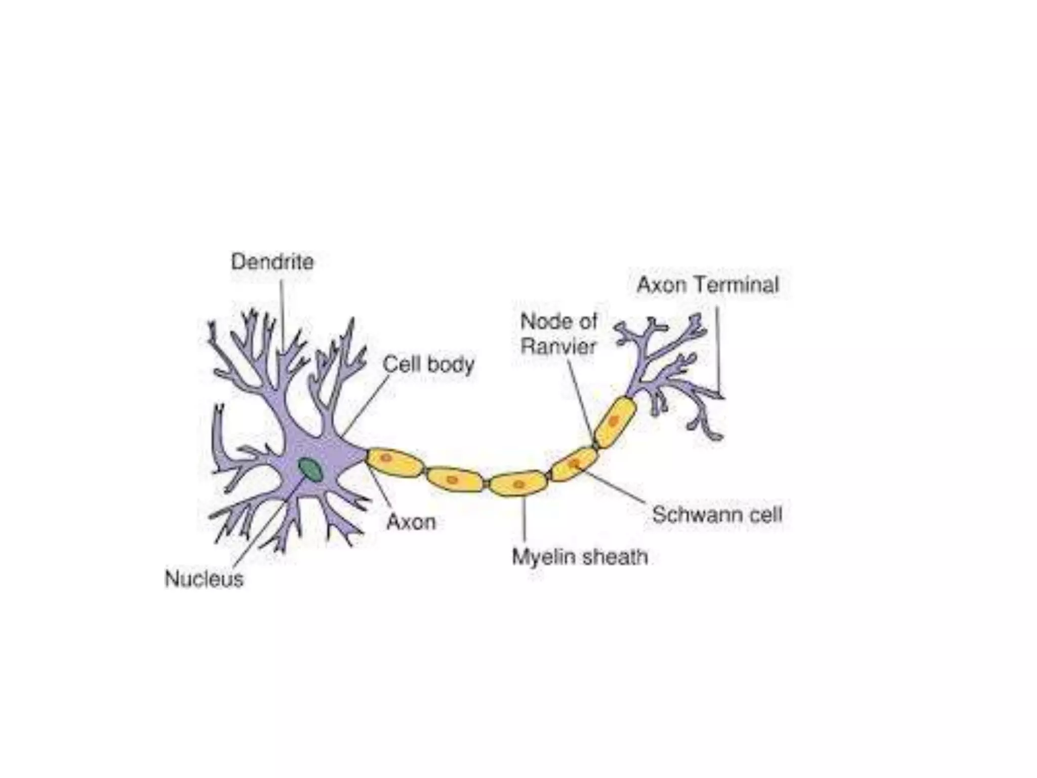
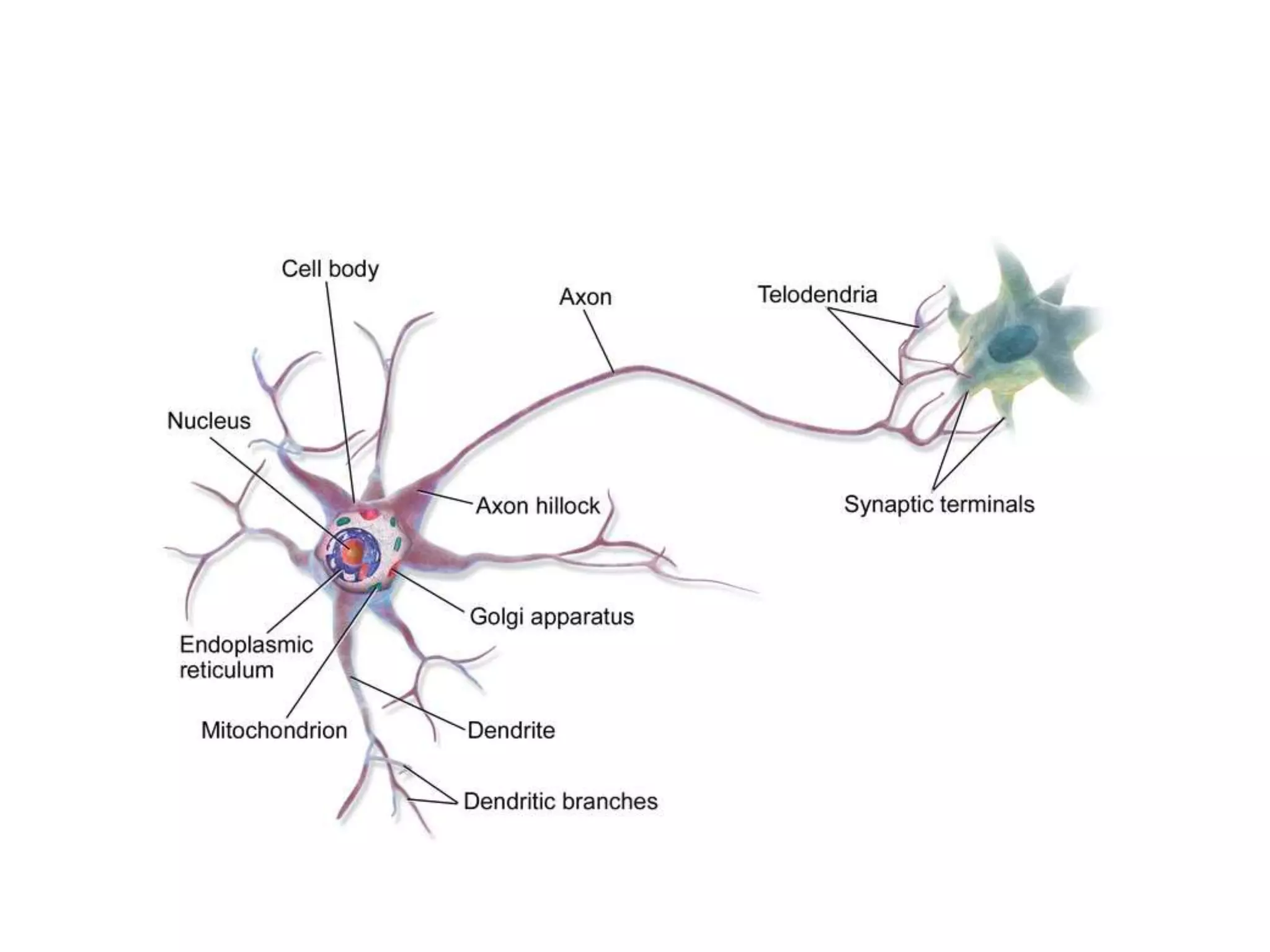
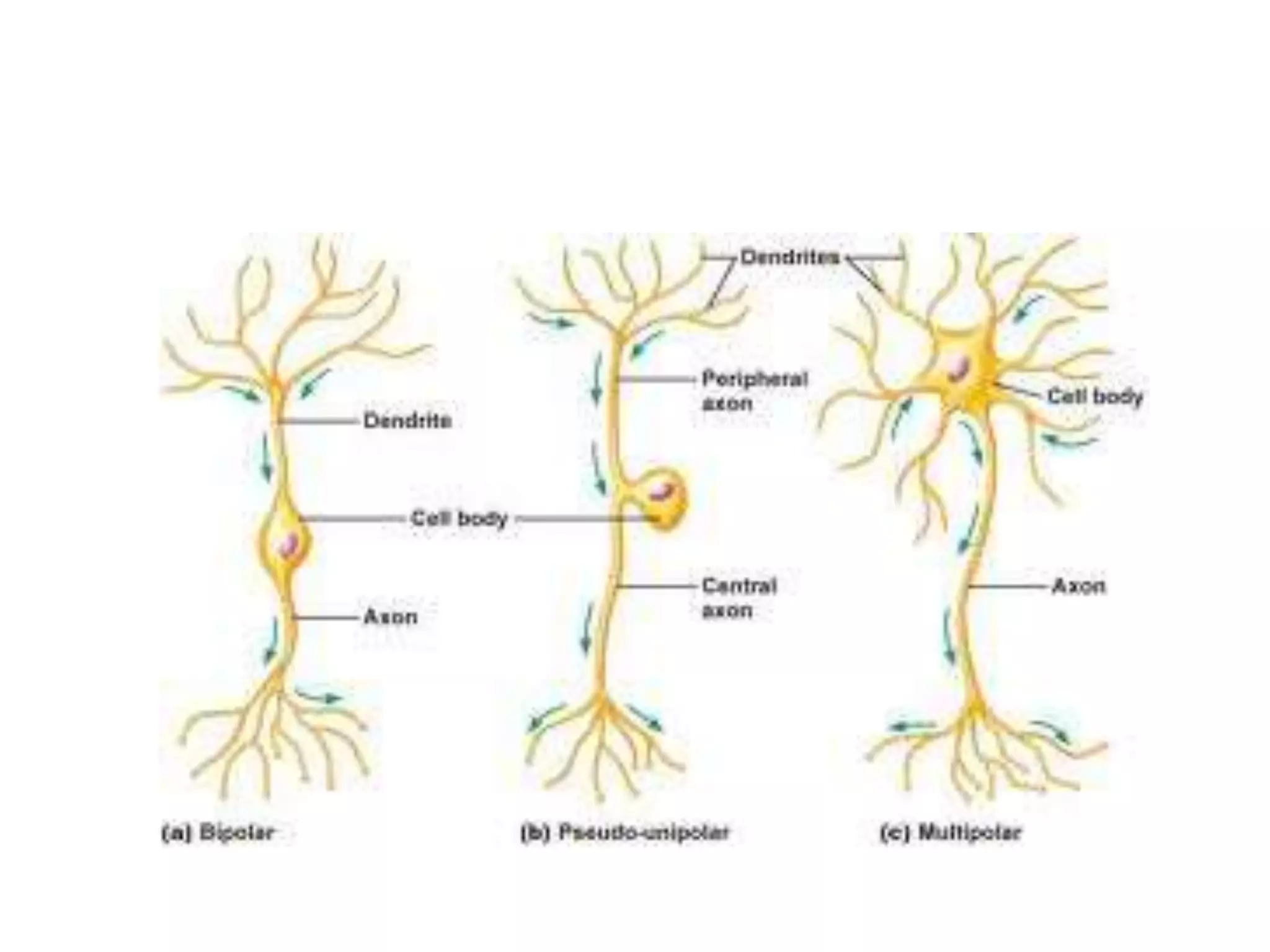
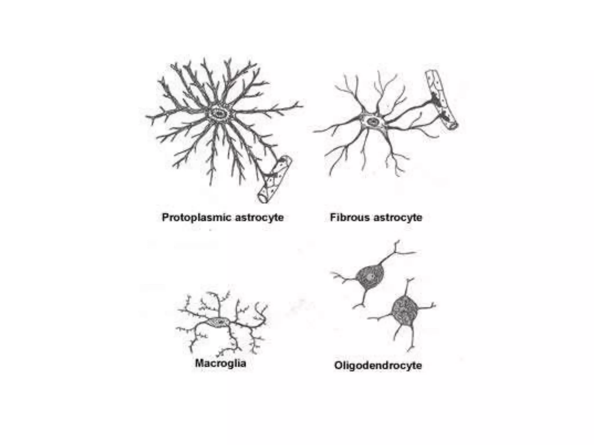
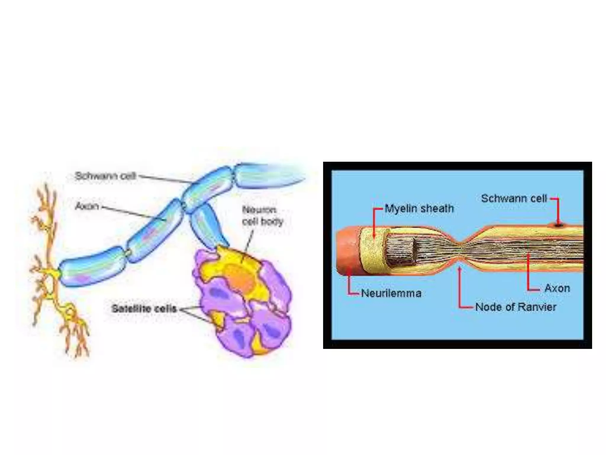
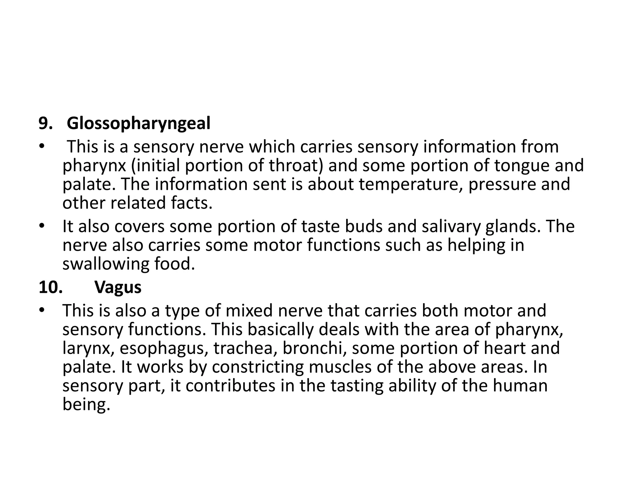
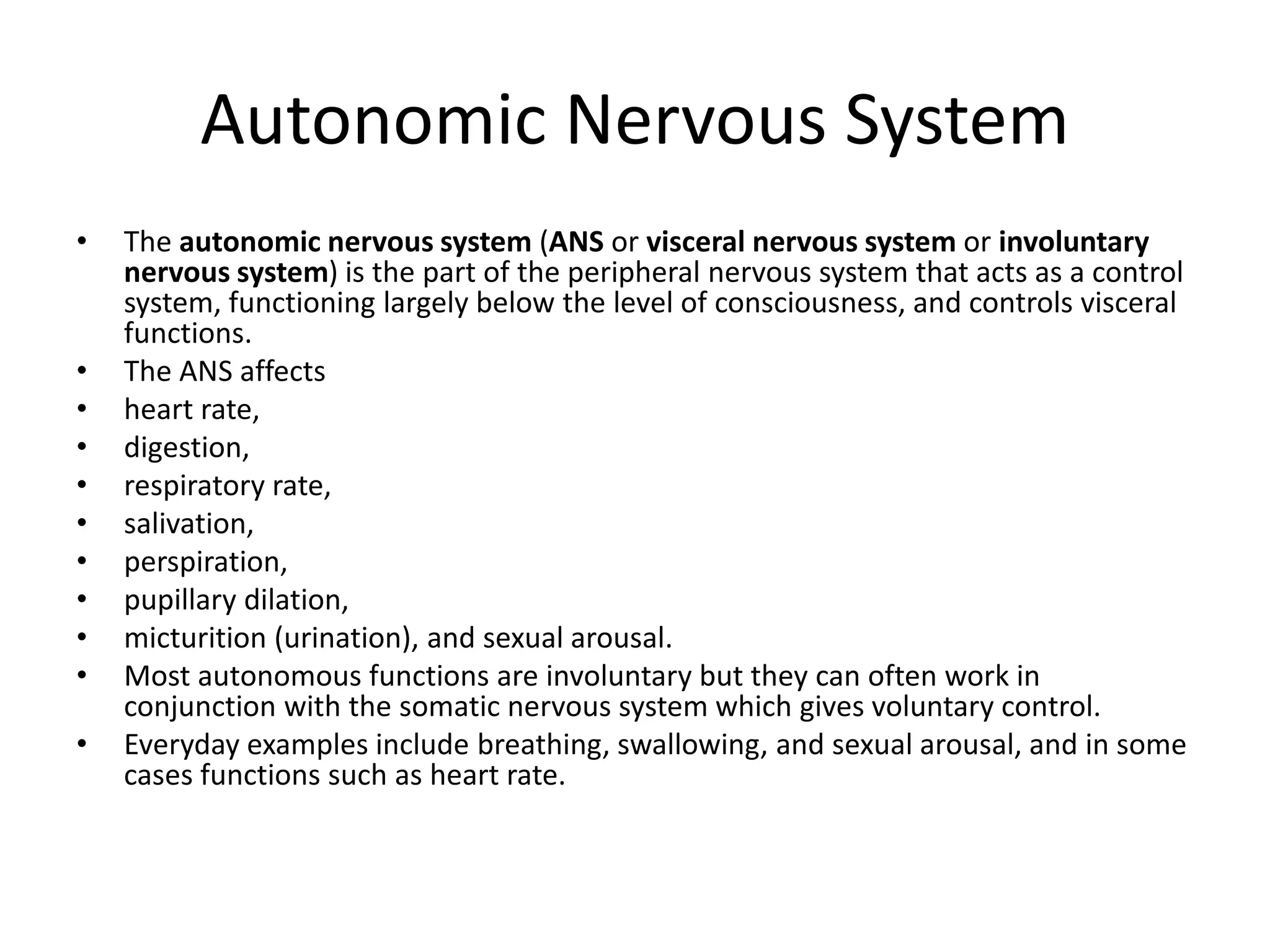
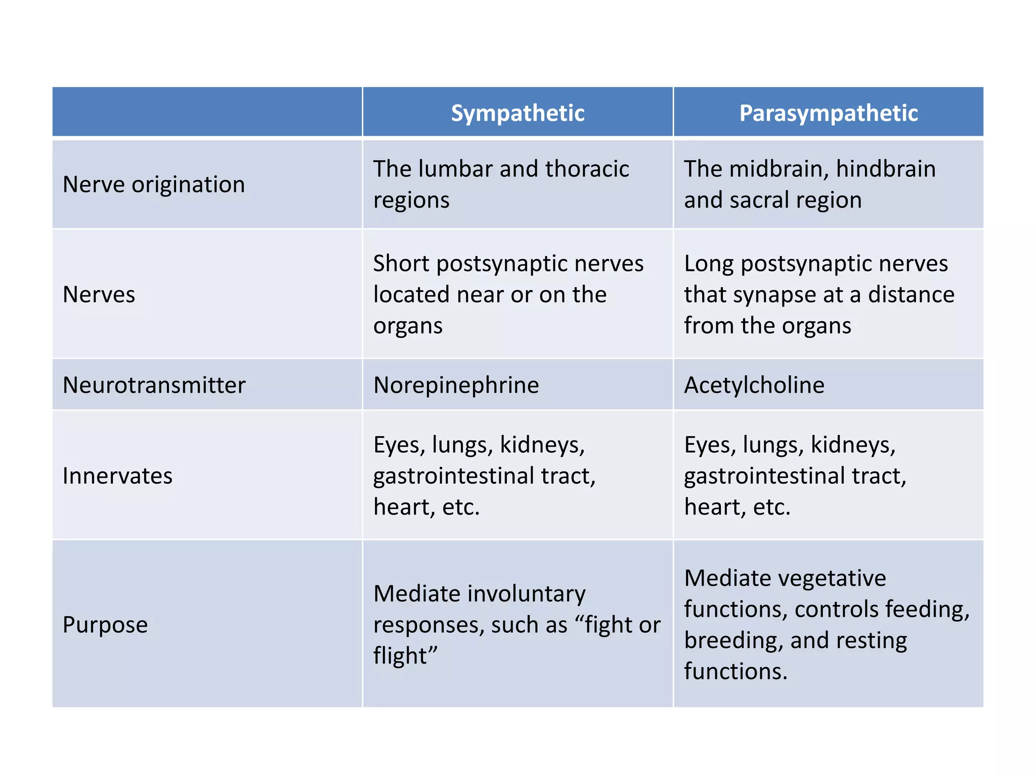
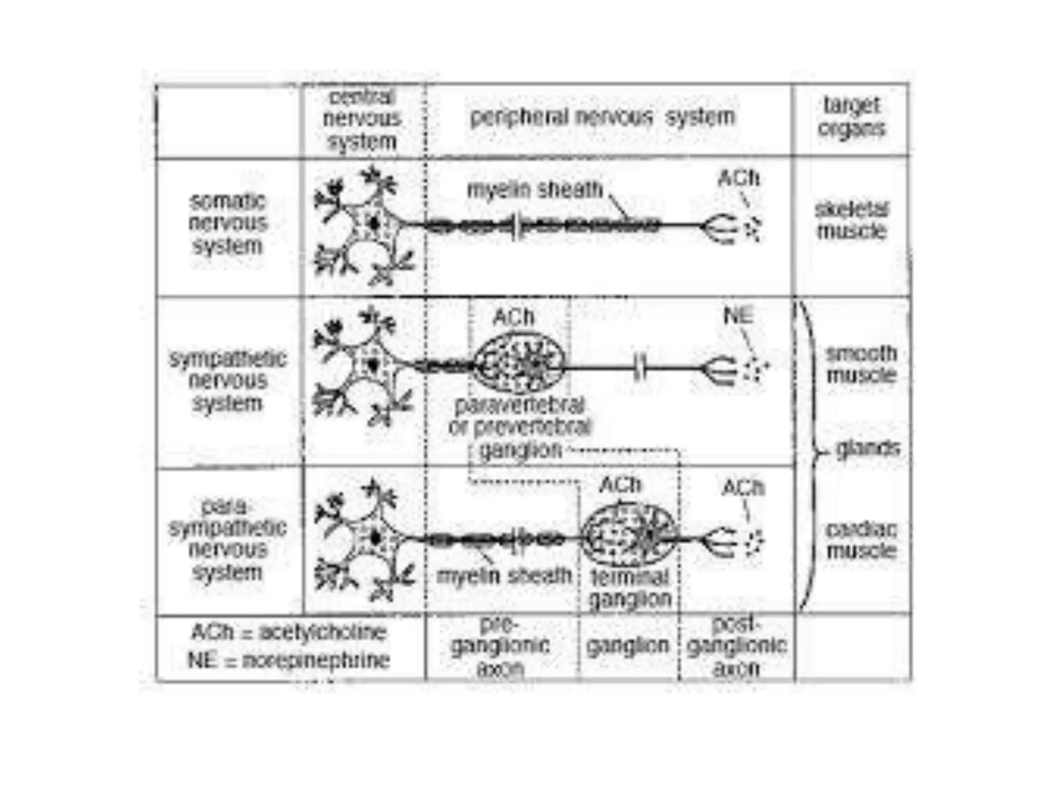
neuron, supportive cells and ANS
Neuron
Definition:-
A neuron ; also known as a neuron or nerve
cell) is an electrically excitable cell that
processes and transmits information through
electrical and chemical signals.
• These signals between neurons occur via synapses,
specialized connections with other cells.
• Neurons can connect to each other to form neural
networks.
• Specialized types of neurons include: sensory neurons
which respond to touch, sound, light and all other stimuli
affecting the cells of the sensory organs, that then send
signals to the spinal cord and brain; motor neurons that
receive signals from the brain and spinal cord, to cause
muscle contractions, and affect glandular outputs, and
interneurons which connect neurons to other neurons
within the same region of the brain or spinal cord, in neural
networks.
Structure of Neuron
• A typical neuron possesses a cell body or (soma), dendrites, and an
axon.
• The term neurite is used to describe either a dendrite or an axon,
particularly in its undifferentiated stage.
• Dendrites are thin structures that arise from the cell body, often
extending for hundreds of micrometres and branching multiple
times, giving rise to a complex "dendritic tree".
• An axon is a special cellular extension that arises from the cell body
at a site called the axon hillock and travels for a distance, as far as 1
meter in humans or even more in other species.
• The cell body of a neuron frequently gives rise to multiple
dendrites, but never to more than one axon, although the axon may
branch hundreds of times before it terminates.
• At the majority of synapses, signals are sent from the axon of one
neuron to a dendrite of another. There are, however, many
exceptions to these rules: neurons that lack dendrites, neurons that
have no axon, synapses that connect an axon to another axon or a
dendrite to another dendrite, etc.
• Each axon has usually few collateral branches
and a number of terminal branches called
telodendria which shows small swellings at
their distal ends called as end bulbs or
boutons.
Morphological classification of
neurons
• Most neurons can be anatomically characterized as:
• Unipolar or pseudounipolar: dendrite and axon
emerging from same process.
• Bipolar: axon and single dendrite on opposite ends of
the soma.
• Multipolar: two or more dendrites, separate from the
axon:
– Golgi I: neurons with long-projecting axonal processes;
examples are pyramidal cells, Purkinje cells, and anterior
horn cells.
– Golgi II: neurons whose axonal process projects locally; the
best example is the granule cell.
Support cells
Support cells are essential to the function and
survival of nerve cells. The CNS and PNS each
have their own specific types of support cells.
Support cells in the CNS:
The general term for support cells in the CNS is
glia or neuroglia (a.k.a. glial cells, neuroglial
cells). There are three types of neuroglial cells.
(1) Oligodendrocytes, the myelin-secreting cells
of the CNS.
(2) Astrocytes, which provide physical and
metabolic support for nerve cells.
(3) Microglia, or microglial cells (a.k.a. mesoglia),
which are the phagocytes of the CNS.
• Oligodendrocytes. As their name implies, oligodendrocytes have few
processes. They are often found in rows between axons. The myelin
sheath around axons is formed by concentric layers of oligodendrocytes
plasma membrane. Each oligodendrocyte gives off several tongue-like
processes that find their way to the axon, where each process wraps itself
around a portion of the axon, forming an internodal segment of myelin.
Each process appears to spiral around its segment of the axon in a
centripetal manner, with the continued insinuation of the leading edge
between the inner surface of newly formed myelin and the axon. One
oligodendrocyte may myelinate one axon or several. The nucleus-
containing region may be at some distance from the axon(s) it is
myelinating. In the CNS, nodes of Ranvier (between myelinated regions)
are larger than those of the PNS, and the larger amount of exposed
axolemma makes saltatory conduction more efficient.
• Unmyelinated axons in the CNS are truly bare, that is they are not
embedded in any glial cell process. (In contrast to the situation in the PNS,
described below.)
• Astrocytes. Astrocytes are the largest of the neuroglial cells.
They have elaborate processes that extend between
neurons and blood vessels. The ends of the processes
expand to form end feet, which cover large areas of the
outer surface of the blood vessel or axolemma. Astrocytes
are believed to play a role in the movement of metabolites
and wastes to and from neurons, and in regulating ionic
concentrations within the neurons. They may be involved in
regulating the tight junctions in the capillaries that form the
blood-brain barrier. Astrocytes also cover the bare areas of
neurons, at nodes of Ranvier and synapses. They may act to
confine neurotransmitters to the synaptic cleft and to
remove excess neurotransmitters.
• Two kinds of astrocytes are identified,
protoplasmic and fibrous astrocytes. Both
types contain prominent bundles of
intermediate filaments, but the filaments are
more numerous in fibrous astrocytes. Fibrous
astrocytes are more prevalent in white matter,
protoplasmic ones in grey matter.
• Microglia. These are the smallest of the glial cells, with short
twisted processes. They are the phagocytes of the CNS, considered
part of the mononuclear phagocytic system (see pg 110 in Ross et
al.). They are believed to originate in bone marrow and enter the
CNS from the blood. In the adult CNS, they are present only in small
numbers, but proliferate and become actively phagocytic in disease
and injury. Their alternate name, mesoglia, reflects their embyonic
origin from mesoderm (the rest of the nervous system, including
the other glial cells, is of neuroectodermal or neural crest origin).
• In routine histological preparations of the CNS, only the nuclei of
glial cells can be identified. In the slides in your collection, you will
not be able to attribute any individual glial cell nucleus to any
specific type of glial cell. You will however be able to distinguish the
neurons from glial cell nuclei. Special preparations are needed to
dentify glial cells, as seen in some of the images shown below.
• Support cells in the PNS
The support cells of the PNS are called satellite cells and
Schwann cells.
Satellite cells.
Satellite cells surround the cell bodies of the neurons in
ganglia (ganglion cells). These small cuboidal cells form a
complete layer around the nerve cell body, but only their
nuclei are visible in routine preparations. They help
maintain a controlled microenvironment around the nerve
cell body, providing electrical insulation and a pathway for
metabolic exchange. In paravertebral and peripheral
ganglia, nerve cell processes must penetrate between
satellite cells to establish a synapse.
• Schwann cells. Schwann cells are responsible for the
myelination of axons in the PNS. A Schwann cell wraps
itself, jelly roll-fashion, in a spiral around a short
segment of an axon. During the wrapping, cytoplasm is
squeezed out of the Schwann cell and the leaflets of
plasma membrance of the concentric layers of the
Schwann cell fuse, forming the layers of the myelin
sheath. An axon's myelin sheath is segmented because
it is formed by numerous Schwann cells arrayed in
sequence along the axon. The junction where two
Schwann cells meet has no myelin and is called (as you
know) the node of Ranvier (the areas covered by
Schwann cells being the internodal regions).
• The lack of Schwann cell cytoplasm in the concentric rings
of the myelin sheath is what makes it lipid-rich. Schwann
cell cytoplasm is however found in several locations. There
is an inner collar of Schwann cell cytoplasm between the
axon and the myelin, and an outer collar around the myelin.
The outer collar is also called the sheath of Schwann or
neurilemma, and contains the nucleus and most of the
organelles of the Schwann cell. The node of Ranvier is also
covered with Schwann cell cytoplasm, and this is the area
where the plasma membranes of adjacent Schwann cells
meet. (These adjacent plasma membranes are not tightly
apposed at the node, so that extracellular fluid has free
acess to the neuronal plasma membrane.) Finally, small
islands of Schwann cell cytoplasm persist within successive
layers of the myelin sheath, these islands are called
Schmidt-Lanterman clefts
• Not all nerve fibres is the PNS are covered in
myelin, some axons are unmyelinated. In contrast
to the situation in the CNS, unmyelinated fibres in
the PNS are not completely bare, but are
enveloped in Schwann cell cytoplasm. The
Schwann cells are elongated in parallel to the
long axis of the axons, which fit into grooves on
the surface of the Schwann cell. One axon or a
group of axons may be enclosed in a single
groove. Schwann cells may have only one or up to
twenty grooves. Single grooves are more
common in the autonomic nervous system
Cranial Nerves
• There are total 12 pairs of cranial nerves that
originate from our brain and brain stem. Each
of them carries different functions related to
different senses of body. Apart from sensory
functions there are also some that work as
motor nerves or mixed nerves.
• Here is a brief description of 12 cranial nerves.
1. Olfactory
• This is a type of sensory nerve that
contributes in the sense of smell in human
being. These basically provide the specific cells
that are termed as olfactory epithelium. It
carries the information from nasal epithelium
to the olfactory center in brain.
2. Optic nerve
• This again is a type of sensory nerve that
transforms information about vision to the
brain. To be specific this supplies information
to the retina in the form of ganglion cells.
3. Oculomoter nerve
• This is a form of motor nerve that supplies to
different centers along midbrain. Its functions
include superiorly uplifting eyelid, superiorly
rotating eyeball, construction of pupil on the
exposure to light and operating several eye
muscles.
4. Trochlear
• This motor nerve also supplies to the midbrain
and performs the function of handling the eye
muscles and turning the eye.
5. Trigeminal
• This is a type of largest cranial nerve in all and
performs many sensory functions related to nose, eyes,
tongue and teeth. It basically is further divided in three
branches that are ophthalmic, maxillary and
mandibular nerve. This is a type of mixed nerve that
performs sensory and motor functions in brain.
6. Abducent
• This is again a type of motor nerve that supplies to the
pons and perform function of turning eye laterally.
7. Facial
• This motor nerve is responsible for different types of facial
expressions. This also performs some functions of sensory
nerve by supplying information about touch on face and
senses of tongue in mouth. It is basically present over brain
stem.
8. Vestibulocochlear
• This motor nerve is basically functional in providing
information related to balance of head and sense of sound
or hearing. It carries vestibular as well as cochlear
information to the brain and is placed near inner ear.
9. Glossopharyngeal
• This is a sensory nerve which carries sensory information from
pharynx (initial portion of throat) and some portion of tongue and
palate. The information sent is about temperature, pressure and
other related facts.
• It also covers some portion of taste buds and salivary glands. The
nerve also carries some motor functions such as helping in
swallowing food.
10. Vagus
• This is also a type of mixed nerve that carries both motor and
sensory functions. This basically deals with the area of pharynx,
larynx, esophagus, trachea, bronchi, some portion of heart and
palate. It works by constricting muscles of the above areas. In
sensory part, it contributes in the tasting ability of the human
being.
11. Spinal accessory nerve
• As the name intimates this motor nerve
supplies information about spinal cord,
trapezius and other surrounding muscles. It
also provides muscle movement of the
shoulders and surrounding neck.
12. Hypoglossal nerve
• This is a typical motor nerve that deals with
the muscles of tongue.
Autonomic Nervous System
• The autonomic nervous system (ANS or visceral nervous system or involuntary
nervous system) is the part of the peripheral nervous system that acts as a control
system, functioning largely below the level of consciousness, and controls visceral
functions.
• The ANS affects
• heart rate,
• digestion,
• respiratory rate,
• salivation,
• perspiration,
• pupillary dilation,
• micturition (urination), and sexual arousal.
• Most autonomous functions are involuntary but they can often work in
conjunction with the somatic nervous system which gives voluntary control.
• Everyday examples include breathing, swallowing, and sexual arousal, and in some
cases functions such as heart rate.
• The ANS is classically divided into two
subsystems: the parasympathetic nervous
system (PSNS) and sympathetic nervous
system (SNS), which operate independently in
some functions and interact co-operatively in
others.
Sympathetic Parasympathetic
Nerve origination
The lumbar and thoracic
regions
The midbrain, hindbrain
and sacral region
Nerves
Short postsynaptic nerves
located near or on the
organs
Long postsynaptic nerves
that synapse at a distance
from the organs
Neurotransmitter Norepinephrine Acetylcholine
Innervates
Eyes, lungs, kidneys,
gastrointestinal tract,
heart, etc.
Eyes, lungs, kidneys,
gastrointestinal tract,
heart, etc.
Purpose
Mediate involuntary
responses, such as “fight or
flight”
Mediate vegetative
functions, controls feeding,
breeding, and resting
functions.
Sympathetic Parasympathetic
Function
Allows the body to adjust
in stressful situations, such
as arousing excitement,
fear, anger, and
embarrassment, increases
the heart rate, thus,
causing an increase in the
blood pressure, dilates the
respiratory bronchioles to
increase uptake of oxygen,
decreases gallbladder
secretions and dilates
blood vessels to increase
blood supply to the skeletal
muscles.
Constriction of pupils,
decreases the heart rate,
thus, causing a drop in the
blood pressure, stimulation
of digestive glands,
stimulation of secretion of
saliva, stimulates the
processes of urination and
defecation, and constricts
the bronchi and thus,
decreasing the diameter of
airway,
NEURON SUPPORTIVE CELLS OR ANS

NEURON SUPPORTIVE CELLS OR ANS

  • 1.
  • 2.
    Neuron Definition:- A neuron ;also known as a neuron or nerve cell) is an electrically excitable cell that processes and transmits information through electrical and chemical signals.
  • 3.
    • These signalsbetween neurons occur via synapses, specialized connections with other cells. • Neurons can connect to each other to form neural networks. • Specialized types of neurons include: sensory neurons which respond to touch, sound, light and all other stimuli affecting the cells of the sensory organs, that then send signals to the spinal cord and brain; motor neurons that receive signals from the brain and spinal cord, to cause muscle contractions, and affect glandular outputs, and interneurons which connect neurons to other neurons within the same region of the brain or spinal cord, in neural networks.
  • 4.
    Structure of Neuron •A typical neuron possesses a cell body or (soma), dendrites, and an axon. • The term neurite is used to describe either a dendrite or an axon, particularly in its undifferentiated stage. • Dendrites are thin structures that arise from the cell body, often extending for hundreds of micrometres and branching multiple times, giving rise to a complex "dendritic tree". • An axon is a special cellular extension that arises from the cell body at a site called the axon hillock and travels for a distance, as far as 1 meter in humans or even more in other species. • The cell body of a neuron frequently gives rise to multiple dendrites, but never to more than one axon, although the axon may branch hundreds of times before it terminates. • At the majority of synapses, signals are sent from the axon of one neuron to a dendrite of another. There are, however, many exceptions to these rules: neurons that lack dendrites, neurons that have no axon, synapses that connect an axon to another axon or a dendrite to another dendrite, etc.
  • 7.
    • Each axonhas usually few collateral branches and a number of terminal branches called telodendria which shows small swellings at their distal ends called as end bulbs or boutons.
  • 8.
    Morphological classification of neurons •Most neurons can be anatomically characterized as: • Unipolar or pseudounipolar: dendrite and axon emerging from same process. • Bipolar: axon and single dendrite on opposite ends of the soma. • Multipolar: two or more dendrites, separate from the axon: – Golgi I: neurons with long-projecting axonal processes; examples are pyramidal cells, Purkinje cells, and anterior horn cells. – Golgi II: neurons whose axonal process projects locally; the best example is the granule cell.
  • 10.
    Support cells Support cellsare essential to the function and survival of nerve cells. The CNS and PNS each have their own specific types of support cells. Support cells in the CNS: The general term for support cells in the CNS is glia or neuroglia (a.k.a. glial cells, neuroglial cells). There are three types of neuroglial cells. (1) Oligodendrocytes, the myelin-secreting cells of the CNS. (2) Astrocytes, which provide physical and metabolic support for nerve cells. (3) Microglia, or microglial cells (a.k.a. mesoglia), which are the phagocytes of the CNS.
  • 11.
    • Oligodendrocytes. Astheir name implies, oligodendrocytes have few processes. They are often found in rows between axons. The myelin sheath around axons is formed by concentric layers of oligodendrocytes plasma membrane. Each oligodendrocyte gives off several tongue-like processes that find their way to the axon, where each process wraps itself around a portion of the axon, forming an internodal segment of myelin. Each process appears to spiral around its segment of the axon in a centripetal manner, with the continued insinuation of the leading edge between the inner surface of newly formed myelin and the axon. One oligodendrocyte may myelinate one axon or several. The nucleus- containing region may be at some distance from the axon(s) it is myelinating. In the CNS, nodes of Ranvier (between myelinated regions) are larger than those of the PNS, and the larger amount of exposed axolemma makes saltatory conduction more efficient. • Unmyelinated axons in the CNS are truly bare, that is they are not embedded in any glial cell process. (In contrast to the situation in the PNS, described below.)
  • 12.
    • Astrocytes. Astrocytesare the largest of the neuroglial cells. They have elaborate processes that extend between neurons and blood vessels. The ends of the processes expand to form end feet, which cover large areas of the outer surface of the blood vessel or axolemma. Astrocytes are believed to play a role in the movement of metabolites and wastes to and from neurons, and in regulating ionic concentrations within the neurons. They may be involved in regulating the tight junctions in the capillaries that form the blood-brain barrier. Astrocytes also cover the bare areas of neurons, at nodes of Ranvier and synapses. They may act to confine neurotransmitters to the synaptic cleft and to remove excess neurotransmitters.
  • 13.
    • Two kindsof astrocytes are identified, protoplasmic and fibrous astrocytes. Both types contain prominent bundles of intermediate filaments, but the filaments are more numerous in fibrous astrocytes. Fibrous astrocytes are more prevalent in white matter, protoplasmic ones in grey matter.
  • 14.
    • Microglia. Theseare the smallest of the glial cells, with short twisted processes. They are the phagocytes of the CNS, considered part of the mononuclear phagocytic system (see pg 110 in Ross et al.). They are believed to originate in bone marrow and enter the CNS from the blood. In the adult CNS, they are present only in small numbers, but proliferate and become actively phagocytic in disease and injury. Their alternate name, mesoglia, reflects their embyonic origin from mesoderm (the rest of the nervous system, including the other glial cells, is of neuroectodermal or neural crest origin). • In routine histological preparations of the CNS, only the nuclei of glial cells can be identified. In the slides in your collection, you will not be able to attribute any individual glial cell nucleus to any specific type of glial cell. You will however be able to distinguish the neurons from glial cell nuclei. Special preparations are needed to dentify glial cells, as seen in some of the images shown below.
  • 16.
    • Support cellsin the PNS The support cells of the PNS are called satellite cells and Schwann cells. Satellite cells. Satellite cells surround the cell bodies of the neurons in ganglia (ganglion cells). These small cuboidal cells form a complete layer around the nerve cell body, but only their nuclei are visible in routine preparations. They help maintain a controlled microenvironment around the nerve cell body, providing electrical insulation and a pathway for metabolic exchange. In paravertebral and peripheral ganglia, nerve cell processes must penetrate between satellite cells to establish a synapse.
  • 17.
    • Schwann cells.Schwann cells are responsible for the myelination of axons in the PNS. A Schwann cell wraps itself, jelly roll-fashion, in a spiral around a short segment of an axon. During the wrapping, cytoplasm is squeezed out of the Schwann cell and the leaflets of plasma membrance of the concentric layers of the Schwann cell fuse, forming the layers of the myelin sheath. An axon's myelin sheath is segmented because it is formed by numerous Schwann cells arrayed in sequence along the axon. The junction where two Schwann cells meet has no myelin and is called (as you know) the node of Ranvier (the areas covered by Schwann cells being the internodal regions).
  • 18.
    • The lackof Schwann cell cytoplasm in the concentric rings of the myelin sheath is what makes it lipid-rich. Schwann cell cytoplasm is however found in several locations. There is an inner collar of Schwann cell cytoplasm between the axon and the myelin, and an outer collar around the myelin. The outer collar is also called the sheath of Schwann or neurilemma, and contains the nucleus and most of the organelles of the Schwann cell. The node of Ranvier is also covered with Schwann cell cytoplasm, and this is the area where the plasma membranes of adjacent Schwann cells meet. (These adjacent plasma membranes are not tightly apposed at the node, so that extracellular fluid has free acess to the neuronal plasma membrane.) Finally, small islands of Schwann cell cytoplasm persist within successive layers of the myelin sheath, these islands are called Schmidt-Lanterman clefts
  • 19.
    • Not allnerve fibres is the PNS are covered in myelin, some axons are unmyelinated. In contrast to the situation in the CNS, unmyelinated fibres in the PNS are not completely bare, but are enveloped in Schwann cell cytoplasm. The Schwann cells are elongated in parallel to the long axis of the axons, which fit into grooves on the surface of the Schwann cell. One axon or a group of axons may be enclosed in a single groove. Schwann cells may have only one or up to twenty grooves. Single grooves are more common in the autonomic nervous system
  • 21.
    Cranial Nerves • Thereare total 12 pairs of cranial nerves that originate from our brain and brain stem. Each of them carries different functions related to different senses of body. Apart from sensory functions there are also some that work as motor nerves or mixed nerves. • Here is a brief description of 12 cranial nerves.
  • 22.
    1. Olfactory • Thisis a type of sensory nerve that contributes in the sense of smell in human being. These basically provide the specific cells that are termed as olfactory epithelium. It carries the information from nasal epithelium to the olfactory center in brain.
  • 23.
    2. Optic nerve •This again is a type of sensory nerve that transforms information about vision to the brain. To be specific this supplies information to the retina in the form of ganglion cells.
  • 25.
    3. Oculomoter nerve •This is a form of motor nerve that supplies to different centers along midbrain. Its functions include superiorly uplifting eyelid, superiorly rotating eyeball, construction of pupil on the exposure to light and operating several eye muscles. 4. Trochlear • This motor nerve also supplies to the midbrain and performs the function of handling the eye muscles and turning the eye.
  • 26.
    5. Trigeminal • Thisis a type of largest cranial nerve in all and performs many sensory functions related to nose, eyes, tongue and teeth. It basically is further divided in three branches that are ophthalmic, maxillary and mandibular nerve. This is a type of mixed nerve that performs sensory and motor functions in brain. 6. Abducent • This is again a type of motor nerve that supplies to the pons and perform function of turning eye laterally.
  • 27.
    7. Facial • Thismotor nerve is responsible for different types of facial expressions. This also performs some functions of sensory nerve by supplying information about touch on face and senses of tongue in mouth. It is basically present over brain stem. 8. Vestibulocochlear • This motor nerve is basically functional in providing information related to balance of head and sense of sound or hearing. It carries vestibular as well as cochlear information to the brain and is placed near inner ear.
  • 28.
    9. Glossopharyngeal • Thisis a sensory nerve which carries sensory information from pharynx (initial portion of throat) and some portion of tongue and palate. The information sent is about temperature, pressure and other related facts. • It also covers some portion of taste buds and salivary glands. The nerve also carries some motor functions such as helping in swallowing food. 10. Vagus • This is also a type of mixed nerve that carries both motor and sensory functions. This basically deals with the area of pharynx, larynx, esophagus, trachea, bronchi, some portion of heart and palate. It works by constricting muscles of the above areas. In sensory part, it contributes in the tasting ability of the human being.
  • 29.
    11. Spinal accessorynerve • As the name intimates this motor nerve supplies information about spinal cord, trapezius and other surrounding muscles. It also provides muscle movement of the shoulders and surrounding neck. 12. Hypoglossal nerve • This is a typical motor nerve that deals with the muscles of tongue.
  • 30.
    Autonomic Nervous System •The autonomic nervous system (ANS or visceral nervous system or involuntary nervous system) is the part of the peripheral nervous system that acts as a control system, functioning largely below the level of consciousness, and controls visceral functions. • The ANS affects • heart rate, • digestion, • respiratory rate, • salivation, • perspiration, • pupillary dilation, • micturition (urination), and sexual arousal. • Most autonomous functions are involuntary but they can often work in conjunction with the somatic nervous system which gives voluntary control. • Everyday examples include breathing, swallowing, and sexual arousal, and in some cases functions such as heart rate.
  • 31.
    • The ANSis classically divided into two subsystems: the parasympathetic nervous system (PSNS) and sympathetic nervous system (SNS), which operate independently in some functions and interact co-operatively in others.
  • 32.
    Sympathetic Parasympathetic Nerve origination Thelumbar and thoracic regions The midbrain, hindbrain and sacral region Nerves Short postsynaptic nerves located near or on the organs Long postsynaptic nerves that synapse at a distance from the organs Neurotransmitter Norepinephrine Acetylcholine Innervates Eyes, lungs, kidneys, gastrointestinal tract, heart, etc. Eyes, lungs, kidneys, gastrointestinal tract, heart, etc. Purpose Mediate involuntary responses, such as “fight or flight” Mediate vegetative functions, controls feeding, breeding, and resting functions.
  • 33.
    Sympathetic Parasympathetic Function Allows thebody to adjust in stressful situations, such as arousing excitement, fear, anger, and embarrassment, increases the heart rate, thus, causing an increase in the blood pressure, dilates the respiratory bronchioles to increase uptake of oxygen, decreases gallbladder secretions and dilates blood vessels to increase blood supply to the skeletal muscles. Constriction of pupils, decreases the heart rate, thus, causing a drop in the blood pressure, stimulation of digestive glands, stimulation of secretion of saliva, stimulates the processes of urination and defecation, and constricts the bronchi and thus, decreasing the diameter of airway,