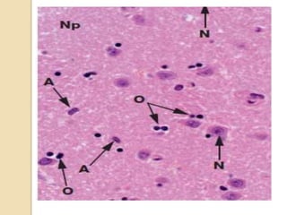
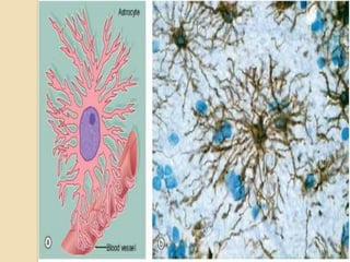
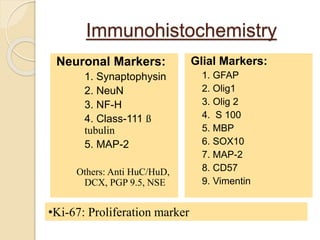
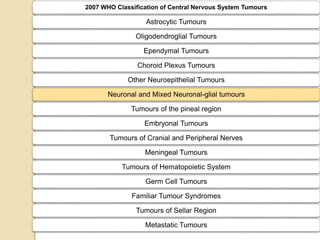
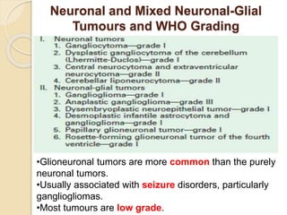
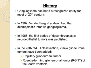
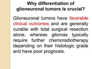
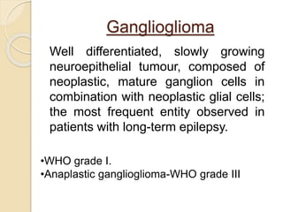
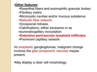
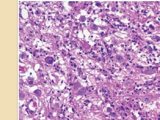
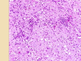
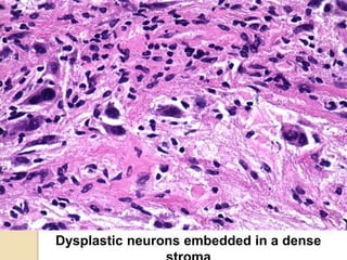
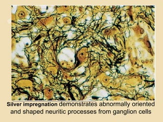
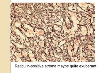
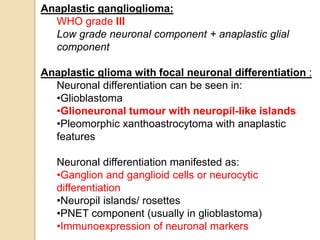
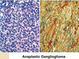
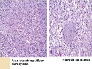
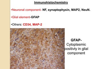
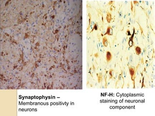
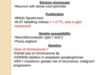
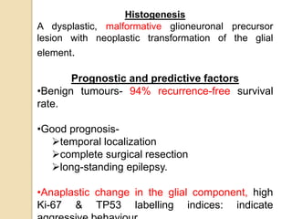
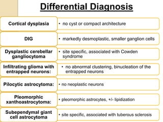
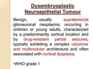
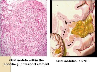
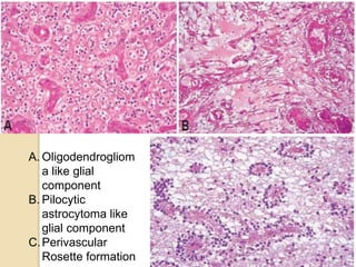
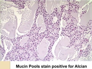
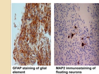
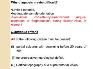
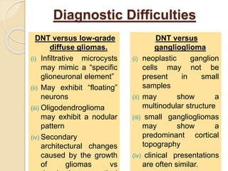
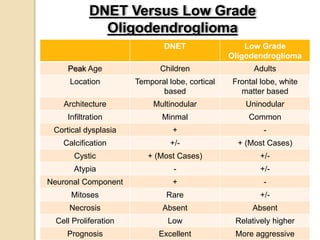
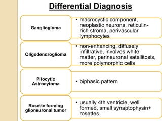
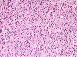
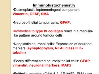
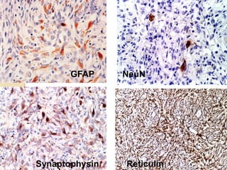
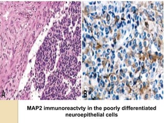
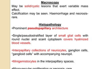
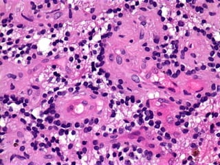
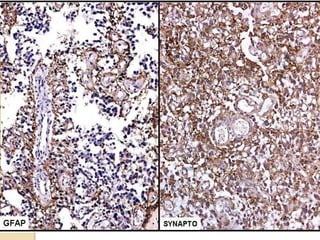
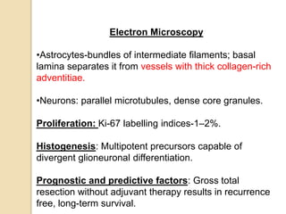
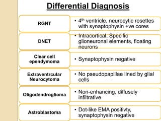
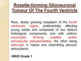
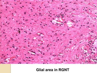
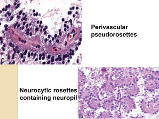
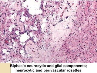
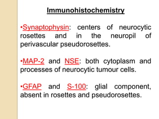
3. Four principal types:
• Oligodendrocytes: responsible for the formation of
myelin sheaths in the CNS.
• Astrocytes: form part of the blood-brain barrier and
also play an important role in repair of CNS tissue.
• Microglia: defence and immunological functions.
• Ependymal: specialised epithelium which lines the
ventricles and spinal canal.
7. 2007 WHO Classification of Central Nervous System Tumours
Astrocytic Tumours
Oligodendroglial Tumours
Ependymal Tumours
Choroid Plexus Tumours
Other Neuroepithelial Tumours
Neuronal and Mixed Neuronal-glial tumours
Tumours of the pineal region
Embryonal Tumours
Tumours of Cranial and Peripheral Nerves
Meningeal Tumours
Tumours of Hematopoietic System
Germ Cell Tumours
Familiar Tumour Syndromes
Tumours of Sellar Region
Metastatic Tumours
8. Neuronal and Mixed Neuronal-Glial
Tumours and WHO Grading
•Glioneuronal tumors are more common than the purely
neuronal tumors.
•Usually associated with seizure disorders, particularly
gangliogliomas.
•Most tumours are low grade.
9. History
Ganglioglioma has been a recognized entity for
most of 20th century.
In 1987, VandenBerg et al described the
desmoplastic infantile ganglioglioma.
In 1988, the first series of dysembryoplastic
neuroepithelial tumors was published.
In the 2007 WHO classfication, 2 new glioneuronal
tumors have been added:
Papillary glioneuronal tumor
Rosette-forming glioneuronal tumor (RGNT) of
the fourth ventricle
10. Why differentiation of
glioneuronal tumors is crucial?
Glioneuronal tumors have favorable
clinical outcomes and are generally
curable with total surgical resection
alone, whereas gliomas typically
require further chemoradiotherapy
depending on their histologic grade
and have poor prognosis.
11. Ganglioglioma
Well differentiated, slowly growing
neuroepithelial tumour, composed of
neoplastic, mature ganglion cells in
combination with neoplastic glial cells;
the most frequent entity observed in
patients with long-term epilepsy.
•WHO grade I.
•Anaplastic ganglioglioma-WHO grade III
12. Incidence
•1.3% of all brain tumours.
Age and Sex Distrbution
•2 months-70 years.
•Male:female ratio-equal distribution or 1.9:1
•In children, mean age at diagnosis was10.3 years.
Localization
•Occur throughout the CNS, especially temporal lobe.
Clinical Features
•Tumours in the cerebrum- Seizures
• Tumours involving brain stem/spinal cord- Crossed
paresis and sphincteric disorder after a mean of
1.5years.
•Most common tumours associated with chronic
temporal lobe epilepsy.
23. Anaplastic ganglioglioma:
WHO grade III
Low grade neuronal component + anaplastic glial
component
Anaplastic glioma with focal neuronal differentiation :
Neuronal differentiation can be seen in:
•Glioblastoma
•Glioneuronal tumour with neuropil-like islands
•Pleomorphic xanthoastrocytoma with anaplastic
features
Neuronal differentiation manifested as:
•Ganglion and ganglioid cells or neurocytic
differentiation
•Neuropil islands/ rosettes
•PNET component (usually in glioblastoma)
•Immunoexpression of neuronal markers
28. Electron microscopy
•Neurons with dense core granules
Proliferation
•Mitotic figures-rare.
•Ki-67 labellling indices-1.1-2.7%, only in glial
component
Genetic susceptibility
•Neurofibromatosis- type 1 and 2
•Peutz-Jeghers
Genetics
•Gain of chromosome 7
•Partial loss of chromosome 9p
•CDKN2A deletion in anaplastic gangliogliomas
•IDH-1 mutations- greater risk of recurrence, malignant
progression
29. Histogenesis
A dysplastic, malformative glioneuronal precursor
lesion with neoplastic transformation of the glial
element.
Prognostic and predictive factors
•Benign tumours- 94% recurrence-free survival
rate.
•Good prognosis-
temporal localization
complete surgical resection
long-standing epilepsy.
•Anaplastic change in the glial component, high
Ki-67 & TP53 labelling indices: indicate
30. • no cyst or compact architectureCortical dysplasia
• markedly desmoplastic, smaller ganglion cellsDIG
• site specific, associated with Cowden
syndrome
Dysplastic cerebellar
gangliocytoma
• no abnormal clustering, binucleation of the
entrapped neurons
Infiltrating glioma with
entrapped neurons:
• no neoplastic neuronsPilocytic astrocytoma:
• pleomorphic astrocytes, +/- lipidization
Pleomorphic
xanthoastrocytoma:
• site specific, associated with tuberous sclerosis
Subependymal giant
cell astrocytoma
Differential Diagnosis
31. Dysembryoplastic
Neuroepithelial Tumour
Benign, usually supratentorial
glioneuronal neoplasms, occurring in
children or young adults, characterized
by a predominantly cortical location and
by drug-resistant partial seizures;
typically exhibiting a complex columnar
and multinodular architecture and often
associated with cortical dysplasia.
•WHO grade 1
32. Incidence
“Typical” DNTs-12% in adults and 13.5% in children
Age and sex distribution
•In 90% of cases, 1st seizure occurs before 20
years of age.
•Diagnosed in the 2nd or 3rd decade of life.
•Males are more frequently affected.
Localization
•Supratentorial cortex, especially temporal lobe
•Can also be found in third ventricle, basal ganglia
etc.
Clinical Features:
Drug-resistant partial seizures, with or without
secondary generalization and no neurological
33. Neuroimaging
•Cortical topography,
absence of mass effect
and no peritumoural
edema
•MRI > CT scan.
•Hyperintense on T2-
weighted and hypointense
on T1-weighted images.
•Deformation of overlying
calvarium
•Ring-shaped contrast
enhancement
34. Macroscopy
•Vary in size.
•Identified at cortical surface, maybe exophytic.
•Leptomeninges are not involved.
•Viscous consistency of the glioneuronal component.
•Maybe associated with multiple/single firmer nodules.
35. Histopathology
•Histological hallmark- ‘Specific glioneuronal
element’, characterized by columns of axons lined
by small oligodendroglia-like cells, oriented
perpendicularly to the cortical surface.
•‘Floating’ neurons with a normal cytology
embedded in a pale, eosinophilic matrix.
•Scattered GFAP-positive stellate astrocytes.
•Fluid extravasation determines if
columnar/alveolar/ compact structure.
•Histological sub-classfication has no clinical or
therapeutic implication.
36. Simple form
•Consists of the unique glioneuronal element.
•Maybe patchy.
Complex form
•Glial nodules + specific glioneuronal element
•Glial components:
•form typical nodules or diffuse pattern
•resemble gliomas or show unusual features
•mimic low-grade gliomas: nuclear atypia,or
necrosis
•microvascular network poor to exuberant.•Frankly hamartomatous, calcified vessels ->
behave as vascular malformations-> haemorrhage.
•Non-specific histological variants (20-50%)-
histologically indistinguishable from low-grade
37. •Adjacent dysplastic
disorganization of the cortex
in 80% cases.
Neuronal populations of
DNTs:
•mature neurons
•may show cytological
anomalies
• no dysplastic ganglion cells
Cortical topography
•Limits of tumour often coincide
with that of the cortex.
•May have disordered neuronal
migration
43. GFAP staining of glial
element
MAP2 immunostaining of
floating neurons
44. Why diagnosis maybe difficult?
•Limited material.
•Inadequate sample orientation
•Semi-liquid consistency->inadvertent surgical
aspiration or fragmentation during fixation->loss of
element
Diagnostic criteria
All of the following criteria must be present:
(i) partial seizures with beginning before 20 years of
age.
(ii) no progressive neurological deficit.
(iii) Cortical topography of a supratentorial lesion.
45. Diagnostic Difficulties
DNT versus low-grade
diffuse gliomas.
(i) Infiltrative microcysts
may mimic a “specific
glioneuronal element”
(ii) May exhibit “floating”
neurons
(iii) Oligodendroglioma
may exhibit a nodular
pattern
(iv) Secondary
architectural changes
caused by the growth
of gliomas vs
DNT versus
ganglioglioma
(i) neoplastic ganglion
cells may not be
present in small
samples
(ii) may show a
multinodular structure
(iii) small gangliogliomas
may show a
predominant cortical
topography
(iv) clinical presentations
are often similar.
46. DNET Low Grade
Oligodendroglioma
Peak Age Children Adults
Location Temporal lobe, cortical
based
Frontal lobe, white
matter based
Architecture Multinodular Uninodular
Infiltration Minmal Common
Cortical dysplasia + -
Calcification +/- + (Most Cases)
Cystic + (Most Cases) +/-
Atypia - +/-
Neuronal Component + -
Mitoses Rare +/-
Necrosis Absent Absent
Cell Proliferation Low Relatively higher
Prognosis Excellent More aggressive
DNET Versus Low Grade
Oligodendroglioma
47. • macrocystic component,
neoplastic neurons, reticulin-
rich stroma, perivascular
lymphocytes
Gangliogloma
• non-enhancing, diffusely
infiltrative, involves white
matter, perineuronal satellitosis,
more polymorphic cells
Oligodendroglioma
• biphasic patternPilocytic
Astrocytoma
• usually 4th ventricle, well
formed, small synaptophysin+
rosettes
Rosette forming
glioneuronal tumor
Differential Diagnosis
48. Proliferation
Ki-67 labelling indices- 0% -8%
Genetic susceptibility
•Neurofibromatosis type 1 (NF1)
•XYY syndrome
Histogenesis
Malformative origin
Prognostic and predictive factors
•Benign.
•No recurrence after surgical removal.
•Risk factors for the development of recurrent seizures after
surgery were: longer pre-operative history of seizures
presence of residual tumour
presence of adjacent cortical dysplasia
49. Desmoplastic Infantile
Ganglioglioma
Large cystic tumours of infants that
involve superficial cerebral cortex and
leptomeninges, often attached to dura,
with good prognosis following surgical
resection; histologically composed of
prominent desmoplastic stroma,
neoplastic astrocytes, a variable
neuronal component and aggregates
of poorly differentiated cells.
•WHO Grade 1
50. Incidence
0.3% of CNS tumours from all ages
Age and sex distribution
•1–24 months
•Male:female ratio of 1.5:1
•Non-infantile cases-5 to 25 years
Localization
•Supratentorial region, involve more than one lobe
•Frontal and parietal>temporal >occipital
Clinical features
•Short duration
•Increasing head circumference, tense and bulging
fontanelles, lethargy, and setting-sun sign.
•May have seizures, focal motor signs or skull bossing
over the tumour.
51. Neuroimaging
•CT scan- Large, hypodense cystic masses with a solid
hyperdense superficial portion that extends to the overlying
meninges. Shows contrast enhancement.
MRI T1-weighted images T2-weghted
images
Solid
component
Isointense, peripheral,
enhancing
Heterogenous
Cystic
component
Hypointense Hyperintense
52. Macroscopy
•Large, measuring up to 13 cm in diameter,
•Deep uni/multiloculated cysts filled with clear or
xanthochromic fluid.
•Solid, superficial portion-primarily extracerebral,
involving leptomeninges and superficial cortex,
commonly attached to the dura, firm or rubbery, and
grey.
•No gross evidence of haemorrhage or necrosis.
53. Histopathology
1. Desmoplastic leptomeningeal component:
•Fibroblast-like spindle-shaped cells arranged
in fascicles/storiform/whorled pattern.
•Reticulin positive network surrounds every
cell.
•Tumour cells- Astrocytes + neoplastic neurons
(atypical ganglionic cells to small polygonal
cells)
2. Poorly differentiated neuroepithelial
component: Cells with small, round, deeply
basophilic nuclei and minimal perikarya.
3. Cortical component: often multinodular, with
54. •Sharp demarcation between the cortical surface
and the desmoplastic tumour.
•Calcifications are common.
•Mononuclear inflammatory cells are not usually
seen.
•Mitotic activity and necrosis are uncommon,
maybe present in poorly differentiated
neuroepithelial cells.
•Microvascular proliferation is not evident.
58. Immunohistochemistry
•Desmoplastic leptomeningeal component:
Vimentin, GFAP, SMA.
•Neuroepithelial tumour cells: GFAP.
•Antibodies to type IV collagen react in a reticulin-
like pattern around tumour cells.
•Neoplastic neuronal cells: Expression of neuronal
markers (synaptophysin, NF-H, class III ß-
tubulin)
•Poorly differentiated neuroepithelial cells: GFAP,
vimentin, neuronal markers, MAP2
61. Electron microscopy
•Extensive basal lamina surrounds individual tumour
cells.
•Neuronal cells-Dense core secretory granules.
Proliferation
•Mitotic activity-rare; restricted to the undifferentiated,
small cell population.
•Ki-67 labelling indices- <2%.
•May predict aggressive behaviour in subtotally resected
cases.
Histogenesis
Embryonal neoplasms programmed to progressive
maturation
Prognostic and predictive factors
•Total resection offer local tumour control.
62. • rare in the pediatric age
• uniform round to oval bland
nuclei, indistinct cell borders,
intranuclear pseudoinclusions,
• psammomma bodies
• no cystic component on
imaging
Fibrous
Meningioma
Differential Diagnosis
63. Papillary Glioneuronal
Tumour
Relatively circumscribed, clinically
indolent and histologically biphasic
cerebral neoplasm composed of flat to
cuboidal, GFAP-positive astrocytes
lining hyalinized vascular
pseudopapillae and synaptophysin-
positive interpapillary collections of
sheets of neurocytes, large neurons and
intermediate-sized “ganglioid” cells.
WHO Grade 1
64. Incidence: Rare neoplasms;
only several dozen reported.
Age and sex distribution:
Any age. No gender
predilection
Localization: Cerebral
hemispheres; esp temporal
lobe.
Clinical features:
•Headache
•Seizures
•Disturbances of vision,
gait, sensation, cognition,
emotional affect
Neuroimaging:
Demarcated, solid to cystic, contrast-enhancing masses
with little mass effect
65. Macroscopy
May be solid/cystic lesions that exert variable mass
effect.
Calcification may be seen. Haemorrhage and necrosis-
rare.
Histopathology
•Prominent pseudopapillary architecture
•Single/pseudostratified layer of small glial cells with
round nuclei and scant cytoplasm covers hyalinized
blood vessels.
•Interpapillary collections of neurocytes, ganglion cells,
“ganglioid cells” with accompanying neuropil.
•Minigemistocytes in the interpapillary spaces.
66.
67.
68. Immunohistochemistry
•Glial cells-GFAP-positive or Olig2-positive, GFAP-
negative
•Neuronal cells- synaptophysin, NSE and class III b-
tubulin, NeuN, membranous immunoreactivity for
NCAM.
•NFP expression is mostly confined to larger ganglioid
and ganglion cells.
•Chromogranin-A expression is lacking.
69.
70. Electron Microscopy
•Astrocytes-bundles of intermediate filaments; basal
lamina separates it from vessels with thick collagen-rich
adventitiae.
•Neurons: parallel microtubules, dense core granules.
Proliferation: Ki-67 labelling indices-1–2%.
Histogenesis: Multipotent precursors capable of
divergent glioneuronal differentiation.
Prognostic and predictive factors: Gross total
resection without adjuvant therapy results in recurrence
free, long-term survival.
71. • 4th ventricle, neurocytic rosettes
with synaptophysin +ve cores
RGNT
• Intracortical, Specific
glioneuronal elements, floating
neurons
DNET
• Synaptophysin negativeClear cell
ependymoma
• No pseudopapillae lined by glial
cells
Extraventrcular
Neurocytoma
• Non-enhancing, diffusely
infiltrative
Oligodendroglioma
• Dot-like EMA positivty,
synaptophysin negative
Astroblastoma
Differential Diagnosis
72. Rosette-forming Glioneuronal
Tumour Of The Fourth Ventricle
Rare, slowly growing neoplasm of the fourth
ventricular region, preferentially affecting
young adults and composed of two distinct
histological components, one with uniform
neurocytes forming rosettes and/or
perivascular pseudorosettes, the other being
astrocytic in nature and resembling pilocytic
astrocytoma.
•WHO Grade 1
73. Incidence: Rare
Age and sex distribution
•12–59 years (mean, 33 years)
•Slight female predilection
Localization
•Arise in the midline, occupy the 4th ventricle and/or
aqueduct
•May involve adjacent brain stem, cerebellar vermis etc
Clinical features
•Headache
•Ataxia
•Cervical pain
•Asymptomatic; incidental imaging findings.
74. Neuromaging
•Relatively circumscribed, solid tumour of the 4th
ventricular region.
•High intensity on T2-weighted images.
•Low intensity on T1-weighted images.
•Focal/multifocal gadolinium enhancement.
•Secondary hydrocephalus may be seen.
75. Macroscopy
•Involves cerebellum and wall or floor of the
fourth ventricle; occasionally with aqueductal
extension.
Histopathology
•Demarcated; may have some peri-lesional
infiltration.
•Neurocytic + glial architecture.
•Low cellularity
•Mitoses and necrosis- absent.
•Vessels may be thin-walled and dilated or
hyalinized.
•Neurocytic rosettes
•Perivascular pseudorosettes
76. •Glial component: (1) dominates, resembles
pilocytic astrocytoma.
(2) May be microcystic,
containing round,
oligodendroglia-like cells
(3) Rosenthal fibers, eosinophilic
granular bodies,
microcalcifications, and
hemosiderin deposits.
•Neurocytic tumour cells: Spherical nuclei with
finely granular chromatin, inconspicuous nucleoli,
scant cytoplasm and delicate cytoplasmic
processes.
•Ganglion cells are occasionally present, but
80. Immunohistochemistry
•Synaptophysin: centers of neurocytic
rosettes and in the neuropil of
perivascular pseudorosettes.
•MAP-2 and NSE: both cytoplasm and
processes of neurocytic tumour cells.
•GFAP and S-100: glial component,
absent in rosettes and pseudorosettes.
82. Electron microscopy
•Rosette-forming neurocytic cells: Cytoplasmic
processes form the centres of rosettes and contain
aligned microtubules and dense core granules.
Proliferation: Ki-67 labelling indices less than 3%.
Histogenesis
Arise from brain tissue surrounding the
infratentorial ventricular system.
Prognostic and predictive factors
Favourable in terms of survival, but disabling
postoperative deficits present.
84. Other tumours that might show
immunoreactivity to both glial and neuronal
markers:
Medulloblastoma
Supratentorial Primitive Neuroectodermal
Tumour
Medulloepithelioma
Oligodendroglioma with neurocytic features.
85. Practical Problems Of Classifying
Mixed Glioneuronal Tumours:
•Recognition of distinctive examples.
•Histologically typical glioblastoma which
show limited immunostaining for a single
neuronal marker, usually synaptophysin: In
such settings, there is no convincing
evidence to suggest that such tumors will
behave differently from conventional
glioblastoma, and designation of such
lesions as mixed glioneuronal neoplasms
is not justified.
86. Conclusion
•Glioneuronal tumours are usually benign and slow
growing neoplasms with WHO grade I.
•Relatively rare neoplasms and may affect any part of the
CNS.
•Tend to cause intractable epilepsy when affecting the
cerebral cortex.
• Along with clinical presentation and neuroimaging; the
histopathology and immunohistochemistry confirms the
diagnosis.
•Surgical resectioning is the treatment of choice with
favorable prognosis and long term cure; adjuvant
treatment is preserved to recurrent tumours or to high
87. References:
WHO Classification of Central Nervous System
Tumours, 2007 edition
Diagnostic Histopathology of Tumours, C. Fletcher
Practical Differential Diagnosis in Neurosurgical
Practice
Mixed Glioneuronal Tumors-Recently Described
Entities byMark A. Edgar; Marc K. Rosenblum; Arch
Pathol Lab Med. 2007;131:228–233
The Expanding Family of Glioneuronal Tumors,
Daniela S. Allende and Richard A. Prayson; Adv
Anat Pathol 2009;16:33–39
Websites: Web Pathology, Pathology Outlines,
Editor's Notes
Neurones are embedded in a mass of support cells, known as neuroglia, which provides mechanical and metabolic support.N
In this chapter, we review each type of glioneuronal tumours and the incidence and distribution. The clinical presentation and neuroimaging will also be briefed, then we describe the specific histopathologic characteristics, immunohistochemestry and genetic suscipitability and finally demonstrate the outcome of these lesions.
, including cerebrum, brain stem, cerebellum, spinal cord, optic nerves, pituitary and pineal glands. ranging in duration from 1 month-50 years before diagnosis
( Differential diagnosis- oligodendroglioma or DNET)
Ganglion cells, neoplastic astrocytes embedded in fv stroma. Perivascular lymphocytes
No prognostic significance except for PNET component
No specific marker available to differentiate dysplastic/neoplastic neurons from normal counterparts. CD34 not present in neural cells of the adult b
i.e. histological similarities to high-grade gliomas such as increased mitotic activity, prominent microvascular proliferation and necrosis,
Important diagnostic criterion.
important criteria for differentiating between DNTs and gliomas. encompass the thickness of the normal cortex
from some millimeters to several centimeters. Cortical topography, pseudocysts
, Thus requires that the clinical presentation and imaging appearance of the lesion be taken into consideration. lack the specific glioneuronal element and multinodular architecture
Columnar architecture of specific glioneuronal element maybe obscured.
A ganglioglioma should be suspected when the tumour shows perivascular lymphocytic infiltration, a network of reticulin fibers and/or a large
Cystic component. Since gangliogliomas may undergo malignant transformation, their distinction from DNT is important from a prognostic point of view.
: presence of focal cortical dysplasia and of ectopic neurons in the adjacent white matter, the young age at the onset of symptoms and bone deformity adjacent to the tumoursVon Recklinghausen neurofibromatosis (VRNF - Neurofibromatosis type 1-NF1), one of the most common genetic disorders, is autosomal dominant and is caused by mutations of a gene on chromosome 17q that encodes a protein called neurofibromin. Neurofibromin is involved in control of cell proliferation and acts as a tumor supressor. Patients with VRNF have a variety of tumors, including bilateral optic nerve astrocytomas, and plexiform neurofibromas and malignant peripheral nerve tumors. VRNF also causes café au lait spots of the skin, axillary and inguinal freckles, dysplasia of the sphenoid wing and other skeletal abnormalities, fibromuscular dysplasia of arteries, and other lesions. Bilateral acoustic neurofibromatosis (BANF - Neurofibromatosis type 2-NF2) is an autosomal dominant condition characterized by acoustic and spinal schwannomas, meningiomas, ependymomas and lenticular opacities. It is due to inactivation of the NF2 gene on chromosome 22q. This gene encodes a structural protein, schwannomin or merlin, which has tumor suppressor activity