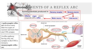
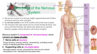
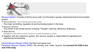
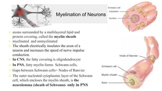
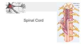
The nervous system consists of two main parts - the central nervous system (CNS) and the peripheral nervous system (PNS). The CNS contains the brain and spinal cord, while the PNS contains nerves that connect the CNS to the rest of the body. The structures that make up the nervous system include neurons, neuroglia, the brain, spinal cord, cranial and spinal nerves. Neurons are specialized to conduct electrical signals, while neuroglia provide support and insulation to the neurons. The spinal cord is protected within the bony vertebral column and surrounded by three layers of meninges.


![FUNCTIONAL CLASSIFICATION
❖ Sensory or afferent neurons:
❖ contain sensory receptors at their distal ends
❖ Carry impulses from sense organs (eyes, ears, etc) to the spinal cord and
brain.
❖ Most sensory neurons are unipolar in structure
❖ Motor or efferent neurons:
❖ convey action potentials away from the CNS to effectors [to muscles and
glands.]
❖ Most motor neurons are multipolar in structure
❖ Interneurons or association neurons:
❖ connect sensory and motor neurons and carry impulses between them.
❖ Most interneurons are multipolar in structure](https://image.slidesharecdn.com/u4-neuronesystem-230507144314-a351fa22/85/U4-Neurone-System-pdf-10-320.jpg)
![Neuroglia of the CNS
1. Astrocytes
1) Astrocytes – anchor neurons to capillaries. Star shaped cells, largest & most
numerous of the neuroglia.
Protoplasmic astrocytes have many short branching processes & are found in gray matter.
Fibrous astrocytes have many long unbranched processes and are located mainly in white
matter.
Functions:
(1)Contain microfilaments that give them considerable strength, which enables them to
support neurons.
(2)Processes of astrocytes wrapped around blood capillaries isolate neurons of the CNS
from various potentially harmful substances. [BBB]
(3)In the embryo, astrocytes secrete chemicals that appear to regulate the growth,
migration, and interconnection among neurons in the brain.
(4)help to maintain the appropriate chemical environment for the generation of nerve
impulses.
(5)Play a role in learning and memory by influencing the formation of neural synapses](https://image.slidesharecdn.com/u4-neuronesystem-230507144314-a351fa22/85/U4-Neurone-System-pdf-12-320.jpg)


![ORGANIZATION OF
THE NERVOUS
SYSTEM
CNS
Brain Spinal Cord
PNS
Somatic Nervous
System [SNS] Autonomic Nervous
System [ANS]
Enteric Nervous
System [ENS]
It is also the source of thoughts,
emotions, and memories
Muscle contraction & Gland
Secretion](https://image.slidesharecdn.com/u4-neuronesystem-230507144314-a351fa22/85/U4-Neurone-System-pdf-19-320.jpg)

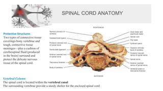
![Meninge
• The spinal meninges surround the spinal cord.
• the dura mater: is composed of dense, irregular connective tissue
• The spinal cord is also protected by a cushion of fat and
connective tissue located in the epidural space –[a space between
the dura mater and the wall of the vertebral canal ]
• The innermost meninx is the pia mater: a thin transparent
connective tissue layer that adheres to the surface of the spinal cord
and brain
• Within the pia mater are many blood vessels](https://image.slidesharecdn.com/u4-neuronesystem-230507144314-a351fa22/85/U4-Neurone-System-pdf-23-320.jpg)

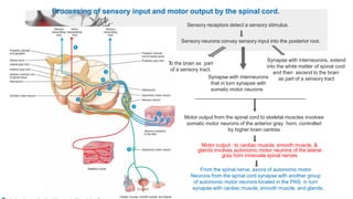
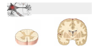
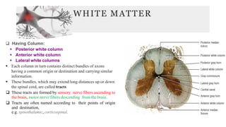
![CROSS SECTION OF SPINAL CORD
[Internal Anatomy]
❑ Divided into two equal parts, anteriorly by a short,
shallow median fissure and posteriorly by a deep
narrow septum, the posterior median sulcus.
❑ Composed of grey matter in the center surrounded
by white matter supported by neuroglia.
❑ Commissures: connections between left and right
halves
❑ central canal: In the center of the gray commissure
is a small space called the central canal](https://image.slidesharecdn.com/u4-neuronesystem-230507144314-a351fa22/85/U4-Neurone-System-pdf-26-320.jpg)

![Connective Tissue Coverings of Spinal Nerves
Individual axons within a nerve, whether myelinated or unmyelinated, are wrapped in endoneurium [the innermost
layer].
Arranged in bundles called fascicles, each of which is
wrapped in perineurium
The outermost covering
over the entire nerve is
the epineurium](https://image.slidesharecdn.com/u4-neuronesystem-230507144314-a351fa22/85/U4-Neurone-System-pdf-31-320.jpg)
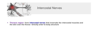
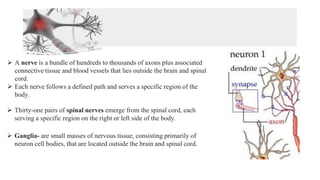
![BRANCHES OF SPINAL NERVES
❑ Dorsal Ramus: innervate deep muscles of the trunk responsible for movements of the vertebral column
and skin near the midline of the back.
❑ Ventral Ramus: serves the muscles and structures of the upper and lower limbs and the skin of the lateral and
anterior
❑ Plaxuses
▪ Remaining spinal nerve[T2-T12] ventral rami (roots of the plexus): they form networks on both the left and
right sides of the body by joining with various numbers of axons from anterior rami of adjacent nerves. Such a
network of axons is called a plexus
o Ventral rami of C1-C4= cervical plexus
o Ventral rami of C5-T1= brachial plexus
o Ventral rami of L1-L5= lumbar plexus
o Ventral rami of L4-S4= sacral plexus
o Ventral rami of S4 & S5= coccygeal plexus
o Communicating Rami: communicate with sympathetic chain of ganglia.](https://image.slidesharecdn.com/u4-neuronesystem-230507144314-a351fa22/85/U4-Neurone-System-pdf-32-320.jpg)