SlideShare a Scribd company logo
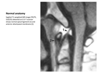
Normal anatomy
Sagittal T1-weighted MR image (TR/TE,
510/25) obtained on 0.3-T scanner
shows normal apical ligament (1) and
anterior atlantoaxial membrane (2).
Normal anatomy in 43-
year-old woman.
Sagittal T2-weighted MR image (TR/TE,
4500/117) obtained on 0.3-T MR
scanner shows normal apical ligament
(1), anterior occipitoatlantal
membrane (2), anterior atlantoaxial
membrane (3), anterior longitudinal
ligament (4), tectorial membrane (5),
dural reflection (6), posterior
occipitoatlantal membrane (7),
posterior atlantoaxial membrane (8),
nuchal ligament (9), flaval ligaments
(10), area of interspinous ligaments
(11), and supraspinous ligament (12).
Normal anatomy in 38-
year-old man
Axial gradientecho or fast low-angle
shot MR image (TR/TE, 420/18; flip
angle, 30°) obtained on 1.0-T MR
scanner shows dens (1), presumed
anterior atlantodental ligaments (2),
alar ligaments (3), transverse ligament
(4), and lateral masses of C1 (5).
Left alar ligament tear in 19-
year-old woman with severe
neck pain after fall on her
head while snowboarding.
Fixed deviation of dens to
right was seen on radiograph
(not shown).
C1-2 rotatory subluxation was
suspected. Axial T2-weighted MR
image (TR/TE, 4000/90) obtained on
1.0-T MR scanner shows isolated tear
of left alar ligament (1) and deviation
of dens (2) toward right with respect
to lateral masses of C2 (3). Transverse
ligament (4) is intact. Sagittal images
(not shown) depict normal alignment
of occipital condyles with C2, thus no
rotatory subluxation is present. CT
performed before MR imaging was
negative for fracture and fixed rotatory
subluxation. These results allowed
confident symptomatic treatment that
led to full recovery.
Occipitoatlantal dislocation in
11-year-old boy who was
neurologically intact after
motor vehicle crash.
Sagittal gradient-echo MR image
(TR/TE, 510/35; flip angle, 20°)
obtained on 0.3-T MR scanner shows
intact (1) and torn (2) portions of
anterior occipitoatlantal membrane,
anterior arch of C1 (3), intact anterior
atlantoaxial membrane (4),
prevertebral edema or hemorrhage
(5), torn tectorial membrane (6), torn
posterior occipitoatlantal membrane
(7), torn posterior atlantoaxial
membrane (8), intact dural reflection
(9), and intact nuchal ligament (10).
Before MR imaging, full extent of
injury and degree of instability were
not appreciated either clinically or
from results of radiographs or CT
scans. Patient underwent surgical
fusion shortly thereafter.
Occipitoatlantal dislocation in
11-year-old boy who was
neurologically intact after
motor vehicle crash
Axial gradient-echo MR image
(510/35; flip angle, 20°) obtained on
0.3-T MR scanner shows torn right alar
ligament (1), displacement of dens (2)
to left with respect to lateral masses of
C2 (3), and intact transverse ligament
(4).
Type II dens fracture in 14-
year-old boy who was
unrestrained passenger in
motor vehicle crash.
Sagittal gradient-echo MR image
(TR/TE, 500/9; flip angle, 15°) obtained
on 1.5-T MR scanner shows intact
occipitoatlantal membrane (1),
anterior dislocation of fractured dens
(2), anterior arch of C1 (3), partial tear
of anterior atlantoaxial membrane (4),
cord contusion (5), intact dura (6),
medullary contusion or edema (7),
torn tectorial membrane (8), intact
posterior occipitoatlantal membrane
(9), posterior arch of C1 (10), torn or
attenuated posterior atlantoaxial
membrane (11), intact dura (12), and
intact flaval ligaments (13).
Type II dens fracture in 14-
year-old boy who was
unrestrained passenger in
motor vehicle crash.
Axial gradient-echo MR image
(250/15; flip angle, 15°) obtained on
1.5-T MR scanner shows right lateral
mass of C1 (1), anteriorly dislocated
dens (2), body of C2 at fracture site (3),
compressed and contused spinal cord
(4), anterior arch of C1 (5), intact alar
ligaments (6), and intact transverse
ligament (7).
Burst fracture of C7 in 30-
year-old woman who was
unrestrained driver in motor
vehicle crash.
Sagittal fast spin-echo inversion-
recovery MR image (TR/TE, 3000/51;
inversion time, 140 msec) obtained on
1.5-T MR scanner shows burst fracture
of C7 (1), prevertebral edema or
hemorrhage (2), flaval (3) and
interspinous ligament tears (4), with
associated distraction of dorsal spines
and spinal cord contusion (5). Also
note signal hyperintensity caused by
bone marrow edema in vertebral
bodies of C6 and T1.
Burst fracture of C4 with
retropulsion in 17-year-old
boy after motor vehicle
crash.
Sagittal gradient-echo MR image
(TR/TE, 650/13; flip angle, 15°)
obtained on 1.5-T MR scanner shows
anterior longitudinal ligament tear (1),
hypointense hemorrhagic cord
contusion (2), posterior longitudinal
ligament tear at C3-4 (3), and flaval
ligament tear at C4-5 (4).
35-year old woman involved in
head-on motor vehicle collision who
presented with severe neck pain,
right arm pain, and numbness.
Radiographs and CT scans (not
shown) showed negative findings.
Four pulse sequences from a 1.0-T MR scanner at
midsagittal level are provided to allow reader to
compare and contrast abnormalities. Findings
include disk extrusion and inferior stripping of
posterior longitudinal ligament at C5-6 (1); disk
extrusion and tear of posterior longitudinal
ligament and annulus fibrosus at C6-7 (2); flaval
ligament tear at C6-7 (3); splaying of dorsal spines
and interspinous ligament tear at C6-7 (4); fracture
of C6 spinous process (5); and mild superior
endplate impaction fractures of T1, T2, and T3
vertebral bodies (6). Solely on basis of results of
MR images, the following day patient was started
in traction and taken to surgery where anterior
diskectomy and fusion at C5-6 and C6-7 were
performed. Patient experienced immediate marked
improvement in symptoms after surgery. Fast spin-
echo inversion-recovery sagittal MR image (TR/TE,
4000/60; inversion time, 140 msec) best shows
bone marrow edema caused by fracture or
trabecular contusion, spinal cord injury, and soft-
tissue edema.
35-year old woman involved in
head-on motor vehicle collision
who presented with severe neck
pain, right arm pain, and
numbness. Radiographs and CT
scans (not shown) showed negative
findings
Four pulse sequences from a 1.0-T MR scanner
at midsagittal level are provided to allow
reader to compare and contrast abnormalities.
Findings include disk extrusion and inferior
stripping of posterior longitudinal ligament at
C5-6 (1); disk extrusion and tear of posterior
longitudinal ligament and annulus fibrosus at
C6-7 (2); flaval ligament tear at C6-7 (3);
splaying of dorsal spines and interspinous
ligament tear at C6-7 (4); fracture of C6
spinous process (5); and mild superior
endplate impaction fractures of T1, T2, and T3
vertebral bodies (6). Solely on basis of results
of MR images, the following day patient was
started in traction and taken to surgery where
anterior diskectomy and fusion at C5-6 and C6-
7 were performed. Patient experienced
immediate marked improvement in symptoms
after surgery. T1-weighted MR image (500/15)
is helpful in showing anatomic detail and
alignment and in detecting fracture.
35-year old woman involved in
head-on motor vehicle collision who
presented with severe neck pain,
right arm pain, and numbness.
Radiographs and CT scans (not
shown) showed negative findings.
Four pulse sequences from a 1.0-T MR scanner at
midsagittal level are provided to allow reader to
compare and contrast abnormalities. Findings
include disk extrusion and inferior stripping of
posterior longitudinal ligament at C5-6 (1); disk
extrusion and tear of posterior longitudinal ligament
and annulus fibrosus at C6-7 (2); flaval ligament tear
at C6-7 (3); splaying of dorsal spines and interspinous
ligament tear at C6-7 (4); fracture of C6 spinous
process (5); and mild superior endplate impaction
fractures of T1, T2, and T3 vertebral bodies (6). Solely
on basis of results of MR images, the following day
patient was started in traction and taken to surgery
where anterior diskectomy and fusion at C5-6 and
C6-7 were performed. Patient experienced
immediate marked improvement in symptoms after
surgery. T2-weighted fast spin-echo MR images
(3500/90), like this one, are often best for showing
ligaments, blood in spinal cord, bone marrow edema,
and soft-tissue edema.
35-year old woman involved in
head-on motor vehicle collision
who presented with severe neck
pain, right arm pain, and
numbness. Radiographs and CT
scans (not shown) showed
negative findings.
Four pulse sequences from a 1.0-T MR scanner
at midsagittal level are provided to allow
reader to compare and contrast abnormalities.
Findings include disk extrusion and inferior
stripping of posterior longitudinal ligament at
C5-6 (1); disk extrusion and tear of posterior
longitudinal ligament and annulus fibrosus at
C6-7 (2); flaval ligament tear at C6-7 (3);
splaying of dorsal spines and interspinous
ligament tear at C6-7 (4); fracture of C6
spinous process (5); and mild superior
endplate impaction fractures of T1, T2, and T3
vertebral bodies (6). Solely on basis of results
of MR images, the following day patient was
started in traction and taken to surgery where
anterior diskectomy and fusion at C5-6 and C6-
7 were performed. Patient experienced
immediate marked improvement in symptoms
after surgery. Gradient-echo MR images
(500/18; flip angle, 30°), like this one, are
often best for showing ligaments and blood in
spinal cord.
Hyperextension injury in 71-
year-old man who fell from
bicycle and presented with
central cord syndrome.
Sagittal T2-weighted MR image (TR/TE,
4500/117) obtained on 0.3-T MR
scanner shows flaval ligament
hypertrophy (1), C5-6 posterior disk
protrusion (2), anterior longitudinal
ligament tear, and partial disruption of
C5-6 intervertebral disk (3).
6-year-old boy with cervical
spine hyperextension injury
during motor vehicle crash.
Sagittal fast spin-echo inversion-
recovery MR image (TR/TE, 3000/51;
inversion time, 140 msec) obtained on
1.5-T MR scanner shows horizontal
fracture through inferior endplate of
C6 (1), posterior longitudinal ligament
tear (2), cord contusion (3), anterior
longitudinal ligament tear (4),
prevertebral hemorrhage or edema
(5), and extradural hemorrhage (6).
MR imaging findings guided therapy
resulting in anterior surgical fusion.
Bilateral interfacetal
dislocation at C4-5 in 62-year-
old man involved in motor
vehicle crash
Sagittal gradient-echo MR image
(TR/TE, 510/35; flip angle, 20°)
obtained on 0.3-T MR scanner shows
prevertebral edema or hemorrhage
(1), posterior longitudinal ligament
tear (2), anterior longitudinal ligament
tear (3), large traumatic posterior disk
extrusion (4), cord contusion and
compression (5), posterior
paravertebral edema or hemorrhage,
and probable interspinous ligament
injury (6).
Bilateral interfacetal
dislocation in 42-year-old
woman involved in motor
vehicle crash.
Sagittal T2-weighted MR image (TR/TE,
4500/117) obtained on 0.3-T MR
scanner shows tear of dura and
posterior atlantoaxial membrane (1),
partial tear of nuchal ligament (2),
distraction of C5-6 spinous process
and torn interspinous ligaments (3),
torn flaval ligaments (4), torn posterior
longitudinal ligament (5), and torn
anterior longitudinal ligament (6).
Teardrop fracture of C7 in
27-year-old man involved
in motor vehicle crash.
Sagittal gradient-echo MR image
(TR/TE, 510/35; flip angle, 20°)
obtained on 0.3-T MR scanner shows
extensive posterior paravertebral
edema or hemorrhage and probable
tearing of interspinous ligaments (1),
partial tear of nuchal ligament (2),
flaval ligament tear (3), partial tear of
posterior longitudinal ligament (4),
anterior superior corner fracture of C7
vertebral body (5), stripping of
anterior longitudinal ligament from
anterior surface of C7 vertebral body
(6), and prevertebral edema or
hemorrhage (7).
Ligament stripping in 450-lb
(202.5-kg) 35-year-old man
ejected from motor vehicle.
Lateral radiographs (not
shown) were nondiagnostic
Sagittal gradient-echo MR image
(TR/TE, 510/35; flip angle, 20°)
obtained on 0.3-T MR scanner shows
anterior longitudinal ligament stripped
completely away from anterior surface
of midthoracic spine vertebral body
(1). Similarly, posterior longitudinal
ligament is stripped away from
posterior vertebral body surface at
level of fracture-subluxation (2).
Adjacent intervertebral disk is
disrupted (3) and thoracic spinal cord
is compressed (4).
11-year-old boy who suffered
flexion-distraction injury
from lap belt during motor
vehicle crash with fractures
at L4 level.
Sagittal gradient-echo MR image
(TR/TE, 500/13; flip angle, 15°)
obtained on 1.5-T MR scanner shows
large presumed cerebrospinal fluid
leak into posterior subcutaneous
tissues (1), distracted fracture
fragments of left L4 articular processes
(similar fracture was also present on
right) (2), and distracted fracture, near
horizontal in orientation, involving
posterosuperior portion of L4
vertebral body (3).
11-year-old boy who suffered
flexion-distraction injury
from lap belt during motor
vehicle crash with fractures
at L4 level.
Sagittal gradient-echo MR image
(500/13; flip angle, 15°) obtained on
1.5-T MR scanner of midline shows
distraction of spinous process of L3
and L4 (1), supraspinous ligament tear
(2), and flaval ligament tear (3).

More Related Content

What's hot

Presentation1.pptx, ct high resolution anatomy of the petrous bone.
Presentation1.pptx, ct high resolution anatomy of the petrous bone.Presentation1.pptx, ct high resolution anatomy of the petrous bone.
Presentation1.pptx, ct high resolution anatomy of the petrous bone.
Abdellah Nazeer
 

What's hot (20)

MR ANATOMY OF WRIST AND ELBOW RV
MR ANATOMY OF WRIST AND ELBOW RVMR ANATOMY OF WRIST AND ELBOW RV
MR ANATOMY OF WRIST AND ELBOW RV
 
Elbow MRI
Elbow MRIElbow MRI
Elbow MRI
 
Presentation1, mr pulse sequences.
Presentation1, mr pulse sequences.Presentation1, mr pulse sequences.
Presentation1, mr pulse sequences.
 
Mdct in cad
Mdct in cadMdct in cad
Mdct in cad
 
Brachial plexus MRI
Brachial plexus MRIBrachial plexus MRI
Brachial plexus MRI
 
Ct perfusion
Ct perfusionCt perfusion
Ct perfusion
 
Role of mri in traumatic brain injury
Role of mri in traumatic brain injuryRole of mri in traumatic brain injury
Role of mri in traumatic brain injury
 
A Radiological Approach to Craniosynostosis
A Radiological Approach to CraniosynostosisA Radiological Approach to Craniosynostosis
A Radiological Approach to Craniosynostosis
 
MR Neurography
MR NeurographyMR Neurography
MR Neurography
 
CT perfusion physics and its application in Neuroimaging
CT perfusion physics and its application in NeuroimagingCT perfusion physics and its application in Neuroimaging
CT perfusion physics and its application in Neuroimaging
 
Recent Advances In MRI
Recent Advances In MRIRecent Advances In MRI
Recent Advances In MRI
 
Presentation1.pptx, ct high resolution anatomy of the petrous bone.
Presentation1.pptx, ct high resolution anatomy of the petrous bone.Presentation1.pptx, ct high resolution anatomy of the petrous bone.
Presentation1.pptx, ct high resolution anatomy of the petrous bone.
 
Ct physics – II
Ct physics – IICt physics – II
Ct physics – II
 
CT SCAN spine
CT SCAN spineCT SCAN spine
CT SCAN spine
 
Mri – spine
Mri – spineMri – spine
Mri – spine
 
Diagnostic Imaging of Intraspinal Masses
Diagnostic Imaging of Intraspinal MassesDiagnostic Imaging of Intraspinal Masses
Diagnostic Imaging of Intraspinal Masses
 
Perfusion and dynamic contrast enhanced mri
Perfusion and dynamic contrast enhanced mriPerfusion and dynamic contrast enhanced mri
Perfusion and dynamic contrast enhanced mri
 
CT perfusion
CT perfusionCT perfusion
CT perfusion
 
Lung MRI
Lung MRILung MRI
Lung MRI
 
CT fluoroscopy
CT fluoroscopyCT fluoroscopy
CT fluoroscopy
 

Viewers also liked

Trigeminal neuralgia
Trigeminal neuralgiaTrigeminal neuralgia
Trigeminal neuralgia
yury
 
Sch.36 surgical management of sphenoid wing meningioma
Sch.36 surgical management of sphenoid wing meningiomaSch.36 surgical management of sphenoid wing meningioma
Sch.36 surgical management of sphenoid wing meningioma
Neurosurgery Vajira
 
Cv Junction Anomaly
Cv Junction AnomalyCv Junction Anomaly
Cv Junction Anomaly
rajasekar
 
It Is Time to Reevaluate the Management of Patients With Brain Metastases
It Is Time to Reevaluate the Management of Patients With Brain MetastasesIt Is Time to Reevaluate the Management of Patients With Brain Metastases
It Is Time to Reevaluate the Management of Patients With Brain Metastases
Apple Samsung
 
Pestana knight diagnosisepilepsy-5.8.2014
Pestana knight diagnosisepilepsy-5.8.2014Pestana knight diagnosisepilepsy-5.8.2014
Pestana knight diagnosisepilepsy-5.8.2014
Cleveland Clinic
 

Viewers also liked (20)

313 AOD and 314 AARS
313 AOD and 314 AARS313 AOD and 314 AARS
313 AOD and 314 AARS
 
13 atlantoaxial subluxation
13 atlantoaxial subluxation13 atlantoaxial subluxation
13 atlantoaxial subluxation
 
Lumbar Injuries
Lumbar InjuriesLumbar Injuries
Lumbar Injuries
 
029 Incision and closure
029 Incision and closure 029 Incision and closure
029 Incision and closure
 
Trigeminal neuralgia
Trigeminal neuralgiaTrigeminal neuralgia
Trigeminal neuralgia
 
178 arachnoid cysts
178 arachnoid cysts178 arachnoid cysts
178 arachnoid cysts
 
009 youmans cerebral edema
009 youmans cerebral edema009 youmans cerebral edema
009 youmans cerebral edema
 
324 Biomechanical basis of TBI
324 Biomechanical basis of TBI324 Biomechanical basis of TBI
324 Biomechanical basis of TBI
 
367 Intracranial internal carotid artery aneurysm
367 Intracranial internal carotid artery aneurysm367 Intracranial internal carotid artery aneurysm
367 Intracranial internal carotid artery aneurysm
 
319 thoracolumbar trauma
319 thoracolumbar trauma319 thoracolumbar trauma
319 thoracolumbar trauma
 
150 Pseudotumor cerebri
150 Pseudotumor cerebri150 Pseudotumor cerebri
150 Pseudotumor cerebri
 
368 ACoA aneurysm
368 ACoA aneurysm368 ACoA aneurysm
368 ACoA aneurysm
 
201 medulloblastoma
201 medulloblastoma201 medulloblastoma
201 medulloblastoma
 
Atlanto-axial subluxation
Atlanto-axial subluxationAtlanto-axial subluxation
Atlanto-axial subluxation
 
Sch.36 surgical management of sphenoid wing meningioma
Sch.36 surgical management of sphenoid wing meningiomaSch.36 surgical management of sphenoid wing meningioma
Sch.36 surgical management of sphenoid wing meningioma
 
Cv Junction Anomaly
Cv Junction AnomalyCv Junction Anomaly
Cv Junction Anomaly
 
imaging of the craniovertebral junction
imaging of the craniovertebral junction imaging of the craniovertebral junction
imaging of the craniovertebral junction
 
It Is Time to Reevaluate the Management of Patients With Brain Metastases
It Is Time to Reevaluate the Management of Patients With Brain MetastasesIt Is Time to Reevaluate the Management of Patients With Brain Metastases
It Is Time to Reevaluate the Management of Patients With Brain Metastases
 
Modernidade
ModernidadeModernidade
Modernidade
 
Pestana knight diagnosisepilepsy-5.8.2014
Pestana knight diagnosisepilepsy-5.8.2014Pestana knight diagnosisepilepsy-5.8.2014
Pestana knight diagnosisepilepsy-5.8.2014
 

Similar to Mr imaging findings in spinal ligamentous injury

Hairy cell leukemia and magnetic resonance imaging in diffuse malignant bone ...
Hairy cell leukemia and magnetic resonance imaging in diffuse malignant bone ...Hairy cell leukemia and magnetic resonance imaging in diffuse malignant bone ...
Hairy cell leukemia and magnetic resonance imaging in diffuse malignant bone ...
TCHF
 
An Expandable Prosthesis with Dual Cage-and-Plate Function in a Single Device...
An Expandable Prosthesis with Dual Cage-and-Plate Function in a Single Device...An Expandable Prosthesis with Dual Cage-and-Plate Function in a Single Device...
An Expandable Prosthesis with Dual Cage-and-Plate Function in a Single Device...
Erwin Chiquete, MD, PhD
 
Thoraco lumbar injuries
Thoraco lumbar injuriesThoraco lumbar injuries
Thoraco lumbar injuries
Drzameer
 
Imaging of spinal trauma
Imaging of spinal traumaImaging of spinal trauma
Imaging of spinal trauma
Vishal Sankpal
 
Dr Deepak Chahar Hip Dislocation JCOT
Dr Deepak Chahar Hip Dislocation JCOTDr Deepak Chahar Hip Dislocation JCOT
Dr Deepak Chahar Hip Dislocation JCOT
Deepak Chahar
 

Similar to Mr imaging findings in spinal ligamentous injury (20)

1 s2.0-s0378603 x16300201-main
1 s2.0-s0378603 x16300201-main1 s2.0-s0378603 x16300201-main
1 s2.0-s0378603 x16300201-main
 
Evidence based medicine pdf
Evidence based medicine pdfEvidence based medicine pdf
Evidence based medicine pdf
 
Aplasia of the Posterior Arches of the Atlas: A Presentation of One Case
Aplasia of the Posterior Arches of the Atlas: A Presentation of One CaseAplasia of the Posterior Arches of the Atlas: A Presentation of One Case
Aplasia of the Posterior Arches of the Atlas: A Presentation of One Case
 
Hairy cell leukemia and magnetic resonance imaging in diffuse malignant bone ...
Hairy cell leukemia and magnetic resonance imaging in diffuse malignant bone ...Hairy cell leukemia and magnetic resonance imaging in diffuse malignant bone ...
Hairy cell leukemia and magnetic resonance imaging in diffuse malignant bone ...
 
Siêu âm gãy xương ức, Bs Tài
Siêu âm gãy xương ức, Bs TàiSiêu âm gãy xương ức, Bs Tài
Siêu âm gãy xương ức, Bs Tài
 
Radiological Investigations of spinal Trauma.pptx
Radiological Investigations of spinal Trauma.pptxRadiological Investigations of spinal Trauma.pptx
Radiological Investigations of spinal Trauma.pptx
 
Ariunaa spine trauma
Ariunaa spine traumaAriunaa spine trauma
Ariunaa spine trauma
 
C2 fracture
C2 fractureC2 fracture
C2 fracture
 
An Expandable Prosthesis with Dual Cage-and-Plate Function in a Single Device...
An Expandable Prosthesis with Dual Cage-and-Plate Function in a Single Device...An Expandable Prosthesis with Dual Cage-and-Plate Function in a Single Device...
An Expandable Prosthesis with Dual Cage-and-Plate Function in a Single Device...
 
Thoraco lumbar injuries
Thoraco lumbar injuriesThoraco lumbar injuries
Thoraco lumbar injuries
 
Thoraco lumbar injuries
Thoraco lumbar injuriesThoraco lumbar injuries
Thoraco lumbar injuries
 
RADIOGRAPHY WITH CT CORRELATION IN PELVIC ACETABULAR TRAUMA
RADIOGRAPHY WITH CT CORRELATION IN PELVIC ACETABULAR TRAUMARADIOGRAPHY WITH CT CORRELATION IN PELVIC ACETABULAR TRAUMA
RADIOGRAPHY WITH CT CORRELATION IN PELVIC ACETABULAR TRAUMA
 
MRI in Tibial Fractures
MRI in Tibial FracturesMRI in Tibial Fractures
MRI in Tibial Fractures
 
Presentation1 radiological film reading of wrist joint.
Presentation1 radiological film reading of wrist joint.Presentation1 radiological film reading of wrist joint.
Presentation1 radiological film reading of wrist joint.
 
CT Cervical Spine
CT Cervical SpineCT Cervical Spine
CT Cervical Spine
 
Imaging of spinal trauma
Imaging of spinal traumaImaging of spinal trauma
Imaging of spinal trauma
 
imagingofspinaltrauMA
imagingofspinaltrauMAimagingofspinaltrauMA
imagingofspinaltrauMA
 
Dr Deepak Chahar Hip Dislocation JCOT
Dr Deepak Chahar Hip Dislocation JCOTDr Deepak Chahar Hip Dislocation JCOT
Dr Deepak Chahar Hip Dislocation JCOT
 
Internal fixation of fractures of the capitellum and trochlea - Retrospective...
Internal fixation of fractures of the capitellum and trochlea - Retrospective...Internal fixation of fractures of the capitellum and trochlea - Retrospective...
Internal fixation of fractures of the capitellum and trochlea - Retrospective...
 
04 radiology in maxillofacial trauma.ppt. new presentation
04 radiology in maxillofacial trauma.ppt. new presentation04 radiology in maxillofacial trauma.ppt. new presentation
04 radiology in maxillofacial trauma.ppt. new presentation
 

More from Battulga Munkhtsetseg (7)

Vascular malformations
Vascular malformationsVascular malformations
Vascular malformations
 
Congenital malformations of brain 2
Congenital malformations of brain 2Congenital malformations of brain 2
Congenital malformations of brain 2
 
Congenitalbrainmalformations 150913122145-lva1-app6891
Congenitalbrainmalformations 150913122145-lva1-app6891Congenitalbrainmalformations 150913122145-lva1-app6891
Congenitalbrainmalformations 150913122145-lva1-app6891
 
ARTERIAL AND VENOUS SUPPLY OF BRAIN
ARTERIAL AND VENOUS SUPPLY OF BRAIN ARTERIAL AND VENOUS SUPPLY OF BRAIN
ARTERIAL AND VENOUS SUPPLY OF BRAIN
 
Brain anatomy ariunaa
Brain anatomy  ariunaaBrain anatomy  ariunaa
Brain anatomy ariunaa
 
Багана нурууны халдварт эмгэгүүд сүүлийн
Багана нурууны халдварт эмгэгүүд сүүлийнБагана нурууны халдварт эмгэгүүд сүүлийн
Багана нурууны халдварт эмгэгүүд сүүлийн
 
Ariunaa congenital spine disorders
Ariunaa congenital spine disordersAriunaa congenital spine disorders
Ariunaa congenital spine disorders
 

Recently uploaded

New Directions in Targeted Therapeutic Approaches for Older Adults With Mantl...
New Directions in Targeted Therapeutic Approaches for Older Adults With Mantl...New Directions in Targeted Therapeutic Approaches for Older Adults With Mantl...
New Directions in Targeted Therapeutic Approaches for Older Adults With Mantl...
i3 Health
 

Recently uploaded (20)

The hemodynamic and autonomic determinants of elevated blood pressure in obes...
The hemodynamic and autonomic determinants of elevated blood pressure in obes...The hemodynamic and autonomic determinants of elevated blood pressure in obes...
The hemodynamic and autonomic determinants of elevated blood pressure in obes...
 
ANATOMY OF THE LOWER URINARY TRACT AND MALE [Autosaved] [Autosaved].pptx
ANATOMY OF THE LOWER URINARY TRACT AND MALE [Autosaved] [Autosaved].pptxANATOMY OF THE LOWER URINARY TRACT AND MALE [Autosaved] [Autosaved].pptx
ANATOMY OF THE LOWER URINARY TRACT AND MALE [Autosaved] [Autosaved].pptx
 
CURRENT HEALTH PROBLEMS AND ITS SOLUTION BY AYURVEDA.pptx
CURRENT HEALTH PROBLEMS AND ITS SOLUTION BY AYURVEDA.pptxCURRENT HEALTH PROBLEMS AND ITS SOLUTION BY AYURVEDA.pptx
CURRENT HEALTH PROBLEMS AND ITS SOLUTION BY AYURVEDA.pptx
 
Prix Galien International 2024 Forum Program
Prix Galien International 2024 Forum ProgramPrix Galien International 2024 Forum Program
Prix Galien International 2024 Forum Program
 
linearity concept of significance, standard deviation, chi square test, stude...
linearity concept of significance, standard deviation, chi square test, stude...linearity concept of significance, standard deviation, chi square test, stude...
linearity concept of significance, standard deviation, chi square test, stude...
 
Arterial health throughout cancer treatment and exercise rehabilitation in wo...
Arterial health throughout cancer treatment and exercise rehabilitation in wo...Arterial health throughout cancer treatment and exercise rehabilitation in wo...
Arterial health throughout cancer treatment and exercise rehabilitation in wo...
 
Antiplatelets in IHD, Dose Duration, DAPT vs SAPT
Antiplatelets in IHD, Dose Duration, DAPT vs SAPTAntiplatelets in IHD, Dose Duration, DAPT vs SAPT
Antiplatelets in IHD, Dose Duration, DAPT vs SAPT
 
Compare home pulse pressure components collected directly from home
Compare home pulse pressure components collected directly from homeCompare home pulse pressure components collected directly from home
Compare home pulse pressure components collected directly from home
 
PT MANAGEMENT OF URINARY INCONTINENCE.pptx
PT MANAGEMENT OF URINARY INCONTINENCE.pptxPT MANAGEMENT OF URINARY INCONTINENCE.pptx
PT MANAGEMENT OF URINARY INCONTINENCE.pptx
 
Factors Affecting child behavior in Pediatric Dentistry
Factors Affecting child behavior in Pediatric DentistryFactors Affecting child behavior in Pediatric Dentistry
Factors Affecting child behavior in Pediatric Dentistry
 
PT MANAGEMENT OF URINARY INCONTINENCE.pptx
PT MANAGEMENT OF URINARY INCONTINENCE.pptxPT MANAGEMENT OF URINARY INCONTINENCE.pptx
PT MANAGEMENT OF URINARY INCONTINENCE.pptx
 
Scientificity and feasibility study of non-invasive central arterial pressure...
Scientificity and feasibility study of non-invasive central arterial pressure...Scientificity and feasibility study of non-invasive central arterial pressure...
Scientificity and feasibility study of non-invasive central arterial pressure...
 
New Directions in Targeted Therapeutic Approaches for Older Adults With Mantl...
New Directions in Targeted Therapeutic Approaches for Older Adults With Mantl...New Directions in Targeted Therapeutic Approaches for Older Adults With Mantl...
New Directions in Targeted Therapeutic Approaches for Older Adults With Mantl...
 
Temporal, Infratemporal & Pterygopalatine BY Dr.RIG.pptx
Temporal, Infratemporal & Pterygopalatine BY Dr.RIG.pptxTemporal, Infratemporal & Pterygopalatine BY Dr.RIG.pptx
Temporal, Infratemporal & Pterygopalatine BY Dr.RIG.pptx
 
US E-cigarette Summit: Taming the nicotine industrial complex
US E-cigarette Summit: Taming the nicotine industrial complexUS E-cigarette Summit: Taming the nicotine industrial complex
US E-cigarette Summit: Taming the nicotine industrial complex
 
Relationship between vascular system disfunction, neurofluid flow and Alzheim...
Relationship between vascular system disfunction, neurofluid flow and Alzheim...Relationship between vascular system disfunction, neurofluid flow and Alzheim...
Relationship between vascular system disfunction, neurofluid flow and Alzheim...
 
Book Trailer: PGMEE in a Nutshell (CEE MD/MS PG Entrance Examination)
Book Trailer: PGMEE in a Nutshell (CEE MD/MS PG Entrance Examination)Book Trailer: PGMEE in a Nutshell (CEE MD/MS PG Entrance Examination)
Book Trailer: PGMEE in a Nutshell (CEE MD/MS PG Entrance Examination)
 
1130525--家醫計畫2.0糖尿病照護研討會-社團法人高雄市醫師公會.pdf
1130525--家醫計畫2.0糖尿病照護研討會-社團法人高雄市醫師公會.pdf1130525--家醫計畫2.0糖尿病照護研討會-社團法人高雄市醫師公會.pdf
1130525--家醫計畫2.0糖尿病照護研討會-社團法人高雄市醫師公會.pdf
 
DECIPHERING COMMON ECG FINDINGS IN ED.pptx
DECIPHERING COMMON ECG FINDINGS IN ED.pptxDECIPHERING COMMON ECG FINDINGS IN ED.pptx
DECIPHERING COMMON ECG FINDINGS IN ED.pptx
 
Antiulcer drugs Advance Pharmacology .pptx
Antiulcer drugs Advance Pharmacology .pptxAntiulcer drugs Advance Pharmacology .pptx
Antiulcer drugs Advance Pharmacology .pptx
 

Mr imaging findings in spinal ligamentous injury

  • 1. Normal anatomy Sagittal T1-weighted MR image (TR/TE, 510/25) obtained on 0.3-T scanner shows normal apical ligament (1) and anterior atlantoaxial membrane (2).
  • 2. Normal anatomy in 43- year-old woman. Sagittal T2-weighted MR image (TR/TE, 4500/117) obtained on 0.3-T MR scanner shows normal apical ligament (1), anterior occipitoatlantal membrane (2), anterior atlantoaxial membrane (3), anterior longitudinal ligament (4), tectorial membrane (5), dural reflection (6), posterior occipitoatlantal membrane (7), posterior atlantoaxial membrane (8), nuchal ligament (9), flaval ligaments (10), area of interspinous ligaments (11), and supraspinous ligament (12).
  • 3. Normal anatomy in 38- year-old man Axial gradientecho or fast low-angle shot MR image (TR/TE, 420/18; flip angle, 30°) obtained on 1.0-T MR scanner shows dens (1), presumed anterior atlantodental ligaments (2), alar ligaments (3), transverse ligament (4), and lateral masses of C1 (5).
  • 4. Left alar ligament tear in 19- year-old woman with severe neck pain after fall on her head while snowboarding. Fixed deviation of dens to right was seen on radiograph (not shown). C1-2 rotatory subluxation was suspected. Axial T2-weighted MR image (TR/TE, 4000/90) obtained on 1.0-T MR scanner shows isolated tear of left alar ligament (1) and deviation of dens (2) toward right with respect to lateral masses of C2 (3). Transverse ligament (4) is intact. Sagittal images (not shown) depict normal alignment of occipital condyles with C2, thus no rotatory subluxation is present. CT performed before MR imaging was negative for fracture and fixed rotatory subluxation. These results allowed confident symptomatic treatment that led to full recovery.
  • 5. Occipitoatlantal dislocation in 11-year-old boy who was neurologically intact after motor vehicle crash. Sagittal gradient-echo MR image (TR/TE, 510/35; flip angle, 20°) obtained on 0.3-T MR scanner shows intact (1) and torn (2) portions of anterior occipitoatlantal membrane, anterior arch of C1 (3), intact anterior atlantoaxial membrane (4), prevertebral edema or hemorrhage (5), torn tectorial membrane (6), torn posterior occipitoatlantal membrane (7), torn posterior atlantoaxial membrane (8), intact dural reflection (9), and intact nuchal ligament (10). Before MR imaging, full extent of injury and degree of instability were not appreciated either clinically or from results of radiographs or CT scans. Patient underwent surgical fusion shortly thereafter.
  • 6. Occipitoatlantal dislocation in 11-year-old boy who was neurologically intact after motor vehicle crash Axial gradient-echo MR image (510/35; flip angle, 20°) obtained on 0.3-T MR scanner shows torn right alar ligament (1), displacement of dens (2) to left with respect to lateral masses of C2 (3), and intact transverse ligament (4).
  • 7. Type II dens fracture in 14- year-old boy who was unrestrained passenger in motor vehicle crash. Sagittal gradient-echo MR image (TR/TE, 500/9; flip angle, 15°) obtained on 1.5-T MR scanner shows intact occipitoatlantal membrane (1), anterior dislocation of fractured dens (2), anterior arch of C1 (3), partial tear of anterior atlantoaxial membrane (4), cord contusion (5), intact dura (6), medullary contusion or edema (7), torn tectorial membrane (8), intact posterior occipitoatlantal membrane (9), posterior arch of C1 (10), torn or attenuated posterior atlantoaxial membrane (11), intact dura (12), and intact flaval ligaments (13).
  • 8. Type II dens fracture in 14- year-old boy who was unrestrained passenger in motor vehicle crash. Axial gradient-echo MR image (250/15; flip angle, 15°) obtained on 1.5-T MR scanner shows right lateral mass of C1 (1), anteriorly dislocated dens (2), body of C2 at fracture site (3), compressed and contused spinal cord (4), anterior arch of C1 (5), intact alar ligaments (6), and intact transverse ligament (7).
  • 9. Burst fracture of C7 in 30- year-old woman who was unrestrained driver in motor vehicle crash. Sagittal fast spin-echo inversion- recovery MR image (TR/TE, 3000/51; inversion time, 140 msec) obtained on 1.5-T MR scanner shows burst fracture of C7 (1), prevertebral edema or hemorrhage (2), flaval (3) and interspinous ligament tears (4), with associated distraction of dorsal spines and spinal cord contusion (5). Also note signal hyperintensity caused by bone marrow edema in vertebral bodies of C6 and T1.
  • 10. Burst fracture of C4 with retropulsion in 17-year-old boy after motor vehicle crash. Sagittal gradient-echo MR image (TR/TE, 650/13; flip angle, 15°) obtained on 1.5-T MR scanner shows anterior longitudinal ligament tear (1), hypointense hemorrhagic cord contusion (2), posterior longitudinal ligament tear at C3-4 (3), and flaval ligament tear at C4-5 (4).
  • 11. 35-year old woman involved in head-on motor vehicle collision who presented with severe neck pain, right arm pain, and numbness. Radiographs and CT scans (not shown) showed negative findings. Four pulse sequences from a 1.0-T MR scanner at midsagittal level are provided to allow reader to compare and contrast abnormalities. Findings include disk extrusion and inferior stripping of posterior longitudinal ligament at C5-6 (1); disk extrusion and tear of posterior longitudinal ligament and annulus fibrosus at C6-7 (2); flaval ligament tear at C6-7 (3); splaying of dorsal spines and interspinous ligament tear at C6-7 (4); fracture of C6 spinous process (5); and mild superior endplate impaction fractures of T1, T2, and T3 vertebral bodies (6). Solely on basis of results of MR images, the following day patient was started in traction and taken to surgery where anterior diskectomy and fusion at C5-6 and C6-7 were performed. Patient experienced immediate marked improvement in symptoms after surgery. Fast spin- echo inversion-recovery sagittal MR image (TR/TE, 4000/60; inversion time, 140 msec) best shows bone marrow edema caused by fracture or trabecular contusion, spinal cord injury, and soft- tissue edema.
  • 12. 35-year old woman involved in head-on motor vehicle collision who presented with severe neck pain, right arm pain, and numbness. Radiographs and CT scans (not shown) showed negative findings Four pulse sequences from a 1.0-T MR scanner at midsagittal level are provided to allow reader to compare and contrast abnormalities. Findings include disk extrusion and inferior stripping of posterior longitudinal ligament at C5-6 (1); disk extrusion and tear of posterior longitudinal ligament and annulus fibrosus at C6-7 (2); flaval ligament tear at C6-7 (3); splaying of dorsal spines and interspinous ligament tear at C6-7 (4); fracture of C6 spinous process (5); and mild superior endplate impaction fractures of T1, T2, and T3 vertebral bodies (6). Solely on basis of results of MR images, the following day patient was started in traction and taken to surgery where anterior diskectomy and fusion at C5-6 and C6- 7 were performed. Patient experienced immediate marked improvement in symptoms after surgery. T1-weighted MR image (500/15) is helpful in showing anatomic detail and alignment and in detecting fracture.
  • 13. 35-year old woman involved in head-on motor vehicle collision who presented with severe neck pain, right arm pain, and numbness. Radiographs and CT scans (not shown) showed negative findings. Four pulse sequences from a 1.0-T MR scanner at midsagittal level are provided to allow reader to compare and contrast abnormalities. Findings include disk extrusion and inferior stripping of posterior longitudinal ligament at C5-6 (1); disk extrusion and tear of posterior longitudinal ligament and annulus fibrosus at C6-7 (2); flaval ligament tear at C6-7 (3); splaying of dorsal spines and interspinous ligament tear at C6-7 (4); fracture of C6 spinous process (5); and mild superior endplate impaction fractures of T1, T2, and T3 vertebral bodies (6). Solely on basis of results of MR images, the following day patient was started in traction and taken to surgery where anterior diskectomy and fusion at C5-6 and C6-7 were performed. Patient experienced immediate marked improvement in symptoms after surgery. T2-weighted fast spin-echo MR images (3500/90), like this one, are often best for showing ligaments, blood in spinal cord, bone marrow edema, and soft-tissue edema.
  • 14. 35-year old woman involved in head-on motor vehicle collision who presented with severe neck pain, right arm pain, and numbness. Radiographs and CT scans (not shown) showed negative findings. Four pulse sequences from a 1.0-T MR scanner at midsagittal level are provided to allow reader to compare and contrast abnormalities. Findings include disk extrusion and inferior stripping of posterior longitudinal ligament at C5-6 (1); disk extrusion and tear of posterior longitudinal ligament and annulus fibrosus at C6-7 (2); flaval ligament tear at C6-7 (3); splaying of dorsal spines and interspinous ligament tear at C6-7 (4); fracture of C6 spinous process (5); and mild superior endplate impaction fractures of T1, T2, and T3 vertebral bodies (6). Solely on basis of results of MR images, the following day patient was started in traction and taken to surgery where anterior diskectomy and fusion at C5-6 and C6- 7 were performed. Patient experienced immediate marked improvement in symptoms after surgery. Gradient-echo MR images (500/18; flip angle, 30°), like this one, are often best for showing ligaments and blood in spinal cord.
  • 15. Hyperextension injury in 71- year-old man who fell from bicycle and presented with central cord syndrome. Sagittal T2-weighted MR image (TR/TE, 4500/117) obtained on 0.3-T MR scanner shows flaval ligament hypertrophy (1), C5-6 posterior disk protrusion (2), anterior longitudinal ligament tear, and partial disruption of C5-6 intervertebral disk (3).
  • 16. 6-year-old boy with cervical spine hyperextension injury during motor vehicle crash. Sagittal fast spin-echo inversion- recovery MR image (TR/TE, 3000/51; inversion time, 140 msec) obtained on 1.5-T MR scanner shows horizontal fracture through inferior endplate of C6 (1), posterior longitudinal ligament tear (2), cord contusion (3), anterior longitudinal ligament tear (4), prevertebral hemorrhage or edema (5), and extradural hemorrhage (6). MR imaging findings guided therapy resulting in anterior surgical fusion.
  • 17. Bilateral interfacetal dislocation at C4-5 in 62-year- old man involved in motor vehicle crash Sagittal gradient-echo MR image (TR/TE, 510/35; flip angle, 20°) obtained on 0.3-T MR scanner shows prevertebral edema or hemorrhage (1), posterior longitudinal ligament tear (2), anterior longitudinal ligament tear (3), large traumatic posterior disk extrusion (4), cord contusion and compression (5), posterior paravertebral edema or hemorrhage, and probable interspinous ligament injury (6).
  • 18. Bilateral interfacetal dislocation in 42-year-old woman involved in motor vehicle crash. Sagittal T2-weighted MR image (TR/TE, 4500/117) obtained on 0.3-T MR scanner shows tear of dura and posterior atlantoaxial membrane (1), partial tear of nuchal ligament (2), distraction of C5-6 spinous process and torn interspinous ligaments (3), torn flaval ligaments (4), torn posterior longitudinal ligament (5), and torn anterior longitudinal ligament (6).
  • 19. Teardrop fracture of C7 in 27-year-old man involved in motor vehicle crash. Sagittal gradient-echo MR image (TR/TE, 510/35; flip angle, 20°) obtained on 0.3-T MR scanner shows extensive posterior paravertebral edema or hemorrhage and probable tearing of interspinous ligaments (1), partial tear of nuchal ligament (2), flaval ligament tear (3), partial tear of posterior longitudinal ligament (4), anterior superior corner fracture of C7 vertebral body (5), stripping of anterior longitudinal ligament from anterior surface of C7 vertebral body (6), and prevertebral edema or hemorrhage (7).
  • 20. Ligament stripping in 450-lb (202.5-kg) 35-year-old man ejected from motor vehicle. Lateral radiographs (not shown) were nondiagnostic Sagittal gradient-echo MR image (TR/TE, 510/35; flip angle, 20°) obtained on 0.3-T MR scanner shows anterior longitudinal ligament stripped completely away from anterior surface of midthoracic spine vertebral body (1). Similarly, posterior longitudinal ligament is stripped away from posterior vertebral body surface at level of fracture-subluxation (2). Adjacent intervertebral disk is disrupted (3) and thoracic spinal cord is compressed (4).
  • 21. 11-year-old boy who suffered flexion-distraction injury from lap belt during motor vehicle crash with fractures at L4 level. Sagittal gradient-echo MR image (TR/TE, 500/13; flip angle, 15°) obtained on 1.5-T MR scanner shows large presumed cerebrospinal fluid leak into posterior subcutaneous tissues (1), distracted fracture fragments of left L4 articular processes (similar fracture was also present on right) (2), and distracted fracture, near horizontal in orientation, involving posterosuperior portion of L4 vertebral body (3).
  • 22. 11-year-old boy who suffered flexion-distraction injury from lap belt during motor vehicle crash with fractures at L4 level. Sagittal gradient-echo MR image (500/13; flip angle, 15°) obtained on 1.5-T MR scanner of midline shows distraction of spinous process of L3 and L4 (1), supraspinous ligament tear (2), and flaval ligament tear (3).