
Recommended
More Related Content
What's hot
What's hot (20)
Similar to Elbow MRI
Similar to Elbow MRI (20)
Recently uploaded
Recently uploaded (20)
Elbow MRI
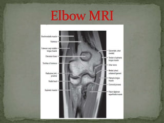
- 2. Use the axis of the epicondyles on a axial localizer to plan the coronal scan. The sagittal images are scaned perpendicular to the coronal scan. In this way you get very persistent images and you will get used to the normal anatomy.
- 5. Typically the radiocapitellar joint is punctured from lateral with the patient prone and the arm flexed 90 degrees overhead (red arrow)
- 6. UCL pathology in throwers. Osteochondral lesions and repair Loose bodies
- 9. This is a finding that you frequently see on coronal images. It looks like an osteochondral lesion, but if you look at the sagittal image you will notice that the coronal image runs through the posterior non-articular portion of the capitellum.
- 12. All these forces make up what is called the "valgus overload syndrome" with very characteristic injuries to the elbow over time. The tension on the medial side causes a tear of the ulnar collateral ligament. Compression on the lateral side causes an osteochondral lesion of the capitellum. The shear forces on the posterior side cause arthrosis.
- 17. A lateral view of the elbow of a patient who fell on the outstretched arm. The radiograph shows joint effusion (red arrows) and a coronoid fracture (yellow arrow).
- 18. Coronal view: Lateral collateral ligament is completely stripped (yellow arrow). radial head is subluxed. marrow edema of the coronoid process due to the fracture (red arrow). Sagittal view: Radial head is a little bit subluxed posteriorly (yellow arrow). Large effusion and capsular disruption posteriorly. Contusion of the posterior side of the capitellum as a result of impaction by the coronoid process (red arrow).
- 19. Osteochondral lesion is the new name for osteochondritis dissecans or OCD. The chronic valgus overload can cause an osteochondral lesion on the lateral side of the elbow. It is the result of repetitive impaction and shear forces.
- 21. The ulnar collateral ligament (UCL) is situated on the medial side and it has three components. The anterior bundle is the strongest component and is the primary restraint against valgus forces. On MR this is the most important structure. The posterior bundle attaches distally in a fan-shape on the olecranon. It forms the floor of the cubital tunnel.
- 23. It consists of the radial collateral, the lateral ulnar collateral and the annular ligament.
- 24. When you look for the radial collateral ligament, first try to identify the common extensor tendon, because right underneath it you will find the radial collateral ligament (yellow arrow). As you go more posteriorly you will see the LUCL - the lateral ulnar collateral ligament, which sweeps behind the radial head (white arrows).
- 25. The common extensor tendon originates at the lateral epicondyle. On a T1W-images the tendon should have a low signal intensity (yellow arrow).
- 26. Lateral epicondylitis is also known as the tennis elbow, although in 95% of cases it is seen in non-tennis players. It is due of chronic stress to the common extensor tendon, which results in partial tearing and tendinosis. Typically, the extensor carpi radialis brevis is the component that is involved.
- 27. Here a typical case. There is thickening and abnormal intrinsic signal on both T1- and T2W- images.
- 28. The common flexor tendon originates at the medial epicondyle. On a T1W-images the tendon should have a low signal intensity (red arrow).
- 30. Here the common flexor tendon is involved. On the sagittal image it is clear that it is only partial tearing. However this can be quite painful. Here we have the coronal T1W- and T2W-images. There is partial tearing, but it is very extensive.
- 31. The medial epicondyle of the affected arm is somewhat more osteopenic. In these cases we usually ask for a comparison view, because it can be very subtle. The diagnosis is a Little leaguer's elbow which results from chronic stress injury. The lucency on the radiograph, which looks like a widened physis, is due to cartilage ingrowth in the metaphysis.
- 32. On the MR the abnormality is very obvious. There is marrow edema in the medial epicondyle and also in the adjacent bone (yellow arrow). Little Leaguer's elbow is also known as medial apophysitis and some call it epiphysiolysis. Notice the normal ulnar collateral ligament (red arrow).
- 33. Here we see the ulnar nerve within the cubital tunnel. The posterior band of the ulnar collateral band forms the floor of the tunnel, while the retinaculum forms the roof.
- 34. Cubital tunnel syndrome is a common peripheral neuropathy. It arises from compression of the ulnar nerve within the cubital tunnel, where the nerve passes beneath the cubital tunnel retinaculum.
- 35. Overuse Subluxation of the ulnar nerve because of congenital laxity in the fibrous tissue Humeral fracture with loose bodies or callus formation Arthritic spur arising from the epicondyle or olecranon, Muscle anomaly (eg: an accessory anconeus muscle) as is present in this case. Soft-tissue mass: ganglion, lipoma, osteochondroma, synovitis secondary to rheumatoid arthritis, infection (eg: tuberculosis), and hemorrhage.
- 36. Thank You
Editor's Notes
- a small piece of fat that you'll see on the sagittal image, that looks like a small loose body or a cartilage defect.
- This structure on the lateral side of the joint is sometimes seen and is a plica.