Download as PDF, PPTX


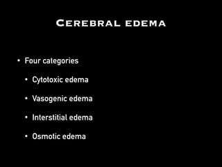


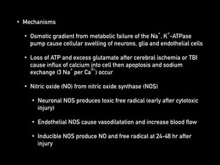
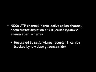
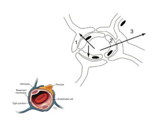
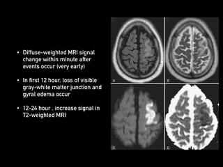

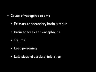
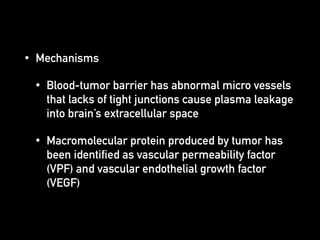

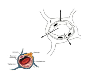

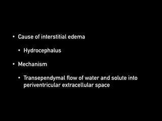
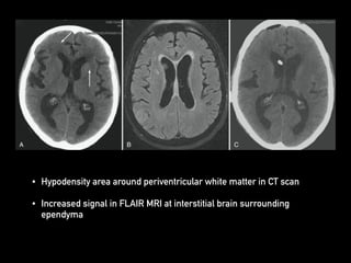

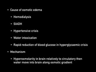

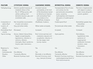


There are four main categories of cerebral edema: cytotoxic edema, vasogenic edema, interstitial edema, and osmotic edema. Cytotoxic edema is caused by cellular swelling from metabolic failure of ion pumps due to conditions like ischemia or trauma. Vasogenic edema results from plasma leakage into brain tissue from damaged blood vessels, often due to tumors. Interstitial edema occurs when fluid moves from ventricles into surrounding brain tissue in hydrocephalus. Osmotic edema is caused by hyperosmolarity differences between the brain and circulation in situations like water intoxication or hemodialysis. Treatment focuses on the underlying cause, while diuretics and osmotic agents can temporarily reduce swelling.


![Management of Cerebral edema 1 [Autosaved].pptx](https://cdn.slidesharecdn.com/ss_thumbnails/managementofcerebraledema1autosaved-231006090123-7bf6a0fb-thumbnail.jpg?width=640&height=640&fit=bounds)


