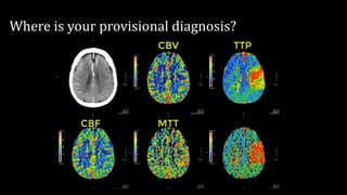
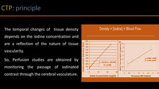
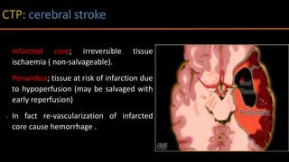
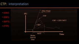
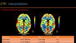
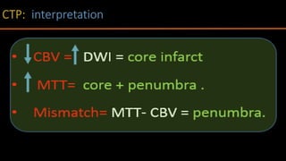
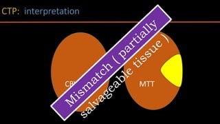
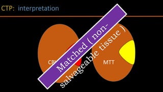
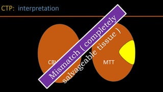
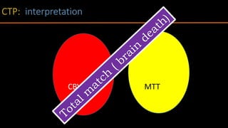
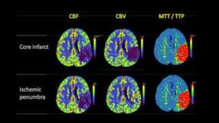
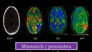
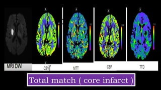
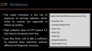
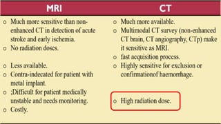
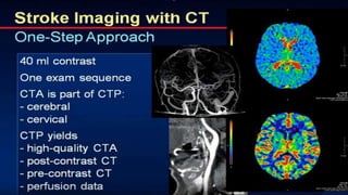
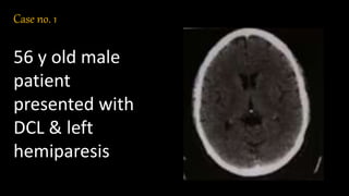
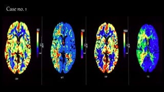
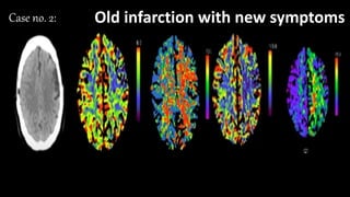
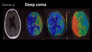
Dr. Mohamed Kamel
Lecturer of radiodiagnosis
Faculty of medicine-Menoufia university
Where is your provisional diagnosis?
Perfusion is the ‘’passage of blood, a blood substitute,
or other fluid through a vessel into an organ or tissue”.
CTP is an imaging technique allows functional
evaluation of tissue vascularity.
The temporal changes of tissue density
depends on the iodine concentration and
are a reflection of the nature of tissue
vascularity.
So, Perfusion studies are obtained by
monitoring the passage of iodinated
contrast through the cerebral vasculature.
• Oncological applications ;brain ,Pancreatic & Rectal tumors:
• Tumor characterization; grading.
• Assisting the therapeutic response, consequently the prognosis.
• Differentiating between recurrent tumor and radiation necrosis.
• Non- oncological applications:
• Acute cerebral Stroke
• To allow qualitative and quantitative evaluation of cerebral perfusion.
• To distinguish infarcted tissue from penumbra.
• Ischaemic Cardiac disease.
• Pancreatitis and pancreatic necrosis.
• Stroke: inadequate blood flow of brain cells causing sudden death in a
localized area.
• Caused by blockage of blood flow ( ischemic )or rupture of an artery
(hemorrhagic).
• Neuroimaging plays a vital role in the workup of acute stroke.
• The main goal of CTP is to allow qualitative and quantitative evaluation of
cerebral perfusion and to differentiate the infarct core from the ischemic
penumbra.
• Infarcted core; irreversible tissue
ischaemia ( non-salvageable).
• Penumbra; tissue at risk of infarction due
to hypoperfusion (may be salvaged with
early reperfusion)
• In fact re-vascularization of infarcted
core cause hemorrhage .
Non
contrast
CT
Ischaemic
< 3 hours IV tPA.
< 6 hours IA
therapies.
< 9 hours
hypertensive Tx,
oxygen
CTA +/- CTP.
MRI (DWI)
+/- MRA – MRP.
Not ischaemic
• Precontrast CT: Hge/ infarction / swelling.
• CTP: penumbra/core infarct.
• CTA: occlusion/ stenosis /variants.
• Repeated acquisitions are obtained at first pass of contrast at ROI ( about 1-2
min), Then, delayed phases ( 2-10 min)
• Thick perfusion slap :-
• 16 MDCT: 2.5 cm.
• 64 MDCT: 4 cm.
• 320 MDCT: full brain coverage.
• Contrast :-
• Vol. 40 ml.
• Conc. 370 mg/ml .
• Rate 4 ml/s.
• Dynamic scan: every 2 s.
• Scan locations: at the level of basal ganglia:- This
includes vascular areas of the brain that frequently
hypo-perfused at acute stroke cases.
• Choose ROI for :-
• Arterial input: ACA.
• Venous output: SSS.
• Time-attenuation curves for arterial ROI, venous ROI
and each pixel are obtained. This is based on the
principle that transient hyperattenuation is directly
proportional to amount of contrast .
• Perfusion parameters are mathematically calculated.
• Color-coded perfusion maps are obtained.
CBV
Cerebral blood volume
CBF
Cerebral blood flow
MTT
Mean transit time
TTP
Time to peak
• (CBV): the blood volume passing through the region of interest measured
(ml/100 g).
• (CBF): the blood flow through the tissue of interest per unit of time
measured in (ml/100 g/min).
• (MTT) : the mean time it takes for blood to circulate through capillaries of a
determined region, passing from arterioles to venule measured in seconds.
• (TTP): The time it takes to reach the maximum attenuation value within
artery measures in seconds.
• (CBV):
• (CBF):
• (MTT) :
• (TTP):
Brain tissue CBF CBV MTT
Grey matter 60 ml/ 100g/min 4 ml/ 100g 4 s
White matter 25 ml/ 100g/min 2 ml/ 100g 4.8 s
• Normal perfusion parameter
Core infarct Pattern:
• CT: MTT - CBF - CBV.
• MRI: DWI - T2WI - FLAIR.
• Penumbra pattern:
• CT : MTT - CBF - CBV.
• MRI: MTT on MRP
•Treatment 3h after event based on mismatch..
• Less mortality risk.
• Less infarct expansion.
• Better functional outcome.
CBV MTT
CBV MTT
CBV MTT
MTTCBV
MTT
Mismatch ( penumbra
NCECT CBF CBV MTT
MTT
Total match ( core infarct )
• The major limitation is the risk of
exposure to ionizing radiation which
limits its routine use especially in
follow-up studies.
• High radiation dose of CTP (about 3.2
Gy) induces temporary hair loss.
• Low KVp (from 120 to 80) is sufficient
to maximal dose reduction without
affection of diagnostic accuracy.
Check list
• Check :-
• Non contrast CT (early findings).
• All 4 CTP series for defects (Cerebral
cortex, basal ganglia and cerebellum).
•Compare CBV to MTT.
• Identify match/ mismatched defects.
•Conclude the core Vs. penumbra (ratio).
Case no. 1
56 y old male
patient
presented with
DCL & left
hemiparesis
•CBV:
•MTT:
•CBF:
•TTP:
• Match/ mismatch
• Core/Penumbra
Case no. 1
Answer
• CBV: Normal.
• MTT: Prolonged at right temporal lobe along the territory of RT MCA.
• CBF: Decreased at right temporal lobe along the territory of RT MCA.
• TTP: Prolonged at right temporal lobe along the territory of RT MCA.
• Match/ mismatch: mismatched CBV/MTT
• Core/Penumbra: penumbra with no core.
•CBV:
•MTT:
•CBF:
•TTP:
• Match/ mismatch
• Core/Penumbra
Case no. 2: Old infarction with new symptoms
Answer
• CBV: decreased at left frontal cortex.
• MTT: Prolonged at left cortical & deep periventricular region.
• CBF: Decreased at left cortical & deep periventricular region.
• TTP: Prolonged at left cortical & deep periventricular region.
• Match/ mismatch: mismatched CBV/MTT
• Core/Penumbra: penumbra with cortical core.
•CBV:
•MTT:
•CBF:
•TTP:
• Match/ mismatch
• Core/Penumbra
Case no. 3: Deep coma
Answer
• CBV: decreased at RT MCA territory .
• MTT: Prolonged at RT MCA territory.
• CBF: Decreased at RT MCA territory .
• TTP: Prolonged at RT MCA territory .
•Match/ mismatch: matched CBV/MTT
•Core/Penumbra: core with no penumbra .
Ct perfusion

Ct perfusion

  • 1.
    Dr. Mohamed Kamel Lecturerof radiodiagnosis Faculty of medicine-Menoufia university
  • 2.
    Where is yourprovisional diagnosis?
  • 3.
    Perfusion is the‘’passage of blood, a blood substitute, or other fluid through a vessel into an organ or tissue”. CTP is an imaging technique allows functional evaluation of tissue vascularity.
  • 4.
    The temporal changesof tissue density depends on the iodine concentration and are a reflection of the nature of tissue vascularity. So, Perfusion studies are obtained by monitoring the passage of iodinated contrast through the cerebral vasculature.
  • 5.
    • Oncological applications;brain ,Pancreatic & Rectal tumors: • Tumor characterization; grading. • Assisting the therapeutic response, consequently the prognosis. • Differentiating between recurrent tumor and radiation necrosis. • Non- oncological applications: • Acute cerebral Stroke • To allow qualitative and quantitative evaluation of cerebral perfusion. • To distinguish infarcted tissue from penumbra. • Ischaemic Cardiac disease. • Pancreatitis and pancreatic necrosis.
  • 9.
    • Stroke: inadequateblood flow of brain cells causing sudden death in a localized area. • Caused by blockage of blood flow ( ischemic )or rupture of an artery (hemorrhagic). • Neuroimaging plays a vital role in the workup of acute stroke. • The main goal of CTP is to allow qualitative and quantitative evaluation of cerebral perfusion and to differentiate the infarct core from the ischemic penumbra.
  • 10.
    • Infarcted core;irreversible tissue ischaemia ( non-salvageable). • Penumbra; tissue at risk of infarction due to hypoperfusion (may be salvaged with early reperfusion) • In fact re-vascularization of infarcted core cause hemorrhage .
  • 13.
    Non contrast CT Ischaemic < 3 hoursIV tPA. < 6 hours IA therapies. < 9 hours hypertensive Tx, oxygen CTA +/- CTP. MRI (DWI) +/- MRA – MRP. Not ischaemic • Precontrast CT: Hge/ infarction / swelling. • CTP: penumbra/core infarct. • CTA: occlusion/ stenosis /variants.
  • 14.
    • Repeated acquisitionsare obtained at first pass of contrast at ROI ( about 1-2 min), Then, delayed phases ( 2-10 min)
  • 15.
    • Thick perfusionslap :- • 16 MDCT: 2.5 cm. • 64 MDCT: 4 cm. • 320 MDCT: full brain coverage. • Contrast :- • Vol. 40 ml. • Conc. 370 mg/ml . • Rate 4 ml/s. • Dynamic scan: every 2 s.
  • 16.
    • Scan locations:at the level of basal ganglia:- This includes vascular areas of the brain that frequently hypo-perfused at acute stroke cases. • Choose ROI for :- • Arterial input: ACA. • Venous output: SSS. • Time-attenuation curves for arterial ROI, venous ROI and each pixel are obtained. This is based on the principle that transient hyperattenuation is directly proportional to amount of contrast .
  • 17.
    • Perfusion parametersare mathematically calculated. • Color-coded perfusion maps are obtained. CBV Cerebral blood volume CBF Cerebral blood flow MTT Mean transit time TTP Time to peak
  • 19.
    • (CBV): theblood volume passing through the region of interest measured (ml/100 g). • (CBF): the blood flow through the tissue of interest per unit of time measured in (ml/100 g/min). • (MTT) : the mean time it takes for blood to circulate through capillaries of a determined region, passing from arterioles to venule measured in seconds. • (TTP): The time it takes to reach the maximum attenuation value within artery measures in seconds.
  • 20.
    • (CBV): • (CBF): •(MTT) : • (TTP):
  • 21.
    Brain tissue CBFCBV MTT Grey matter 60 ml/ 100g/min 4 ml/ 100g 4 s White matter 25 ml/ 100g/min 2 ml/ 100g 4.8 s • Normal perfusion parameter
  • 22.
    Core infarct Pattern: •CT: MTT - CBF - CBV. • MRI: DWI - T2WI - FLAIR. • Penumbra pattern: • CT : MTT - CBF - CBV. • MRI: MTT on MRP
  • 23.
    •Treatment 3h afterevent based on mismatch.. • Less mortality risk. • Less infarct expansion. • Better functional outcome.
  • 24.
  • 25.
  • 26.
  • 27.
  • 29.
  • 30.
    MTT Total match (core infarct )
  • 32.
    • The majorlimitation is the risk of exposure to ionizing radiation which limits its routine use especially in follow-up studies. • High radiation dose of CTP (about 3.2 Gy) induces temporary hair loss. • Low KVp (from 120 to 80) is sufficient to maximal dose reduction without affection of diagnostic accuracy.
  • 37.
    Check list • Check:- • Non contrast CT (early findings). • All 4 CTP series for defects (Cerebral cortex, basal ganglia and cerebellum). •Compare CBV to MTT. • Identify match/ mismatched defects. •Conclude the core Vs. penumbra (ratio).
  • 39.
    Case no. 1 56y old male patient presented with DCL & left hemiparesis
  • 40.
  • 41.
    Answer • CBV: Normal. •MTT: Prolonged at right temporal lobe along the territory of RT MCA. • CBF: Decreased at right temporal lobe along the territory of RT MCA. • TTP: Prolonged at right temporal lobe along the territory of RT MCA. • Match/ mismatch: mismatched CBV/MTT • Core/Penumbra: penumbra with no core.
  • 42.
    •CBV: •MTT: •CBF: •TTP: • Match/ mismatch •Core/Penumbra Case no. 2: Old infarction with new symptoms
  • 43.
    Answer • CBV: decreasedat left frontal cortex. • MTT: Prolonged at left cortical & deep periventricular region. • CBF: Decreased at left cortical & deep periventricular region. • TTP: Prolonged at left cortical & deep periventricular region. • Match/ mismatch: mismatched CBV/MTT • Core/Penumbra: penumbra with cortical core.
  • 44.
  • 45.
    Answer • CBV: decreasedat RT MCA territory . • MTT: Prolonged at RT MCA territory. • CBF: Decreased at RT MCA territory . • TTP: Prolonged at RT MCA territory . •Match/ mismatch: matched CBV/MTT •Core/Penumbra: core with no penumbra .