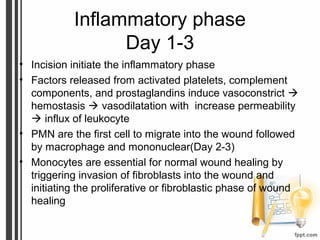
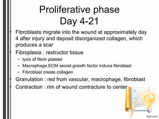
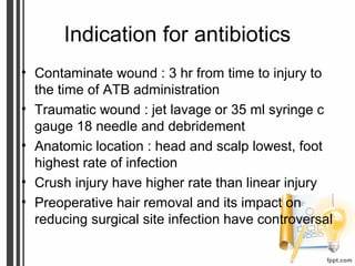
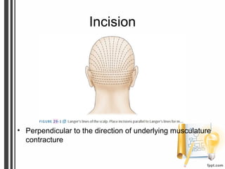
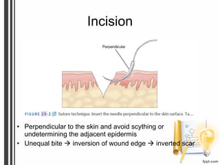
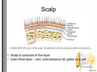
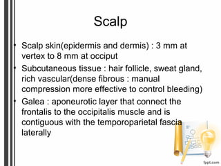
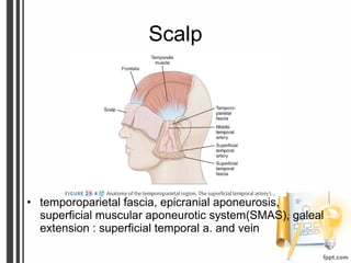
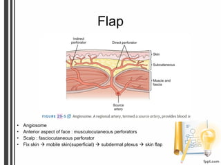
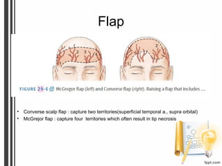
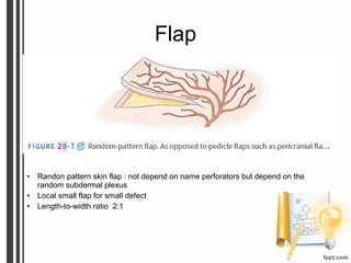
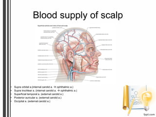
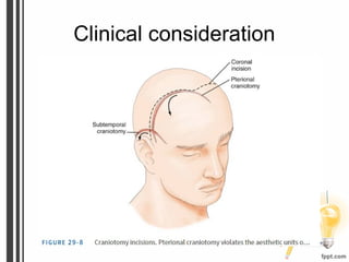
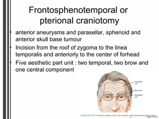
Incision and Closure
Youmans Chapter 29
Outline
• Wound healing
• Indication for antibiotics
• Incision
• Surgical anatomy
• Clinical consideration
• Closure
Wound healing
• Inflammatory phase
• Proliferative phase
• Maturation phase
Inflammatory phase
Day 1-3
• Incision initiate the inflammatory phase
• Factors released from activated platelets, complement
components, and prostaglandins induce vasoconstrict 
hemostasis  vasodilatation with increase permeability
 influx of leukocyte
• PMN are the first cell to migrate into the wound followed
by macrophage and mononuclear(Day 2-3)
• Monocytes are essential for normal wound healing by
triggering invasion of fibroblasts into the wound and
initiating the proliferative or fibroblastic phase of wound
healing
Proliferative phase
Day 4-21
• Fibroblasts migrate into the wound at approximately day
4 after injury and deposit disorganized collagen, which
produces a scar
• Fibroplasia : restructor tissue
– lysis of fibrin platelet
– Macrophage,ECM secret growth factor induce fibroblast
– Fibroblast create collagen
• Granulation : red from vascular, macrophage, fibroblast
• Contraction : rim of wound contracture to center
Maturation phase
21 day – 1-2 Yrs
• ECM remodeling
Indication for antibiotics
• Contaminate wound : 3 hr from time to injury to
the time of ATB administration
• Traumatic wound : jet lavage or 35 ml syringe c
gauge 18 needle and debridement
• Anatomic location : head and scalp lowest, foot
highest rate of infection
• Crush injury have higher rate than linear injury
• Preoperative hair removal and its impact on
reducing surgical site infection have controversal
Incision
• Perpendicular to the direction of underlying musculature
contracture
Incision
• Perpendicular to the skin and avoid scything or
undetermining the adjacent epidermis
• Unequal bite  inversion of wound edge  inverted scar
Surgical anatomy
Scalp
• Scalp is composd of five layer
• outer three layer – skin, subcutaneous fat, galea as a unit
Scalp
• Scalp skin(epidermis and dermis) : 3 mm at
vertex to 8 mm at occiput
• Subcutaneous tissue : hair follicle, sweat gland,
rich vascular(dense fibrous : manual
compression more effective to control bleeding)
• Galea : aponeurotic layer that connect the
frontalis to the occipitalis muscle and is
contiguous with the temporoparietal fascia
laterally
Scalp
• temporoparietal fascia, epicranial aponeurosis,
superficial muscular aponeurotic system(SMAS), galeal
extension : superficial temporal a. and vein
Flap
• Angiosome
• Anterior aspect of face : musculocutaneous perforators
• Scalp : fasciocutaneous perforator
• Fix skin  mobile skin(superficial)  subdermal plexus  skin flap
Flap
• Converse scalp flap : capture two territories(superficial temporal a., supra orbital)
• McGrejor flap : capture four territories which often result in tip necrosis
Flap
• Randon pattern skin flap : not depend on name perforators but depend on the
random subdermal plexus
• Local small flap for small defect
• Length-to-width ratio 2:1
Blood supply of scalp
• Supra orbital a.(internal carotid a.  ophthalmic a.)
• Supra trochlear a. (internal carotid a.  ophthalmic a.)
• Superficial temporal a. (external carotid a.)
• Posterior auricular a. (external carotid a.)
• Occipital a. (external carotid a.)
Blood supply of scalp
• Anterior : supraorbital and supratrochlear a.
– Supraorbital a. exit through supraorbital
notch,superficial to orbital rim 1.5 cm and lateral to
midline 3 cm
– supratrochlear a.,1 cm superior to medial palpebral
ligament, 1.5 cm lateral to midline
• Posterior territory : medial and two lateral occipital
a.,5 cm lateral to occipital protuberance
Blood supply of scalp
• Posterolateral territory : posterior auricular a.,2 cm
inferior to the auditory canal
• Lateral scalp territory : frontal and parietal branch of
the superficial temporal a.(superficial temporal a.
give branch to middle temporal a.the blood supply to
the temporalris muscle and deep temporal fascia)
Facial nerve
• Facial n. lies within a small fat pad between the layers of
the deep temporal fascia,posterior to the superficial
temporal a. and vein,at level of zygomatic arche
• 2.5 cm anterior to the tragus
• 1.5 cm lateral to the orbital rim
• Lie within deep temporalis fascia
• Should be preserve when elevating a pericarnial flap
Clinical consideration
Frontosphenotemporal or
pterional craniotomy
• anterior aneurysms and parasellar, sphenoid and
anterior skull base tumour
• Incision from the roof of zygoma to the linea
temporalis and anteriorly to the center of forhead
• Five aesthetic part unit : two temporal, two brow and
one central component
Frontosphenotemporal or
pterional craniotomy
• Coronal incision,providing better exposure and
cosmetic result because it preservs all the aesthetic
units of the head.
• Longer incision, necessitate operative time
• Should be preserve frontal branch of the facial nerve
• Detach of the muscle from it superior insertion result
in retracton of m. inferiorly and muscle wasting
• Leaving a cuff of temporalis m. superiorly attached
to skull to provide, reapproximating m. fiber and
resultant in muscle wasting
Subtemporal craniotomy
• Hoarseshoe-shape flap
• Tentorial, clivus and basilar a lesion
• Becareful, superficial temporal a. and posterior
auricle a.
Midline suboccipital craniotomy
• Inconspicious scar, advantage of vascular between
two bellies
• Reliable closure is complicated by many factor
– Radiation
– Embolization
– Previous scalp surgery
– > 60 Years
Closure
• Primary closure : simple, atraumatic,
noncontaminated wound
• Goal
– Obliterate dead space
– Distribute tension along deep suture line
– Maintain suture strength until tissue tension strength
adequate
• Interrupted stitch,it avoid compromising the vessel
within the galea suppling the scalp but longer
operative time
• Metallic staples may used in region of compromised
vascularity to improve potential
Closure
Monofilament Multifilament
Infection less more
Tension Less tension More tension
Galea,scalp
Nylon

029 Incision and closure

  • 1.
  • 2.
    Outline • Wound healing •Indication for antibiotics • Incision • Surgical anatomy • Clinical consideration • Closure
  • 3.
    Wound healing • Inflammatoryphase • Proliferative phase • Maturation phase
  • 4.
    Inflammatory phase Day 1-3 •Incision initiate the inflammatory phase • Factors released from activated platelets, complement components, and prostaglandins induce vasoconstrict  hemostasis  vasodilatation with increase permeability  influx of leukocyte • PMN are the first cell to migrate into the wound followed by macrophage and mononuclear(Day 2-3) • Monocytes are essential for normal wound healing by triggering invasion of fibroblasts into the wound and initiating the proliferative or fibroblastic phase of wound healing
  • 5.
    Proliferative phase Day 4-21 •Fibroblasts migrate into the wound at approximately day 4 after injury and deposit disorganized collagen, which produces a scar • Fibroplasia : restructor tissue – lysis of fibrin platelet – Macrophage,ECM secret growth factor induce fibroblast – Fibroblast create collagen • Granulation : red from vascular, macrophage, fibroblast • Contraction : rim of wound contracture to center
  • 6.
    Maturation phase 21 day– 1-2 Yrs • ECM remodeling
  • 7.
    Indication for antibiotics •Contaminate wound : 3 hr from time to injury to the time of ATB administration • Traumatic wound : jet lavage or 35 ml syringe c gauge 18 needle and debridement • Anatomic location : head and scalp lowest, foot highest rate of infection • Crush injury have higher rate than linear injury • Preoperative hair removal and its impact on reducing surgical site infection have controversal
  • 8.
    Incision • Perpendicular tothe direction of underlying musculature contracture
  • 9.
    Incision • Perpendicular tothe skin and avoid scything or undetermining the adjacent epidermis • Unequal bite  inversion of wound edge  inverted scar
  • 10.
  • 11.
    Scalp • Scalp iscomposd of five layer • outer three layer – skin, subcutaneous fat, galea as a unit
  • 12.
    Scalp • Scalp skin(epidermisand dermis) : 3 mm at vertex to 8 mm at occiput • Subcutaneous tissue : hair follicle, sweat gland, rich vascular(dense fibrous : manual compression more effective to control bleeding) • Galea : aponeurotic layer that connect the frontalis to the occipitalis muscle and is contiguous with the temporoparietal fascia laterally
  • 13.
    Scalp • temporoparietal fascia,epicranial aponeurosis, superficial muscular aponeurotic system(SMAS), galeal extension : superficial temporal a. and vein
  • 14.
    Flap • Angiosome • Anterioraspect of face : musculocutaneous perforators • Scalp : fasciocutaneous perforator • Fix skin  mobile skin(superficial)  subdermal plexus  skin flap
  • 15.
    Flap • Converse scalpflap : capture two territories(superficial temporal a., supra orbital) • McGrejor flap : capture four territories which often result in tip necrosis
  • 16.
    Flap • Randon patternskin flap : not depend on name perforators but depend on the random subdermal plexus • Local small flap for small defect • Length-to-width ratio 2:1
  • 17.
    Blood supply ofscalp • Supra orbital a.(internal carotid a.  ophthalmic a.) • Supra trochlear a. (internal carotid a.  ophthalmic a.) • Superficial temporal a. (external carotid a.) • Posterior auricular a. (external carotid a.) • Occipital a. (external carotid a.)
  • 18.
    Blood supply ofscalp • Anterior : supraorbital and supratrochlear a. – Supraorbital a. exit through supraorbital notch,superficial to orbital rim 1.5 cm and lateral to midline 3 cm – supratrochlear a.,1 cm superior to medial palpebral ligament, 1.5 cm lateral to midline • Posterior territory : medial and two lateral occipital a.,5 cm lateral to occipital protuberance
  • 19.
    Blood supply ofscalp • Posterolateral territory : posterior auricular a.,2 cm inferior to the auditory canal • Lateral scalp territory : frontal and parietal branch of the superficial temporal a.(superficial temporal a. give branch to middle temporal a.the blood supply to the temporalris muscle and deep temporal fascia)
  • 20.
    Facial nerve • Facialn. lies within a small fat pad between the layers of the deep temporal fascia,posterior to the superficial temporal a. and vein,at level of zygomatic arche • 2.5 cm anterior to the tragus • 1.5 cm lateral to the orbital rim • Lie within deep temporalis fascia • Should be preserve when elevating a pericarnial flap
  • 21.
  • 22.
    Frontosphenotemporal or pterional craniotomy •anterior aneurysms and parasellar, sphenoid and anterior skull base tumour • Incision from the roof of zygoma to the linea temporalis and anteriorly to the center of forhead • Five aesthetic part unit : two temporal, two brow and one central component
  • 23.
    Frontosphenotemporal or pterional craniotomy •Coronal incision,providing better exposure and cosmetic result because it preservs all the aesthetic units of the head. • Longer incision, necessitate operative time • Should be preserve frontal branch of the facial nerve • Detach of the muscle from it superior insertion result in retracton of m. inferiorly and muscle wasting • Leaving a cuff of temporalis m. superiorly attached to skull to provide, reapproximating m. fiber and resultant in muscle wasting
  • 24.
    Subtemporal craniotomy • Hoarseshoe-shapeflap • Tentorial, clivus and basilar a lesion • Becareful, superficial temporal a. and posterior auricle a.
  • 25.
    Midline suboccipital craniotomy •Inconspicious scar, advantage of vascular between two bellies • Reliable closure is complicated by many factor – Radiation – Embolization – Previous scalp surgery – > 60 Years
  • 26.
    Closure • Primary closure: simple, atraumatic, noncontaminated wound • Goal – Obliterate dead space – Distribute tension along deep suture line – Maintain suture strength until tissue tension strength adequate • Interrupted stitch,it avoid compromising the vessel within the galea suppling the scalp but longer operative time • Metallic staples may used in region of compromised vascularity to improve potential
  • 27.
    Closure Monofilament Multifilament Infection lessmore Tension Less tension More tension Galea,scalp Nylon

Editor's Notes

  • #5 PMN จะเข้ามาเก็บกินเนื้อตายและหลั่ง cytokine เพื่อให้ macrophages เข้ามายังแผล Macrophage ที่เข้ามาจะหลั่ง growth factor หลายชนิดทำให้ fibroblast และ endothelial cell เข้ามาที่แผลและแบ่งตัวเพิ่มมากขึ้น
  • #13 Contiguous ติดกัน