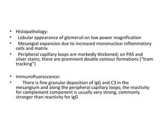
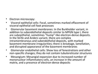
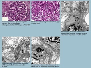
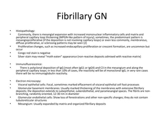
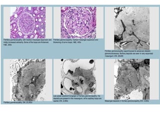
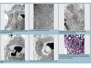
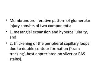
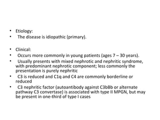
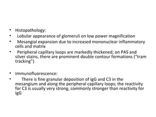
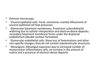
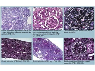
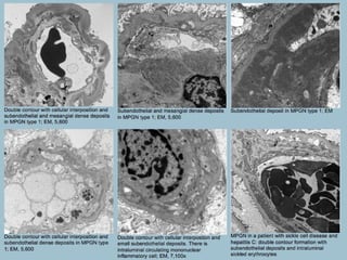
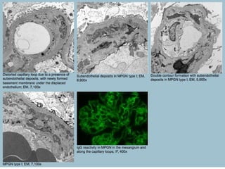
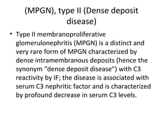
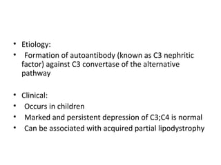
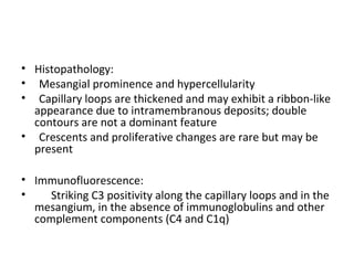
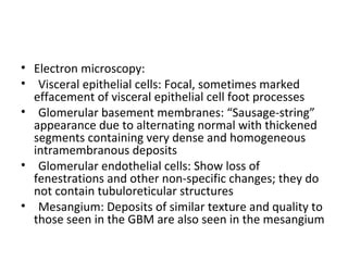
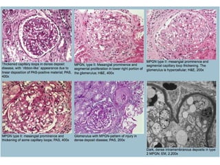
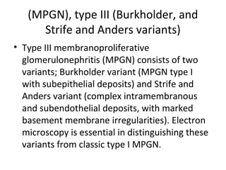
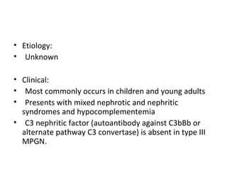
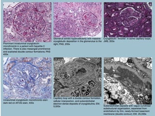
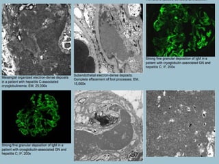
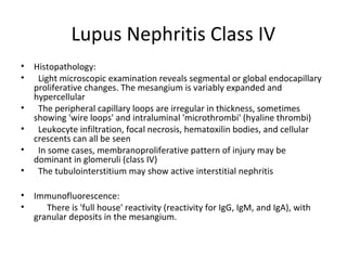
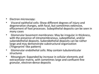
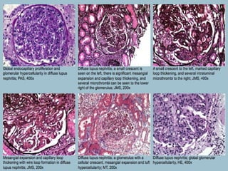
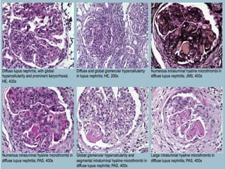
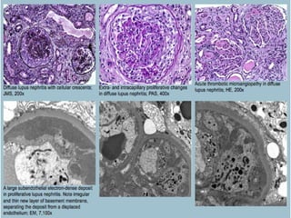
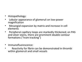
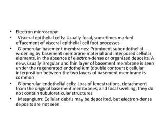
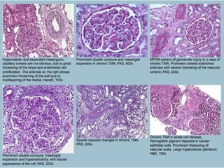
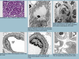
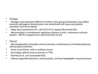
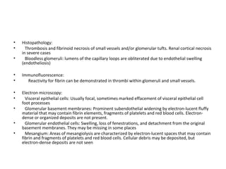
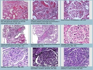
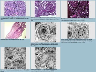
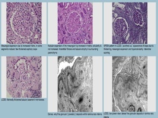
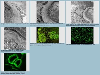
This document discusses several types of glomerular diseases that present with a membranoproliferative pattern of injury on histology, including membranoproliferative glomerulonephritis (MPGN) types I, II, and III, cryoglobulin-associated glomerulonephritis, lupus nephritis class IV, and thrombotic microangiopathies (both acute and chronic). It provides details on the etiology, clinical features, histopathology, immunofluorescence, and electron microscopy findings of each condition.
most of the glomerular diseases , either primary or secondary..touching all the aspects including light microscopy, electron microscopy and immunoflourescence.
Rapidly progressive glomerulonephritisajith joseph
Rapidly progressive glomerulonephritis (RPGN) is a syndrome of the kidney that is characterized by a rapid loss of renal function,[4][5] (usually a 50% decline in the glomerular filtration rate (GFR) within 3 months)[5] with glomerular crescent formation seen in at least 50%[5] or 75%[4] of glomeruli seen on kidney biopsies. If left untreated, it rapidly progresses into acute renal failure[6] and death within months. In 50% of cases, RPGN is associated with an underlying disease such as Goodpasture syndrome, systemic lupus erythematosus or granulomatosis with polyangiitis; the remaining cases are idiopathic. Regardless of the underlying cause, RPGN involves severe injury to the kidneys' glomeruli, with many of the glomeruli containing characteristic glomerular crescents (crescent-shaped scars)
Churg-Strauss syndrome is a disorder marked by blood vessel inflammation. This condition is also known as eosinophilic granulomatosis with polyangiitis (EGPA).
download link : https://www.dropbox.com/s/xc0fpdul47g1gu8/IgA%20Nephropathy.ppt?m
Join us on our facebook group: NephroTube...............Follow our blog: www.nephrotube.blogspot.com
This presentation is made to teach aboudt approach to glomerulonephritis in children. Final diagnosis of this case was membranoproliferative glomerulonephritis.
Market Research Reports, Inc. has announced the addition of “Mammalian Target of Rapamycin (Mtor) Inhibitors, Pipeline Insights, 2016” research report to their offering. See more at - http://mrr.cm/JRH
The target of rapamycin (TOR) is a key modulator of ageing in different organisms, ranging from yeast to rodents, and it is likely that this function has been conserved also in humans (mTOR, mammalian target of rapamycin). The signaling pathway of mTOR senses and integrates a variety of environmental inputs to regulate not only the organismal growth but also its homeostasis. This pathway regulates many important cellular processes that are implicated in the appearance of age-related diseases including cancer, obesity, type 2 diabetes, and neurodegeneration in late life. Here, are been reviewed the recent knowledge in the understanding of the mTOR pathway and its role in ageing and disease. Special emphasis in mTORC1 will be done due to this particular complex can be modulated by some compounds such as rapamycin. Moreover, are going to be discussed pharmacological approaches to treat human pathologies related with mTORC1 deregulation.
Membranous GN
MOST COMMON cause is idiopathic (85%); peak age 30-50; male:female, 2:1
May be secondary to:
Drugs-captopril, penicillamine, gold, mercury, trimethadione, NSAIDS
Infections-malaria (P. malariae), leprosy, schistosomiasis, syphilis, hepatitis B and C, filariasis, hydatid disease and enterococcal endocarditis
Diseases-malignancy (Carcinoma of breast, lung, colon, stomach, and esophagus) melanoma, renal cell CA, SLE, sarcoidosis, diabetes, thyroiditis, sickle cell anemia, Crohn’s disease
This presentation i have made to understand the approach to a kidney biopsy in depth. kidney biopsy is not done in all centers and that's why its difficult to understand it. i have put some cases also to understand it better.
These simplified slides by Dr. Sidra Arshad present an overview of the non-respiratory functions of the respiratory tract.
Learning objectives:
1. Enlist the non-respiratory functions of the respiratory tract
2. Briefly explain how these functions are carried out
3. Discuss the significance of dead space
4. Differentiate between minute ventilation and alveolar ventilation
5. Describe the cough and sneeze reflexes
Study Resources:
1. Chapter 39, Guyton and Hall Textbook of Medical Physiology, 14th edition
2. Chapter 34, Ganong’s Review of Medical Physiology, 26th edition
3. Chapter 17, Human Physiology by Lauralee Sherwood, 9th edition
4. Non-respiratory functions of the lungs https://academic.oup.com/bjaed/article/13/3/98/278874
Rasamanikya is a excellent preparation in the field of Rasashastra, it is used in various Kushtha Roga, Shwasa, Vicharchika, Bhagandara, Vatarakta, and Phiranga Roga. In this article Preparation& Comparative analytical profile for both Formulationon i.e Rasamanikya prepared by Kushmanda swarasa & Churnodhaka Shodita Haratala. The study aims to provide insights into the comparative efficacy and analytical aspects of these formulations for enhanced therapeutic outcomes.
Basavarajeeyam is a Sreshta Sangraha grantha (Compiled book ), written by Neelkanta kotturu Basavaraja Virachita. It contains 25 Prakaranas, First 24 Chapters related to Rogas& 25th to Rasadravyas.
Recomendações da OMS sobre cuidados maternos e neonatais para uma experiência pós-natal positiva.
Em consonância com os ODS – Objetivos do Desenvolvimento Sustentável e a Estratégia Global para a Saúde das Mulheres, Crianças e Adolescentes, e aplicando uma abordagem baseada nos direitos humanos, os esforços de cuidados pós-natais devem expandir-se para além da cobertura e da simples sobrevivência, de modo a incluir cuidados de qualidade.
Estas diretrizes visam melhorar a qualidade dos cuidados pós-natais essenciais e de rotina prestados às mulheres e aos recém-nascidos, com o objetivo final de melhorar a saúde e o bem-estar materno e neonatal.
Uma “experiência pós-natal positiva” é um resultado importante para todas as mulheres que dão à luz e para os seus recém-nascidos, estabelecendo as bases para a melhoria da saúde e do bem-estar a curto e longo prazo. Uma experiência pós-natal positiva é definida como aquela em que as mulheres, pessoas que gestam, os recém-nascidos, os casais, os pais, os cuidadores e as famílias recebem informação consistente, garantia e apoio de profissionais de saúde motivados; e onde um sistema de saúde flexível e com recursos reconheça as necessidades das mulheres e dos bebês e respeite o seu contexto cultural.
Estas diretrizes consolidadas apresentam algumas recomendações novas e já bem fundamentadas sobre cuidados pós-natais de rotina para mulheres e neonatos que recebem cuidados no pós-parto em unidades de saúde ou na comunidade, independentemente dos recursos disponíveis.
É fornecido um conjunto abrangente de recomendações para cuidados durante o período puerperal, com ênfase nos cuidados essenciais que todas as mulheres e recém-nascidos devem receber, e com a devida atenção à qualidade dos cuidados; isto é, a entrega e a experiência do cuidado recebido. Estas diretrizes atualizam e ampliam as recomendações da OMS de 2014 sobre cuidados pós-natais da mãe e do recém-nascido e complementam as atuais diretrizes da OMS sobre a gestão de complicações pós-natais.
O estabelecimento da amamentação e o manejo das principais intercorrências é contemplada.
Recomendamos muito.
Vamos discutir essas recomendações no nosso curso de pós-graduação em Aleitamento no Instituto Ciclos.
Esta publicação só está disponível em inglês até o momento.
Prof. Marcus Renato de Carvalho
www.agostodourado.com
Antimicrobial stewardship to prevent antimicrobial resistanceGovindRankawat1
India is among the nations with the highest burden of bacterial infections.
India is one of the largest consumers of antibiotics worldwide.
India carries one of the largest burdens of drug‑resistant pathogens worldwide.
Highest burden of multidrug‑resistant tuberculosis,
Alarmingly high resistance among Gram‑negative and Gram‑positive bacteria even to newer antimicrobials such as carbapenems.
NDM‑1 ( New Delhi Metallo Beta lactamase 1, an enzyme which inactivates majority of Beta lactam antibiotics including carbapenems) was reported in 2008
Knee anatomy and clinical tests 2024.pdfvimalpl1234
This includes all relevant anatomy and clinical tests compiled from standard textbooks, Campbell,netter etc..It is comprehensive and best suited for orthopaedicians and orthopaedic residents.
- Video recording of this lecture in English language: https://youtu.be/kqbnxVAZs-0
- Video recording of this lecture in Arabic language: https://youtu.be/SINlygW1Mpc
- Link to download the book free: https://nephrotube.blogspot.com/p/nephrotube-nephrology-books.html
- Link to NephroTube website: www.NephroTube.com
- Link to NephroTube social media accounts: https://nephrotube.blogspot.com/p/join-nephrotube-on-social-media.html
Integrating Ayurveda into Parkinson’s Management: A Holistic ApproachAyurveda ForAll
Explore the benefits of combining Ayurveda with conventional Parkinson's treatments. Learn how a holistic approach can manage symptoms, enhance well-being, and balance body energies. Discover the steps to safely integrate Ayurvedic practices into your Parkinson’s care plan, including expert guidance on diet, herbal remedies, and lifestyle modifications.
2. • Membranoproliferative pattern of glomerular
injury consists of two components:
• 1. mesangial expansion and hypercellularity,
and
• 2. thickening of the peripheral capillary loops
due to double contour formation ('tram-
tracking', best appreciated on silver or PAS
stains).
3. • This pattern of glomerular injury can be appreciated in three types of disorders:
• 1. IMMUNE COMPLEX-MEDIATED DISEASES
• a. Idiopathic or primary forms (MPGN I, II, III)
• b. Secondary forms (chronic infections, autoimmune diseases)
• 2. THROMBOTIC MICROANGIOPATHIES
• 3. PARAPROTEIN DEPOSITION DISEASES
• a. monoclonal immunoglobulin deposition disease, such as light chain DD
• b. fibrillary and immunotactoid glomerulopathy
• c. cryoglobulin-associated GN
• d. Waldenström macroglobulinemia
• e. POEMS syndrome (polyneuropathy, organomegaly, endocrinopathy, m-protein,
skin change)
4. Membranoproliferative
glomerulonephritis (MPGN), idiopathic,
type I
• Idiopathic membranoproliferative glomerulonephritis is an immune
complex-mediated disease of unknown etiology with a
membranoproliferative pattern of glomerular injury (mesangial
hypercellularity and expansion and double contour formations in
peripheral capillary loops). The diagnosis of primary MPGN is one of
exclusion, since similar morphologic findings can be seen in
secondary forms with an MPGN pattern of injury (autoimmune
diseases and chronic infections, see classification). There are three
types (I, II, and III) of primary MPGN.
• Type I MPGN is the most common, “classic” type of primary MPGN,
with subendothelial and mesangial deposits and with strong C3 and
less intense IgG immunofluorescence reactivity.
5. • Etiology:
• The disease is idiopathic (primary).
• Clinical:
• Occurs more commonly in young patients (ages 7 – 30 years).
• Usually presents with mixed nephrotic and nephritic syndrome,
with predominant nephrotic component; less commonly the
presentation is purely nephritic
• C3 is reduced and C1q and C4 are commonly borderline or
reduced
• C3 nephritic factor (autoantibody against C3bBb or alternate
pathway C3 convertase) is associated with type II MPGN, but may
be present in one-third of type I cases
6. • Histopathology:
• Lobular appearance of glomeruli on low power magnification
• Mesangial expansion due to increased mononuclear inflammatory
cells and matrix
• Peripheral capillary loops are markedly thickened; on PAS and
silver stains, there are prominent double contour formations (“tram
tracking”)
• Immunofluorescence:
• There is fine granular deposition of IgG and C3 in the
mesangium and along the peripheral capillary loops; the reactivity
for C3 is usually very strong, commonly stronger than reactivity for
IgG
7. • Electron microscopy:
• Visceral epithelial cells: Focal, sometimes marked effacement of
visceral epithelial cell foot processes
• Glomerular basement membranes: Prominent subendothelial
widening due to cellular interposition and electron-dense deposits;
secondary basement membrane forms under the displaced
endothelium (double contour formation)
• Glomerular endothelial cells: Show loss of fenestrations and other
non-specific changes; they do not contain tubuloreticular structures
• Mesangium: Mesangial expansion due to increased number of
mononuclear inflammatory cells, an increase in the amount of
matrix and a presence of electron-dense deposits
8.
9.
10.
11.
12. (MPGN), type II (Dense deposit
disease)
• Type II membranoproliferative
glomerulonephritis (MPGN) is a distinct and
very rare form of MPGN characterized by
dense intramembranous deposits (hence the
synonym “dense deposit disease”) with C3
reactivity by IF; the disease is associated with
serum C3 nephritic factor and is characterized
by profound decrease in serum C3 levels.
13. • Etiology:
• Formation of autoantibody (known as C3 nephritic
factor) against C3 convertase of the alternative
pathway
• Clinical:
• Occurs in children
• Marked and persistent depression of C3;C4 is normal
• Can be associated with acquired partial lipodystrophy
14. • Histopathology:
• Mesangial prominence and hypercellularity
• Capillary loops are thickened and may exhibit a ribbon-like
appearance due to intramembranous deposits; double
contours are not a dominant feature
• Crescents and proliferative changes are rare but may be
present
• Immunofluorescence:
• Striking C3 positivity along the capillary loops and in the
mesangium, in the absence of immunoglobulins and other
complement components (C4 and C1q)
15. • Electron microscopy:
• Visceral epithelial cells: Focal, sometimes marked
effacement of visceral epithelial cell foot processes
• Glomerular basement membranes: “Sausage-string”
appearance due to alternating normal with thickened
segments containing very dense and homogeneous
intramembranous deposits
• Glomerular endothelial cells: Show loss of
fenestrations and other non-specific changes; they do
not contain tubuloreticular structures
• Mesangium: Deposits of similar texture and quality to
those seen in the GBM are also seen in the mesangium
16.
17.
18.
19. (MPGN), type III (Burkholder, and
Strife and Anders variants)
• Type III membranoproliferative
glomerulonephritis (MPGN) consists of two
variants; Burkholder variant (MPGN type I
with subepithelial deposits) and Strife and
Anders variant (complex intramembranous
and subendothelial deposits, with marked
basement membrane irregularities). Electron
microscopy is essential in distinguishing these
variants from classic type I MPGN.
20. • Etiology:
• Unknown
• Clinical:
• Most commonly occurs in children and young adults
• Presents with mixed nephrotic and nephritic
syndromes and hypocomplementemia
• C3 nephritic factor (autoantibody against C3bBb or
alternate pathway C3 convertase) is absent in type III
MPGN.
21. • Histopathology:
• Lobular appearance of glomeruli on low power magnification
• Mesangial expansion due to increased mononuclear inflammatory
cells and matrix
• Peripheral capillary loops are markedly thickened; on PAS and
silver stains, there are prominent double contour formations (“tram
tracking”)
• Immunofluorescence:
• There is fine granular deposition of IgG and C3 in the
mesangium and along the peripheral capillary loops; the reactivity
for complement component is usually very strong, commonly
stronger than reactivity for IgG
22. • Electron microscopy:
• Visceral epithelial cells: Focal, sometimes marked effacement of
visceral epithelial cell foot processes
• Glomerular basement membranes: In the Burkholder variant, in
addition to subendothelial deposits similar to MPGN type I, there
are subepithelial, sometimes “hump”-like electron-dense deposits.
In the Strife and Anders variant, there are complex
intramembranous and subendothelial deposits, with marked
basement membrane irregularities; there is breakage, lamellation,
and disrupted appearance of the basement membranes
• Glomerular endothelial cells: Show loss of fenestrations and other
non-specific changes; they do not contain tubuloreticular structures
• Mesangium: Mesangial expansion due to increased number of
mononuclear inflammatory cells, an increase in the amount of
matrix, and a presence of electron-dense deposits
25. • Etiology:
• Production of cryoglobulins can result from neoplastic or non-neoplastic
monoclonal or polyclonal B-cell proliferation, associated with dysproteinemia,
chronic infections, or autoimmune diseases. (1)Type 1 cryoglobulinemia (with
singe monoclonal Ig) is associated with B-cell lymphoproliferative disorders
(multiple myeloma, lymphomas, Waldenstrom macroglobulinemia) (2)Type 2
(mixed monoclonal and polyclonal Ig) is associated with hepatitis C, other
infections (EBV, bacterial endocarditis, hepatitis B), autoimmune diseases (SLE, SS,
RA), or paraproteinemias (3)Type 3 (mixed polyclonal Ig) is associated with chronic
infections and autoimmune disorders
• Clinical:
• Hematuria, proteinuria, renal failure
• Systemic vasculitis (purpura, arthralgias, arthritis, Raynaud’s phenomenon,
peripheral neuropathy, abdominal pain)
• Underlying systemic diseases; chronic infection (hepatitis C), autoimmune
diseases, dysproteinemia
• Low complements
26. • Histopathology:
• Lobular appearance of glomeruli on low power magnification.
• Commonly, mesangial expansion with increased mononuclear
inflammatory cells and matrix and peripheral capillary loop thickening
(MPGN-like pattern of injury); rarely, there is diffuse or focal proliferative
pattern of glomerulonephritis
• Subendothelial or intraluminal “microthrombi” that are composed of
cryoglobulins
• Vasculitis may be present in some cases
• Immunofluorescence:
• In type 1 cryoglobulinemia, there will be a monoclonal
immunoglobulin (often IgG/kappa). Monoclonal IgM is seen in
cryoglobulinemia associated with Waldenstrom macroglobulinemia. In
type 2, associated with hepatitis C, there is staining for IgG and IgM, C3,
C1q, kappa and lambda light chains, but the reactivity is stronger for
IgM/kappa.
27. • Electron microscopy:
• Visceral epithelial cells: Focal effacement of visceral
epithelial cell foot processes
• Glomerular basement membranes: May show mild
irregularities. Subendothelial space is expanded by
sometimes large deposits, with or without substructural
organization; the deposits can exhibit curvilinear or
microtubular organization
• Glomerular endothelial cells: Loss of fenestrations and
other non-specific changes; they do not contain
tubuloreticular structures
• Mesangium: Usually expanded by matrix and deposits; an
increase in cells may be also noted
28.
29.
30. Lupus Nephritis Class IV
• Histopathology:
• Light microscopic examination reveals segmental or global endocapillary
proliferative changes. The mesangium is variably expanded and
hypercellular
• The peripheral capillary loops are irregular in thickness, sometimes
showing 'wire loops' and intraluminal 'microthrombi' (hyaline thrombi)
• Leukocyte infiltration, focal necrosis, hematoxilin bodies, and cellular
crescents can all be seen
• In some cases, membranoproliferative pattern of injury may be
dominant in glomeruli (class IV)
• The tubulointerstitium may show active interstitial nephritis
• Immunofluorescence:
• There is 'full house' reactivity (reactivity for IgG, IgM, and IgA), with
granular deposits in the mesangium.
31. • Electron microscopy:
• Visceral epithelial cells: Show different degrees of injury and
degenerative changes, with focal, but sometimes extensive,
effacement of foot processes. Subepithelial deposits can be seen in
many cases
• Glomerular basement membranes: May be irregular in thickness,
with the presence of intramembranous, subepithelial, and/or
subendothelial deposits. Subendothelial deposits can be rather
large and may demonstrate substructural organization
('fingerprint'-like pattern)
• Glomerular endothelial cells: May contain tubuloreticular
structures
• Mesangium: Expanded by increase in cellular elements and
extracellular matrix, with sometimes large and confluent fine
granular, electron-dense deposits
35. Thrombotic microangiopathy, chronic
(CTMA)
• Thrombotic microangiopathies are a diverse
group of disorders that affect small vasculature
and/or glomerular capillary walls. In chronic
TMAs, there are no active thrombotic lesions, but
there is a membranoproliferative type of
glomerular injury, with widespread glomerular
capillary loop double contour formations, in the
absence of immune complex or paraprotein
deposition. Chronic TMAs present with chronic
renal insufficiency.
36. • Etiology:
• Etiology varies between different entities in this group of disorders
(see classification). Common pathogenic denominators are
endothelial cell injury and platelet activation and consumption in
acute TMAs; alternating injury and repair lead to complex
remodeling of vascular and glomerular capillary wall elements, as
seen in chronic TMAs
• Clinical:
• Progressive chronic renal failure
• Clinical history reveals thrombophilia (acquired or inherited),
autoimmune disease, previous episode of HUS/TTP, chemotherapy/
immunosuppressive regimens, malignancy, or other factors that
may have resulted in vascular injury
37. • Histopathology:
• Lobular appearance of glomeruli on low-power
magnification
• Mesangial expansion by matrix and increase in cell
elements
• Peripheral capillary loops are markedly thickened; on PAS
and silver stains, there are prominent double contour
formations (“tram tracking”)
• Immunofluorescence:
• Reactivity for fibrin can be demonstrated in thrombi
within glomeruli and small vessels
38. • Electron microscopy:
• Visceral epithelial cells: Usually focal, sometimes marked
effacement of visceral epithelial cell foot processes
• Glomerular basement membranes: Prominent subendothelial
widening by basement membrane material and interposed cellular
elements, in the absence of electron-dense or organized deposits. A
new, usually irregular and thin layer of basement membrane is seen
under the regenerated endothelium (double contours); cellular
interposition between the two layers of basement membrane is
common
• Glomerular endothelial cells: Loss of fenestrations, detachment
from the original basement membranes, and focal swelling; they do
not contain tubuloreticular structures
• Mesangium: Cellular debris may be deposited, but electron-dense
deposits are not seen
39.
40.
41. Thrombotic microangiopathy, acute
(ATMA)
• Thrombotic microangiopathies are a diverse
group of disorders that affect small vasculature
and/or glomeruli. Acute TMAs area a
histopathologic term that defines glomerular,
arterial and arteriolar lesions, characterized by
patchy distribution, bloodless glomeruli,
mesangiolysis, intimal cell proliferation,
thickening and necrosis of the vascular walls,
thrombi, and narrowed lumens. Clinically, acute
TMAs present with microangiopathic hemolytic
anemia, microvascular thrombosis, and
thrombocytopenia.
42. • Etiology:
• Etiology varies between different entities in this group of disorders (see table);
common pathogenic denominators are endothelial cell injury and platelet
activation and consumption
• Shiga-toxin (verotoxin) of E. coli O157:H7 in typical (diarrheal) HUS
• Abnormalities in complement regulators (factors H and I, membrane cofactor
protein - MCP) in atypical (non-diarrheal) HUS {1} {2}
• Clinical:
• Microangiopathic hemolytic anemia (anemia, schistocytosis, thrombocytopenia)
with purpura and fever
• Acute renal failure, with or without anuria
• Neurologic deficits (more common in TTP)
• Diarrhea (in E. coli associated HUS)
• Indirect hyperbilirubinemia, reticulocytosis, and low heptoglobin may be present.
43. • Histopathology:
• Thrombosis and fibrinoid necrosis of small vessels and/or glomerular tufts. Renal cortical necrosis
in severe cases
• Bloodless glomeruli: lumens of the capillary loops are obliterated due to endothelial swelling
(endotheliosis)
• Immunofluorescence:
• Reactivity for fibrin can be demonstrated in thrombi within glomeruli and small vessels.
• Electron microscopy:
• Visceral epithelial cells: Usually focal, sometimes marked effacement of visceral epithelial cell
foot processes
• Glomerular basement membranes: Prominent subendothelial widening by electron-lucent fluffy
material that may contain fibrin elements, fragments of platelets and red blood cells. Electron-
dense or organized deposits are not present.
• Glomerular endothelial cells: Swelling, loss of fenestrations, and detachment from the original
basement membranes. They may be missing in some places
• Mesangium: Areas of mesangiolysis are characterized by electron-lucent spaces that may contain
fibrin and fragments of platelets and red blood cells. Cellular debris may be deposited, but
electron-dense deposits are not seen
44.
45.
46. Fibrillary GN
• Histopathology:
• Commonly, there is mesangial expansion with increased mononuclear inflammatory cells and matrix and
peripheral capillary loop thickening (MPGN-like pattern of injury); sometimes, the predominant pattern is
mesangioproliferative (if the deposition is not involving capillary loops) or even less commonly, membranous,
diffuse proliferative, or sclerosing patterns may be seen {2}
• Proliferative changes, such as increased endocapillary proliferation or crescent formation, are uncommon but
occur
• Congo red stain is negative
• Silver stain may reveal “moth eaten” appearance (non-reactive deposits admixed with reactive matrix)
• Immunofluorescence:
• There is polyclonal deposition of IgG (most often IgG1 or IgG4) and C3 in the mesangium and along the
peripheral capillary loops; in less than 10% of cases, the reactivity will be of monoclonal IgG; in very rare cases
there will be no immunoglobulin reactivity.
• Electron microscopy:
• Visceral epithelial cells: Focal, sometimes marked effacement of visceral epithelial cell foot processes
• Glomerular basement membranes: Usually marked thickening of the membranes with extensive fibrillary
deposits; the deposition extends to subepithelial, subendothelial, and paramesangial spaces. The fibrils are non-
branching, randomly oriented, 12-30 nm in diameter
• Glomerular endothelial cells: Show loss of fenestrations and other non-specific changes; they do not contain
tubuloreticular structures
• Mesangium: Usually expanded by matrix and organized fibrillary deposits
47.
48.
49. Immunotactoid glomerulopathy
• Histopathology:
• Commonly, there is mesangial expansion with increased mononuclear
inflammatory cells and matrix and peripheral capillary loop thickening
(MPGN-like pattern of injury); rarely, the predominant pattern can be
mesangioproliferative (if the deposition is not involving capillary loops) or,
even less commonly, predominantly membranous
• Proliferative changes, such as increased endocapillary proliferation or
crescent formation, can be seen on rare occasions
• Congo red stain is negative
• Silver stain may reveal “moth eaten” appearance (non-reactive deposits
admixed with reactive matrix)
• Immunofluorescence:
• Monoclonal (kappa or lambda) reactivity of immunoglobulins (usually
IgG)
50. • Electron microscopy:
• Visceral epithelial cells: Focal, sometimes marked effacement of
visceral epithelial cell foot processes
• Glomerular basement membranes: Microtubular deposits,
frequently in parallel arrangements, can be seen in subepithelial
and intramembranous locations, extending to the paramesangial
and mesangial compartment; the basement membrane can be
affected with deposits in various degrees. The fibrils or
microtubules are non-branching, usually greater than 30 nm (10-90
nm) in diameter
• Glomerular endothelial cells: Show loss of fenestrations and other
non-specific changes; they do not contain tubuloreticular structures
• Mesangium: Usually expanded by matrix and organized
microtubular deposits
51.
52. Monoclonal immunoglobulin
deposition disease (MIDD)
• Histopathology:
• Marked mesangial expansion, with formation of distinct nodules
(nodular glomerulosclerosis)
• Nodules may show hypercellularity, some may be hypocellular and
laminated
• No fibrin caps, capsular drops, or prominent hyalinosis
• The tubular basement membranes show marked thickening, with
refractile appearance
• The vessel walls may be thickened
• Congo red stain is negative
• Immunofluorescence:
• There is immunofluorescence reactivity restricted to abnormal
truncated protein (one of the light chains - kappa or lambda, or a single
heavy chain - gamma, mu, alpha)
53. • Electron microscopy:
• Visceral epithelial cells: Focal, sometimes marked effacement of
visceral epithelial cell foot processes
• Glomerular basement membranes: Markedly thickened, with
band-like, sometimes laminated deposition of powdery, very dense
fine granular material. The deposits show no substructural
organization. Usual place of deposition is within lamina rara interna
and inner portion of lamina densa; similarly, in tubular basement
membranes, the deposits are found on the interstitial side of the
membrane
• Glomerular endothelial cells: Loss of fenestrations and other non-
specific changes
• Mesangium: Deposition of powdery, very dense fine granular
material; the deposits show no substructural organization