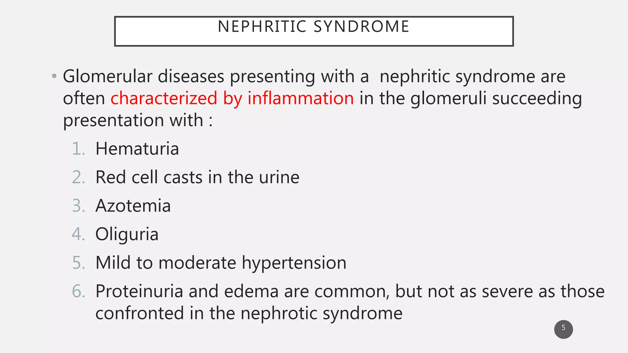
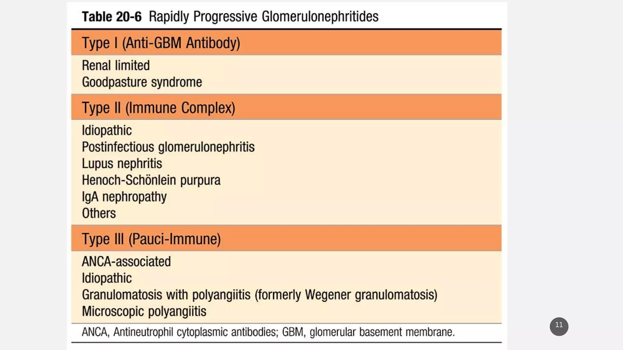
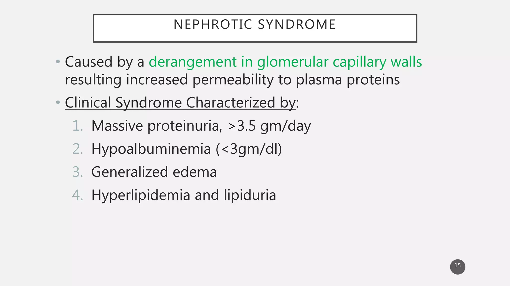
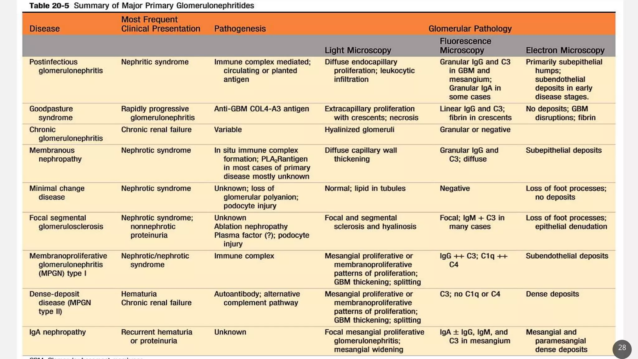
NEPHRITIC AND NEPHROTIC
SYNDROME
PBL 6
8 sept 2017
1
OBJECTIVES
• Nephritic and nephrotic syndrome
- pathophysiology
- clinical features
2
KIDNEY DISEASE
Affect four basic morphologic components
• Glomeruli
• Tubules
• Interstitium
• Blood vessels
3
GLOMERULUS
4
NEPHRITIC SYNDROME
• Glomerular diseases presenting with a nephritic syndrome are
often characterized by inflammation in the glomeruli succeeding
presentation with :
1. Hematuria
2. Red cell casts in the urine
3. Azotemia
4. Oliguria
5. Mild to moderate hypertension
6. Proteinuria and edema are common, but not as severe as those
confronted in the nephrotic syndrome
5
Acute Proliferative (poststreptococcal, Postinfectious)
Glomerulonephritis
Rapidly progressive (Crescentic ) Glomerulonephritis
6
ACUTE PROLIFERATIVE GLOMERULONEPHRITIS
Characterized histologically by diffuse proliferation of glomerular
cells associated with influx of leukocytes
# Pathophysiology
Deposition of immune complexes (in situ) containing streptococcal
antigens and specific antibodies leading to complement activation
and inflammation.
• Begins after 2-6 weeks of
Nephritogenic Streptococcal Infection
- Throat(M type : 1,3,4,3,25,49,12) or
- Skin(M type: 47,49,55,2,60,57)
7
MORPHOLOGY
8
a. Normal glomerulus
b. Glomerular
hypercellularity
(Leukocyte and glomerular
cells)
c. Electron dense sub-
epithelial Hump
d. Coarse granular deposits
of C3
CLINICAL FEATURES
• Frequently seen in children
• Present with peripheral and periorbital edema
• Cola-color urine
• Hypertension
• Positive strep. titres
• Decrease C3 complement level due
consumption
9
RAPIDLY PROGRESSIVE (CRESCENTIC) GLOMERULONEPHRITIS
• It is a syndrome associated with severe glomerular injury, but does
not denote a specific etiologic form of glomerulonephritis
• Most common histologic picture is the presence of crescents in most
of the glomeruli
10
11
# Pathophysiology
Several distinct pathogenic mechanism
1. Anti-GBM antibody mediated diseases (immunologic basis)
• Characterized by linear deposits of IgG and, in many cases, C3 in GBM
• Anti GBM when cross-react with pulmonary alveolar basement
membrane produces clinical picture of pulmonary hemorrhage with renal
failure (Goodpasture syndrome)
2. Disease caused by immune complex deposition
• RPGN as a complication of any of immune nephritides : eg. Lupus
nephritis, PSGN
• Granular pattern of staining (shows immune complex deposition)
3. Pauci- immune RPGN
• Defined as lack of detectable anti-GBM antibodies or immune complex
by immunofluorescence and electron microscopy 12
MORPHOLOGY
Fig. Endothelial and mesangial
proliferation (PAS stain )
Fig. glomerulus in immunofluorescence
(crescent)
13
Crescent : proliferation of parietal layer and migration of monocyte
, macrophage and fibrin strand between cellular layer
Collapsed
glomerular
tuft,
Leukocytes in
bowman's
capsule
CLINICAL FEATURES
• Rbc cast ,
• Rapidly deteriorating renal function (days to
weeks)
• Hematuria/hemoptysis
• Pulmonary hemorrhage(GPS)
14
NEPHROTIC SYNDROME
• Caused by a derangement in glomerular capillary walls
resulting increased permeability to plasma proteins
• Clinical Syndrome Characterized by:
1. Massive proteinuria, >3.5 gm/day
2. Hypoalbuminemia (<3gm/dl)
3. Generalized edema
4. Hyperlipidemia and lipiduria
15
16
MINIMAL CHANGE DISEASE
• Characterized by diffuse effacement of foot process of podocytes,
detectable only by electron microscopy, in glomeruli that appear
virtually normal by light microscopy
• Absence of immune complex deposit in the glomerulus, but
immunologic basis as :
- clinical association with respiratory infection and prophylactic
immunization
- response to corticosteroids
- Increase incidence in Hodgkin lymphoma
17
MORPHOLOGY
18
A. Normal appearance in LM B. Effacement of foot processes (arrows) and no deposits in
EM
MEMBRANOUS NEPHROPATHY
• Characterized by diffuse thickening of glomerular capillary wall
due to the accumulation of Ig deposits along the sub-epithelial
side of basement membrane
- Caused by auto Abs that cross react with Ag express by
podocytes.
• 75% cases are primary, secondarily due to :
1. Drugs
2. Malignant Tumors
3. SLE
4. Infections 19
MORPHOLOGY
20
A. Diffuse thickening of capillary wall, spikes on
silver stain (arrow)
B. Electron dense sub-epithelial deposit on
epithelial BM side
C. Granular immunofluorescent deposits of IgG
D. Diagrammatic representation
FOCAL SEGMENTAL GLOMERULOSCLEROSIS (FSGN)
• Characterized by sclerosis of some, but not all glomeruli (focal); and
in the affected glomeruli, only a portion of capillary tuft is involved
(segmental)
• Epidemiology:
• Most common cause of nephrotic syndrome in African American
• Primarily :idiopathic
• Secondarily as:
1) HIV nephropathy, Heroin nephropathy
2) IgA nephropathy
3) Maladaptation after nephron loss
4) Inherited congenital forms 21
PATHOPHYSIOLOGY
• Characteristic degeneration and focal disruption of podocyte with
effacement of foot processes resembling diffuse epithelial cell change
• Epithelial damage as hallmark of FSGS
• Genetic basis of pathology :
- Mutation in NPHS 1 (Nephrin)
- Mutation in NPHS2 gene (Podocin)
- defect in alfa-actinin4
- mutation in TRPC6
22
MORPHOLOGY
23Fig. Trichome stain and PAS stain
Collagen deposition on
vascular side
CLINICAL FEATURES
Basic features of nephrotic syndrome which
may also lead to the renal insufficiency and
chronic renal disease
24
MEMBRANOPROLIFERATIVE GLOMERULONEPHRITIS (MPGN)
• Either may be combined with nephrotic-nephritic picture and with
proteinuria or hematuria
• Two types
Type I : Deposition of immune complex containing IgG and
complement
Type II (Dense deposit disease) : Activation of complement (C3)
appears to be of significance
• Characterized by alternation in GBM, proliferation of glomerular cell,
leukocyte infiltration and presence of deposits in mesangial regions
and glomerular capillary walls
25
MORPHOLOGY
26
- Accentuated lobular appearance due to
proliferation of mesangial cells and
increase mesangial matrix
- GBM thickened and shows a tram-track
appearance (due to duplication of
basement membrane ), in jones silver stain
ISOLATED GLOMERULAR NEPHRITIS
• IgA nephropathy (berger disease) :
Characterized by IgA-containing complex
mesangial deposit
Most common cause of glomerulonephritis
worldwide
• Alport syndrome :
a form of hereditary nephritis caused by
mutation in genes encoding GBM type IV
collagen
Hematuria and proteinuria 27
28
REFERENCES
• Robbins and Cotran Pathologic basis of
disease,9e,vol2
• Robbins and Cotran atlas of pathology, 3e
29
Thank you
30

nephritic and nephrotic syndrome

  • 1.
  • 2.
    OBJECTIVES • Nephritic andnephrotic syndrome - pathophysiology - clinical features 2
  • 3.
    KIDNEY DISEASE Affect fourbasic morphologic components • Glomeruli • Tubules • Interstitium • Blood vessels 3
  • 4.
  • 5.
    NEPHRITIC SYNDROME • Glomerulardiseases presenting with a nephritic syndrome are often characterized by inflammation in the glomeruli succeeding presentation with : 1. Hematuria 2. Red cell casts in the urine 3. Azotemia 4. Oliguria 5. Mild to moderate hypertension 6. Proteinuria and edema are common, but not as severe as those confronted in the nephrotic syndrome 5
  • 6.
    Acute Proliferative (poststreptococcal,Postinfectious) Glomerulonephritis Rapidly progressive (Crescentic ) Glomerulonephritis 6
  • 7.
    ACUTE PROLIFERATIVE GLOMERULONEPHRITIS Characterizedhistologically by diffuse proliferation of glomerular cells associated with influx of leukocytes # Pathophysiology Deposition of immune complexes (in situ) containing streptococcal antigens and specific antibodies leading to complement activation and inflammation. • Begins after 2-6 weeks of Nephritogenic Streptococcal Infection - Throat(M type : 1,3,4,3,25,49,12) or - Skin(M type: 47,49,55,2,60,57) 7
  • 8.
    MORPHOLOGY 8 a. Normal glomerulus b.Glomerular hypercellularity (Leukocyte and glomerular cells) c. Electron dense sub- epithelial Hump d. Coarse granular deposits of C3
  • 9.
    CLINICAL FEATURES • Frequentlyseen in children • Present with peripheral and periorbital edema • Cola-color urine • Hypertension • Positive strep. titres • Decrease C3 complement level due consumption 9
  • 10.
    RAPIDLY PROGRESSIVE (CRESCENTIC)GLOMERULONEPHRITIS • It is a syndrome associated with severe glomerular injury, but does not denote a specific etiologic form of glomerulonephritis • Most common histologic picture is the presence of crescents in most of the glomeruli 10
  • 11.
  • 12.
    # Pathophysiology Several distinctpathogenic mechanism 1. Anti-GBM antibody mediated diseases (immunologic basis) • Characterized by linear deposits of IgG and, in many cases, C3 in GBM • Anti GBM when cross-react with pulmonary alveolar basement membrane produces clinical picture of pulmonary hemorrhage with renal failure (Goodpasture syndrome) 2. Disease caused by immune complex deposition • RPGN as a complication of any of immune nephritides : eg. Lupus nephritis, PSGN • Granular pattern of staining (shows immune complex deposition) 3. Pauci- immune RPGN • Defined as lack of detectable anti-GBM antibodies or immune complex by immunofluorescence and electron microscopy 12
  • 13.
    MORPHOLOGY Fig. Endothelial andmesangial proliferation (PAS stain ) Fig. glomerulus in immunofluorescence (crescent) 13 Crescent : proliferation of parietal layer and migration of monocyte , macrophage and fibrin strand between cellular layer Collapsed glomerular tuft, Leukocytes in bowman's capsule
  • 14.
    CLINICAL FEATURES • Rbccast , • Rapidly deteriorating renal function (days to weeks) • Hematuria/hemoptysis • Pulmonary hemorrhage(GPS) 14
  • 15.
    NEPHROTIC SYNDROME • Causedby a derangement in glomerular capillary walls resulting increased permeability to plasma proteins • Clinical Syndrome Characterized by: 1. Massive proteinuria, >3.5 gm/day 2. Hypoalbuminemia (<3gm/dl) 3. Generalized edema 4. Hyperlipidemia and lipiduria 15
  • 16.
  • 17.
    MINIMAL CHANGE DISEASE •Characterized by diffuse effacement of foot process of podocytes, detectable only by electron microscopy, in glomeruli that appear virtually normal by light microscopy • Absence of immune complex deposit in the glomerulus, but immunologic basis as : - clinical association with respiratory infection and prophylactic immunization - response to corticosteroids - Increase incidence in Hodgkin lymphoma 17
  • 18.
    MORPHOLOGY 18 A. Normal appearancein LM B. Effacement of foot processes (arrows) and no deposits in EM
  • 19.
    MEMBRANOUS NEPHROPATHY • Characterizedby diffuse thickening of glomerular capillary wall due to the accumulation of Ig deposits along the sub-epithelial side of basement membrane - Caused by auto Abs that cross react with Ag express by podocytes. • 75% cases are primary, secondarily due to : 1. Drugs 2. Malignant Tumors 3. SLE 4. Infections 19
  • 20.
    MORPHOLOGY 20 A. Diffuse thickeningof capillary wall, spikes on silver stain (arrow) B. Electron dense sub-epithelial deposit on epithelial BM side C. Granular immunofluorescent deposits of IgG D. Diagrammatic representation
  • 21.
    FOCAL SEGMENTAL GLOMERULOSCLEROSIS(FSGN) • Characterized by sclerosis of some, but not all glomeruli (focal); and in the affected glomeruli, only a portion of capillary tuft is involved (segmental) • Epidemiology: • Most common cause of nephrotic syndrome in African American • Primarily :idiopathic • Secondarily as: 1) HIV nephropathy, Heroin nephropathy 2) IgA nephropathy 3) Maladaptation after nephron loss 4) Inherited congenital forms 21
  • 22.
    PATHOPHYSIOLOGY • Characteristic degenerationand focal disruption of podocyte with effacement of foot processes resembling diffuse epithelial cell change • Epithelial damage as hallmark of FSGS • Genetic basis of pathology : - Mutation in NPHS 1 (Nephrin) - Mutation in NPHS2 gene (Podocin) - defect in alfa-actinin4 - mutation in TRPC6 22
  • 23.
    MORPHOLOGY 23Fig. Trichome stainand PAS stain Collagen deposition on vascular side
  • 24.
    CLINICAL FEATURES Basic featuresof nephrotic syndrome which may also lead to the renal insufficiency and chronic renal disease 24
  • 25.
    MEMBRANOPROLIFERATIVE GLOMERULONEPHRITIS (MPGN) •Either may be combined with nephrotic-nephritic picture and with proteinuria or hematuria • Two types Type I : Deposition of immune complex containing IgG and complement Type II (Dense deposit disease) : Activation of complement (C3) appears to be of significance • Characterized by alternation in GBM, proliferation of glomerular cell, leukocyte infiltration and presence of deposits in mesangial regions and glomerular capillary walls 25
  • 26.
    MORPHOLOGY 26 - Accentuated lobularappearance due to proliferation of mesangial cells and increase mesangial matrix - GBM thickened and shows a tram-track appearance (due to duplication of basement membrane ), in jones silver stain
  • 27.
    ISOLATED GLOMERULAR NEPHRITIS •IgA nephropathy (berger disease) : Characterized by IgA-containing complex mesangial deposit Most common cause of glomerulonephritis worldwide • Alport syndrome : a form of hereditary nephritis caused by mutation in genes encoding GBM type IV collagen Hematuria and proteinuria 27
  • 28.
  • 29.
    REFERENCES • Robbins andCotran Pathologic basis of disease,9e,vol2 • Robbins and Cotran atlas of pathology, 3e 29
  • 30.