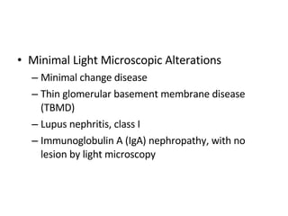
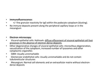
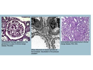
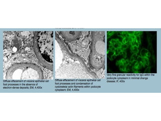
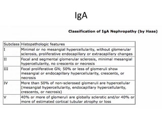
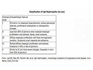
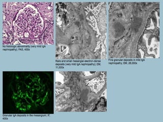
Minimal LM canges
Minimal Light Microscopic Alterations Minimal change disease Thin glomerular basement membrane disease  (TBMD) Lupus nephritis, class I Immunoglobulin A (IgA) nephropathy, with no lesion by light microscopy
Minimal Change Disease Histopathology:  LM usually discloses no significant abnormalities GBM- unremarkable thickness and texture Mesangium +/- mild expansion Podocytes +/- slightly prominent but no proliferative changes (pseudocrescents)  Proximal tubules may contain prominent protein reabsorption granules The interstitium is usually delicate; foamy macrophages may be seen on rare occasions Acute interstitial nephritis may be seen in association with drug-induced (particularly NSAID-induced) minimal change disease Interstitial fibrosis and tubular atrophy may be seen in elderly patients with comorbid states
Immunofluorescence:  +/- fine granular reactivity for IgG within the podocyte cytoplasm (dusting).  No immune deposits present along the peripheral capillary loops or in the mesangium Electron microscopy:   Visceral epithelial cells: Hallmark-  diffuse effacement of visceral epithelial cell foot processes in the absence of electron-dense deposits .  Other degenerative changes of visceral epithelial cells: microvillous degeneration, vacuolization of the cytoplasm, increased number of lysosomes and other cytoplasmic organelles GBM: Usually unremarkable Glomerular endothelial cells: Usually unremarkable and do not contain tubuloreticular structures Mesangium: Normal cell elements and an extracellular matrix without electron-dense deposits
 
 
IgA
 
Histopathology:  Glomeruli are normal, or they show minimal mesangial expansion The glomerular basement membranes are of unremarkable thickness and texture The mesangium may be minimally expanded, but is normocellular The tubulointerstitium is usually unremarkable This form of IgA is quite common and can be detected incidentally on renal biopsy, superimposed to any other renal disease
Immunofluorescence:  Dominant reactivity for IgA in the mesangium; C3 may be equally or less reactive. There is usually stronger reactivity for lambda than for kappa light chains in the mesangial deposits Electron microscopy:  Visceral epithelial cells: Unremarkable, with well-preserved foot processes Glomerular basement membranes: May be thin; there is higher incidence of thin glomerular basement membrane disease in IgA nephropathy than in any other glomerular disease. Glomerular endothelial cells: Usually unremarkable and tubuloreticular structures are not seen. Mesangium: Shows normal cell elements and an extracellular matrix with scattered small fine granular electron-dense deposits
 
Class I lupus nephritis Histopathology:  Light microscopic examination usually discloses no significant abnormalities The glomerular basement membranes are of unremarkable thickness and texture The mesangium may be mildly expanded, but is normocellular The tubulointerstitium is usually unremarkable
Immunofluorescence:  'Full house' reactivity  Electron microscopy:  Visceral epithelial cells: Unremarkable and foot processes are well preserved Glomerular basement membranes: Normal appearance and texture Glomerular endothelial cells: May contain tubuloreticular structures Mesangium: Shows normal cell elements and an extracellular matrix with scattered fine granula electron-dense deposits
 
Thin Glomerular BMD Histopathology:  The capillary loops are of normal contour and may appear delicate Normocellular mesangium The tubules and interstitium are usually unremarkable; foamy macrophages may be seen on rare occasions in the interstitium More pronounced interstitial fibrosis and tubular atrophy may be seen in elderly patients with comorbid states
 
Electron microscopy:  Visceral epithelial cells: The visceral epithelial cells and their foot processes are well preserved. Glomerular basement membranes: Morphometric measurements disclose diffuse thinning, with the mean thickness below the lower normal limit of 264 nm. Electron-dense deposits are not seen along the capillary loops Glomerular endothelial cells: Unremarkable and do not contain tubuloreticular structures Mesangium: Normal cell elements and an extracellular matrix without electron-dense deposits
 

Minimal Light Microscopic Changes

  • 1.
  • 2.
    Minimal Light MicroscopicAlterations Minimal change disease Thin glomerular basement membrane disease (TBMD) Lupus nephritis, class I Immunoglobulin A (IgA) nephropathy, with no lesion by light microscopy
  • 3.
    Minimal Change DiseaseHistopathology: LM usually discloses no significant abnormalities GBM- unremarkable thickness and texture Mesangium +/- mild expansion Podocytes +/- slightly prominent but no proliferative changes (pseudocrescents) Proximal tubules may contain prominent protein reabsorption granules The interstitium is usually delicate; foamy macrophages may be seen on rare occasions Acute interstitial nephritis may be seen in association with drug-induced (particularly NSAID-induced) minimal change disease Interstitial fibrosis and tubular atrophy may be seen in elderly patients with comorbid states
  • 4.
    Immunofluorescence: +/-fine granular reactivity for IgG within the podocyte cytoplasm (dusting). No immune deposits present along the peripheral capillary loops or in the mesangium Electron microscopy: Visceral epithelial cells: Hallmark- diffuse effacement of visceral epithelial cell foot processes in the absence of electron-dense deposits . Other degenerative changes of visceral epithelial cells: microvillous degeneration, vacuolization of the cytoplasm, increased number of lysosomes and other cytoplasmic organelles GBM: Usually unremarkable Glomerular endothelial cells: Usually unremarkable and do not contain tubuloreticular structures Mesangium: Normal cell elements and an extracellular matrix without electron-dense deposits
  • 5.
  • 6.
  • 7.
  • 8.
  • 9.
    Histopathology: Glomeruliare normal, or they show minimal mesangial expansion The glomerular basement membranes are of unremarkable thickness and texture The mesangium may be minimally expanded, but is normocellular The tubulointerstitium is usually unremarkable This form of IgA is quite common and can be detected incidentally on renal biopsy, superimposed to any other renal disease
  • 10.
    Immunofluorescence: Dominantreactivity for IgA in the mesangium; C3 may be equally or less reactive. There is usually stronger reactivity for lambda than for kappa light chains in the mesangial deposits Electron microscopy: Visceral epithelial cells: Unremarkable, with well-preserved foot processes Glomerular basement membranes: May be thin; there is higher incidence of thin glomerular basement membrane disease in IgA nephropathy than in any other glomerular disease. Glomerular endothelial cells: Usually unremarkable and tubuloreticular structures are not seen. Mesangium: Shows normal cell elements and an extracellular matrix with scattered small fine granular electron-dense deposits
  • 11.
  • 12.
    Class I lupusnephritis Histopathology: Light microscopic examination usually discloses no significant abnormalities The glomerular basement membranes are of unremarkable thickness and texture The mesangium may be mildly expanded, but is normocellular The tubulointerstitium is usually unremarkable
  • 13.
    Immunofluorescence: 'Fullhouse' reactivity Electron microscopy: Visceral epithelial cells: Unremarkable and foot processes are well preserved Glomerular basement membranes: Normal appearance and texture Glomerular endothelial cells: May contain tubuloreticular structures Mesangium: Shows normal cell elements and an extracellular matrix with scattered fine granula electron-dense deposits
  • 14.
  • 15.
    Thin Glomerular BMDHistopathology: The capillary loops are of normal contour and may appear delicate Normocellular mesangium The tubules and interstitium are usually unremarkable; foamy macrophages may be seen on rare occasions in the interstitium More pronounced interstitial fibrosis and tubular atrophy may be seen in elderly patients with comorbid states
  • 16.
  • 17.
    Electron microscopy: Visceral epithelial cells: The visceral epithelial cells and their foot processes are well preserved. Glomerular basement membranes: Morphometric measurements disclose diffuse thinning, with the mean thickness below the lower normal limit of 264 nm. Electron-dense deposits are not seen along the capillary loops Glomerular endothelial cells: Unremarkable and do not contain tubuloreticular structures Mesangium: Normal cell elements and an extracellular matrix without electron-dense deposits
  • 18.