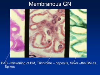
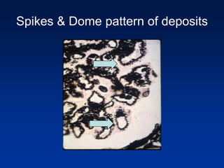
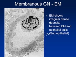
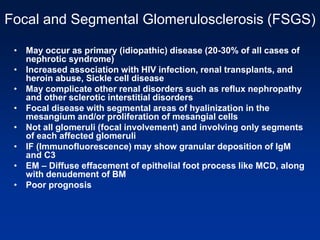
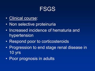
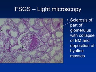
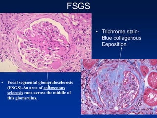
Membranous GN
• MOST COMMON cause is idiopathic (85%); peak age
30-50; male:female, 2:1
• May be secondary to:
– Drugs-captopril, penicillamine, gold, mercury,
trimethadione, NSAIDS
– Infections-malaria (P. malariae), leprosy,
schistosomiasis, syphilis, hepatitis B and C,
filariasis, hydatid disease and enterococcal
endocarditis
– Diseases-malignancy (Carcinoma of breast, lung,
colon, stomach, and esophagus) melanoma, renal
cell CA, SLE, sarcoidosis, diabetes, thyroiditis, sickle
cell anemia, Crohn’s disease
Membranous GN
• Pathogenesis is immune complex deposition and/or
planted antigens in GBM
• Basement membrane is thickened and abnormally
permeable
• Epimembranous spikes & dome appearance with silver
stains
• Subepithelial deposits of IgG and Complement
• No inflammatory infiltrate
• Direct action of C5b-C9 on podocytes  activation of
epithelial and mesangial cells  proteases & oxidants
 capillary wall injury  leaking
• Any adult who presents with nephrotic syndrome
should be suspected of having an underlying
malignancy or SLE
Membranous GN
Membranous GN
• Light
microscopy
exhibiting
uniform
thickening of
the
glomerular
capillary wall
Membranous GN
PAS –thickening of BM, Trichrome – deposits, Silver –the BM as
Spikes
Spikes & Dome pattern of deposits
Membranous GN - EM
• EM shows
irregular dense
deposits
between BM and
epithelial cells
(Sub epithelial)
Membranous GN – Clinical course
• Adults develop as nephrotic syndrome
• Chronic immune complex nephritis
• Persistent proteinuria (over 60% cases)
• Few cases may have hematuria or hypertension
• Does not respond well to corticosteroid therapy
• Clinical course is variable – remain indolent,
may remit or become progressive (40%) disease
to renal failure after 2 to 20 yrs
• 10-30% cases have more benign with partial or
complete remission of proteinuria
Focal and Segmental Glomerulosclerosis (FSGS)
• May occur as primary (idiopathic) disease (20-30% of all cases of
nephrotic syndrome)
• Increased association with HIV infection, renal transplants, and
heroin abuse, Sickle cell disease
• May complicate other renal disorders such as reflux nephropathy
and other sclerotic interstitial disorders
• Focal disease with segmental areas of hyalinization in the
mesangium and/or proliferation of mesangial cells
• Not all glomeruli (focal involvement) and involving only segments
of each affected glomeruli
• IF (Immunofluorescence) may show granular deposition of IgM
and C3
• EM – Diffuse effacement of epithelial foot process like MCD, along
with denudement of BM
• Poor prognosis
FSGS
• Clinical course:
• Non selective proteinuria
• Increased incidence of hematuria and
hypertension
• Respond poor to corticosteroids
• Progression to end stage renal disease in
10 yrs
• Poor prognosis in adults
FSGS – Light microscopy
• Sclerosis of
part of
glomerulus
with collapse
of BM and
deposition of
hyaline
masses
FSGS
• Focal segmental glomerulosclerosis
(FSGS)-An area of collagenous
sclerosis runs across the middle of
this glomerulus.
• Trichrome stain-
Blue collagenous
Deposition
FSGS – PAS stain
• Lipid
vacuoles
• Hyaline
masses
HIV associated FSGS
• Collapsing variant of FSGS – With
collapse and involvement of entire tuft
• Tubules – Show cystic dilatation filled with
proteinaceous material
• Tubuloreticular inclusions in endothelial
cells
HIV associated FSGS
• Left – Silver stain sclerotic involvement of the entire tuft
• Right – Cystic dilatation of tubules with proteinaceous
material
Membranoproliferative (Mesangiocapillary)
Glomerulonephritis
• Characterized by proliferation of mesangial
and endothelial cells and basement
membrane thickening-occurs in adolescents
and young adults
• Type I: subendothelial deposit type-80%
– Immune complex; complement deposits;
transient low complement levels (both early
& late complement)
– Relative benign course compared to type II
and III
Membranoproliferative (Mesangiocapillary)
Glomerulonephritis
• Type II: linear dense deposit disease-20%
– complement activation (alternate pathway) by C3
nephritic factor (anti-C3 convertase) without
immune complexes; persistent low complement
(60%)
– Low C3 levels but early complements C1 & C4 are
normal in serum
– Composition of dense deposits unknown
(deposits found in lamina densa of BM)
• Type II may be associated with partial
lipodystrophy which predates the onset of
renal symptoms by several months to years
Membranoproliferative
Glomerulonephritis
• Type III has features of Type I MPGN
(subendothelial deposits) and membranous
nephropathy (subepithelial deposits)
• All types exhibit “tram-track” appearance or
splitting of the GBM due to extension of the
mesangial matrix beneath the capillary loops
(More common with type I) by light
microscopy
• Prognosis is poor with type II and type III
• Secondary MPGN - Associated with
a variety of chronic infections
(bacterial endocarditis, HIV,
Hepatitis B and C), systemic
immune complex diseases (SLE,
cryoglobulinemia) and malignancies
(leukemias, lymphomas)
Membranoproliferative GN
MPGN Type I
• Note the lobular appearance.There is thickening of capillary walls. There is
also hypercellularity. Much of this hypercellularity is mesangial proliferation,
and some of the capillary wall thickening is caused by mesangial
interposition into the subendothelial zone of the capillary loops. This is the
basis for the term mesangiocapillary glomerulonephritis
• The silver stain shows the typical” tram
track” appearance of membranoproliferative
glomerulonephritis.
MPGN-I
• This electron
micrograph shows
the urinary space,
the effaced foot
processes, the
original basement
membrane, and
conspicuous
subendothelial
deposits
MPGN-II
• EM shows GBM as
well as mesangial
deposits. These
dense deposits are
not subepithelial or
subendothelial, but
rather are within the
basement
membrane
MPGN
• Clinical course:
• Nephrotic or Nephritic or Nephritic-
nephrotic syndrome
• 40% of pts develop chronic renal failure
within 10yrs
• 30% variable renal insufficiently
• 30% persistent nephrotic syndrome
without renal failure.
• Generally poor prognosis
IgA Nephropathy(Berger’s disease)
• Abnormal (polymeric) forms of IgA in these
patients are trapped in the mesangium of the
glomerulus which results in glomerular damage
probably as a result of alternate complement
pathway activation.
• The abnormal IgA is a produced in susceptible
patients after infections ,usually upper
respiratory and may be produced in patients with
intestinal disorders such as celiac disease and
with liver disease (IgA not eliminated).
IgA nephropathy
• It is a common cause of Benign recurrent
hematuria and most common type of
glomerulonephritis world wide
• Characteristically seen in young men or
children(80%) with microscopic hematuria
within one to 1 1/2 days following an upper
respiratory infection (no skin lesions)
• Clinical course variable maintaining renal
function for decades and slow progression to
CRF
IgA Nephropathy
. The IgA is deposited mainly in
mesangium, which then
increases mesangial cellularity
as shown at the arrow.
• IF shows IgA
deposits in
mesangium
IgA Nephropathy
Henoch-Schonlein Purpura
(HSP)
• Both have mesangial deposits of IgA
• HSP is systemic with skin (purpuric rash),
GI tract (abdominal pain) joint (arthritis)
and renal involvement
• Relationship between the two diseases
remains uncertain
Chronic Glomerulonephritis
• Represents end stage of glomerular disease
• Primary GN leading to CGN:
• RPGN – 90%
• FSGS – 50-80%
• MPGN – 50%
• Membranous GN – 50%
• IgA nephropathy – 30-50%
• PSGN (Poststreptococcal GN) – 1-2%
Chronic GN
• Gross – Symmetrically contracted and has
diffuse granularity of the surface. Cut section
shows cortical thinning
• Micro – Early cases will show evidence of
primary disease
• Late stages- Hyaline masses accumulate
transforming glomeruli into acellular eosinophilic
mass
• Also changes related to hypertension can be
seen – arteriolar sclerosis
CGN
• Gross –contracted and has diffuse
granularity of the surface. Cut section
shows cortical thinning
Chronic GN
Hyaline masses accumulate transforming glomeruli into
acellular eosinophilic mass
Chronic GN
• Clinical course :
• Slowly progress to uremia and death
• Proteinuria, hypertension and azotemia
• Uremic complications – Pericarditis,
gastroenteritis
• Secondary hyperparathyroidism
• Cerebral & CVS manifestations
• Pt will require dialysis or renal transplant
Hereditary Nephritis
• Alport syndrome is a familial nephropathy
usually X linked dominantly transmitted.
• Mutation involves the synthesis of alpha 5 chain
of the type IV collagen
• It is marked by progressive fibrosing renal
disease and by eye disorders (lens dislocation
and posterior cataract) and by nerve deafness.
• A characteristic EM finding is irregular thickening
of the glomerular basement membrane and
reduplication of the lamina densa.
Alport’s syndrome
• The renal tubular cells appear foamy because of the
accumulation of neutral fats and
mucopolysaccharides (Foam cells)
Alport’s syndrome - EM
The glomeruli show irregular thickening and
splitting of basement membranes.
Hereditary Nephritis
• Thin Membrane Disease –
• This disease is fairly common and
manifest as benign familial asymptomatic
hematuria.
• Mild proteinuria occurs and renal function
is maintained and has good prognosis
• Electron microscopy shows marked
thinning of the glomerular basement
membrane.
Thin membrane disease
• Marked thinning
of the Basement
membrane
shown by arrows
Glomerular Lesions associated
with systemic diseases
Henoch Schoenlein’s Purpura
• Most common in children (3-8yrs) with
strong background of atopy and follows
after respiratory infection
• skin purpura (leukocytoclastic vasculitis)
involving arms and legs, buttocks
• GI involvement - abdominal pain, GI
bleeding
• Non migratory arthralgias
• Renal involvement
Henoch Schoenlein’s Purpura
• Renal manifestation – hematuria, proteinuria
• Histology – Early stages mesangial proliferation
and later show cresents (RPGN)
• IF - IgA is deposited in mesangium of affected
glomeruli (therefore are closely related to IgA
nephropathy)
• Skin biopsies – Necrotizing vasculitis of small
vessels of dermis
• Vasculitis also seen in GI (Rare in kidney)
• Children affected have excellent prognosis
HSP
• In HSP, there is a
characteristic
purpuric rash
Diabetes Mellitus
• Approximately 35-45% of Type I insulin-
dependent diabetics develop nephropathy
in 17 +/- years; 20% of Type II develop
renal disease
• Associated renal disease (Diabetic
nephropathy)
– Complications of diabetic vascular
disease
– Diabetic glomerular damage
– Increased susceptibility to infection and
papillary necrosis
Diabetic nephropathy
• Pathogenesis:
1) Related to metabolic defect(hyperglycemia)
2) Due to hyperglycemia – endothelial cells
produce increased type IV collagen and
fibronectin but decreased proteoglycan
(Heparen sulphate) in BM
3) Non-enzymatic glycosylation
products(AGE’s binding to vessel wall and
BM collagen) interfering with glomerular
barrier function
Pathogenesis:
4) Hemodynamic chages : Particularly in type I
DM, hyperglycemia causes increased GFR,
increased capillary pressure and glomerular
hypertrophy.(later leading to sclerosis)
Mesangium proliferation and expansion is seen
in diabetics is due to growth factors &
cytokines released by binding of AGE to many
cells
• Diabetic Vascular Disease
– Atherosclerosis
– Hyaline arteriosclerosis of efferent arteriole and
afferent arteriole eventually affects the GFR
Diabetic glomerular disease
• Capillary basement membrane thickening:
– This is seen in all diabetics with / Without proteinuria
• Diffuse glomerulosclerosis: Changes in mesangium
leads to proliferation and expansion of mesangial
matrix
• Nodular glomerulosclerosis:
– Also called Kimmelstiel Wilson disease-
pathognomic for diabetic nephropathy
– Lesions have ovoid or spherical hyaline masses
situated at periphery ( lie within mesangial core and
surrounded by surrounding patent capillary loops)
producing nodules
• Nodular changes resemble “christmas-tree” balls
within the mesangium of the glomeruli
Diabetic Nephropathy
• diffuse
glomerulosclerosis
and hyaline
arteriolosclerosis
• Nodular
glomerulosclerosis
Diabetic nephropathy
• Capsular
drop lesion
• Represents
leakage of
plasma
proteins
from the
damaged
capillaries
Diabetic Glomerular Disease
– Microalbuminuria (first lab abnormality-30-300
mg/day of albumin) seen in early course of
disease
– Late course – nephrotic range proteinuria
– Endstage renal failure with loss of GFR within 5
years, along with hypertension
– Severe disease - Dialysis & renal transplant
Renal amyloidosis
• The amorphous pink depositis of amyloid may be found in and
around arteries, in interstitium, or in glomeruli. A Congo red stain
will demonstrate the pink material to be amyloid.
Amyloidosis
• Amyloid fibrils
may be
subepithlial,
subendothelial
or within BM
Systemic Lupus Erythematosus
Glomerulonephritis
• Kidney involvement occurs in 60-70% of cases
• Lupus nephritis is subdivided into 5 WHO classes
– Class I-Normal
– Class II-mesangial GN
– Class III-focal proliferative GN
– Class IV-diffuse proliferative GN (50% of
cases)
• 50% present with nephritic syndrome and up to
50% with nephrotic syndrome
• Most aggressive renal lesion-30% progress
to terminal renal failure
– Class V-membranous GN
Diffuse Proliferative (Class IV) lupus
nephritis
• Characterized by:
– increased numbers of endothelial and mesangial cells
– “wire-looping” of the capillaries (due to subendothelial deposits-
composed of either circulating immune complexes of DNA + anti-
DNA antibodies or DNA deposited in the basement membrane that
attracts anti-DNA antibodies and complement)
– Foci of neutrophilic infiltration with fibrinoid necrosis (70%) and
hematoxylin bodies (30% of cases)
– IF stains shows a “lumpy-bumpy” granular pattern (IgG, C3, fibrin,
sometimes other immunoglobulins)
– There is also some immune complex damage to tubules and
interstitium
– Tubuloreticular structures (cellular inclusions induced by interferon)
are seen in endothelial cells, monocytes, and lymphocytes
– Only HIV is associated with similar numbers of these inclusions
Class IV Lupus glomerulonephritis
Diffuse proliferative
Lupus Glomerulonephritis
Wire loop lesion
• The thickened capillary loops resemble "wire loops". This light
microscopic appearance is very suggestive of SLE
Lupus Nephritis
• Subendothelial
deposits in lupus
• Immune complex
deposition may be
subepithelial(mem
branous type)
• Subendothelial in
diffuse proliferative
type
Fibrillary & Immunotactoid
glomerulonephritis
• Fibrillary GN : Fibrillary deposits in
mesangium and do not stain with congo
red resemble MPGN histology by light
microscopy
• EM – Size of fibrils are 20-30nm (amyloid
is 10nm size)
• Clinical – Hematuria, nephrotic syndrome
• Immunotactoid GN: microtubular deposits
of size 30-50nm
Essential mixed cryoglobinemia
• Deposits of cryoglobulins (IgG-IgM
complexes)
• Cutaneous vasculitis, reynauds
phenomenon, synovitis, focal or diffuse
GN)
• Cryoglobulins associated with Hepatitis C
infection may show MPGN
Focal Proliferative
Glomerulonephritis
• This entity ,as the name implies, is characterized
by focal involvement of a portion of the glomeruli
in the kidney.
• It can be seen with Lupus, subacute bacterial
endocarditis and with Wegener’s syndrome
• It may also occur with any of the above
described glomerulonephridites, especially IgA
nephropathy.
• It may occur unrelated to other systemic or renal
disease (Idiopathic focal GN).
• Note that it is proliferative (often with fibrin and
necrosis) and not sclerosing.
Focal proliferative GN
• Note segmental
proliferation of
cells with
necrosis and
fibrin deposition.
The remainder
of glomerulus is
uninvolved

Renal pathology ii

  • 1.
    Membranous GN • MOSTCOMMON cause is idiopathic (85%); peak age 30-50; male:female, 2:1 • May be secondary to: – Drugs-captopril, penicillamine, gold, mercury, trimethadione, NSAIDS – Infections-malaria (P. malariae), leprosy, schistosomiasis, syphilis, hepatitis B and C, filariasis, hydatid disease and enterococcal endocarditis – Diseases-malignancy (Carcinoma of breast, lung, colon, stomach, and esophagus) melanoma, renal cell CA, SLE, sarcoidosis, diabetes, thyroiditis, sickle cell anemia, Crohn’s disease
  • 2.
    Membranous GN • Pathogenesisis immune complex deposition and/or planted antigens in GBM • Basement membrane is thickened and abnormally permeable • Epimembranous spikes & dome appearance with silver stains • Subepithelial deposits of IgG and Complement • No inflammatory infiltrate • Direct action of C5b-C9 on podocytes  activation of epithelial and mesangial cells  proteases & oxidants  capillary wall injury  leaking • Any adult who presents with nephrotic syndrome should be suspected of having an underlying malignancy or SLE
  • 3.
  • 4.
  • 5.
    Membranous GN PAS –thickeningof BM, Trichrome – deposits, Silver –the BM as Spikes
  • 6.
    Spikes & Domepattern of deposits
  • 7.
    Membranous GN -EM • EM shows irregular dense deposits between BM and epithelial cells (Sub epithelial)
  • 8.
    Membranous GN –Clinical course • Adults develop as nephrotic syndrome • Chronic immune complex nephritis • Persistent proteinuria (over 60% cases) • Few cases may have hematuria or hypertension • Does not respond well to corticosteroid therapy • Clinical course is variable – remain indolent, may remit or become progressive (40%) disease to renal failure after 2 to 20 yrs • 10-30% cases have more benign with partial or complete remission of proteinuria
  • 9.
    Focal and SegmentalGlomerulosclerosis (FSGS) • May occur as primary (idiopathic) disease (20-30% of all cases of nephrotic syndrome) • Increased association with HIV infection, renal transplants, and heroin abuse, Sickle cell disease • May complicate other renal disorders such as reflux nephropathy and other sclerotic interstitial disorders • Focal disease with segmental areas of hyalinization in the mesangium and/or proliferation of mesangial cells • Not all glomeruli (focal involvement) and involving only segments of each affected glomeruli • IF (Immunofluorescence) may show granular deposition of IgM and C3 • EM – Diffuse effacement of epithelial foot process like MCD, along with denudement of BM • Poor prognosis
  • 10.
    FSGS • Clinical course: •Non selective proteinuria • Increased incidence of hematuria and hypertension • Respond poor to corticosteroids • Progression to end stage renal disease in 10 yrs • Poor prognosis in adults
  • 11.
    FSGS – Lightmicroscopy • Sclerosis of part of glomerulus with collapse of BM and deposition of hyaline masses
  • 12.
    FSGS • Focal segmentalglomerulosclerosis (FSGS)-An area of collagenous sclerosis runs across the middle of this glomerulus. • Trichrome stain- Blue collagenous Deposition
  • 13.
    FSGS – PASstain • Lipid vacuoles • Hyaline masses
  • 14.
    HIV associated FSGS •Collapsing variant of FSGS – With collapse and involvement of entire tuft • Tubules – Show cystic dilatation filled with proteinaceous material • Tubuloreticular inclusions in endothelial cells
  • 15.
    HIV associated FSGS •Left – Silver stain sclerotic involvement of the entire tuft • Right – Cystic dilatation of tubules with proteinaceous material
  • 16.
    Membranoproliferative (Mesangiocapillary) Glomerulonephritis • Characterizedby proliferation of mesangial and endothelial cells and basement membrane thickening-occurs in adolescents and young adults • Type I: subendothelial deposit type-80% – Immune complex; complement deposits; transient low complement levels (both early & late complement) – Relative benign course compared to type II and III
  • 17.
    Membranoproliferative (Mesangiocapillary) Glomerulonephritis • TypeII: linear dense deposit disease-20% – complement activation (alternate pathway) by C3 nephritic factor (anti-C3 convertase) without immune complexes; persistent low complement (60%) – Low C3 levels but early complements C1 & C4 are normal in serum – Composition of dense deposits unknown (deposits found in lamina densa of BM) • Type II may be associated with partial lipodystrophy which predates the onset of renal symptoms by several months to years
  • 18.
    Membranoproliferative Glomerulonephritis • Type IIIhas features of Type I MPGN (subendothelial deposits) and membranous nephropathy (subepithelial deposits) • All types exhibit “tram-track” appearance or splitting of the GBM due to extension of the mesangial matrix beneath the capillary loops (More common with type I) by light microscopy • Prognosis is poor with type II and type III
  • 19.
    • Secondary MPGN- Associated with a variety of chronic infections (bacterial endocarditis, HIV, Hepatitis B and C), systemic immune complex diseases (SLE, cryoglobulinemia) and malignancies (leukemias, lymphomas)
  • 20.
  • 21.
    MPGN Type I •Note the lobular appearance.There is thickening of capillary walls. There is also hypercellularity. Much of this hypercellularity is mesangial proliferation, and some of the capillary wall thickening is caused by mesangial interposition into the subendothelial zone of the capillary loops. This is the basis for the term mesangiocapillary glomerulonephritis
  • 22.
    • The silverstain shows the typical” tram track” appearance of membranoproliferative glomerulonephritis.
  • 23.
    MPGN-I • This electron micrographshows the urinary space, the effaced foot processes, the original basement membrane, and conspicuous subendothelial deposits
  • 24.
    MPGN-II • EM showsGBM as well as mesangial deposits. These dense deposits are not subepithelial or subendothelial, but rather are within the basement membrane
  • 25.
    MPGN • Clinical course: •Nephrotic or Nephritic or Nephritic- nephrotic syndrome • 40% of pts develop chronic renal failure within 10yrs • 30% variable renal insufficiently • 30% persistent nephrotic syndrome without renal failure. • Generally poor prognosis
  • 26.
    IgA Nephropathy(Berger’s disease) •Abnormal (polymeric) forms of IgA in these patients are trapped in the mesangium of the glomerulus which results in glomerular damage probably as a result of alternate complement pathway activation. • The abnormal IgA is a produced in susceptible patients after infections ,usually upper respiratory and may be produced in patients with intestinal disorders such as celiac disease and with liver disease (IgA not eliminated).
  • 27.
    IgA nephropathy • Itis a common cause of Benign recurrent hematuria and most common type of glomerulonephritis world wide • Characteristically seen in young men or children(80%) with microscopic hematuria within one to 1 1/2 days following an upper respiratory infection (no skin lesions) • Clinical course variable maintaining renal function for decades and slow progression to CRF
  • 28.
    IgA Nephropathy . TheIgA is deposited mainly in mesangium, which then increases mesangial cellularity as shown at the arrow. • IF shows IgA deposits in mesangium
  • 29.
    IgA Nephropathy Henoch-Schonlein Purpura (HSP) •Both have mesangial deposits of IgA • HSP is systemic with skin (purpuric rash), GI tract (abdominal pain) joint (arthritis) and renal involvement • Relationship between the two diseases remains uncertain
  • 30.
    Chronic Glomerulonephritis • Representsend stage of glomerular disease • Primary GN leading to CGN: • RPGN – 90% • FSGS – 50-80% • MPGN – 50% • Membranous GN – 50% • IgA nephropathy – 30-50% • PSGN (Poststreptococcal GN) – 1-2%
  • 31.
    Chronic GN • Gross– Symmetrically contracted and has diffuse granularity of the surface. Cut section shows cortical thinning • Micro – Early cases will show evidence of primary disease • Late stages- Hyaline masses accumulate transforming glomeruli into acellular eosinophilic mass • Also changes related to hypertension can be seen – arteriolar sclerosis
  • 33.
    CGN • Gross –contractedand has diffuse granularity of the surface. Cut section shows cortical thinning
  • 34.
    Chronic GN Hyaline massesaccumulate transforming glomeruli into acellular eosinophilic mass
  • 35.
    Chronic GN • Clinicalcourse : • Slowly progress to uremia and death • Proteinuria, hypertension and azotemia • Uremic complications – Pericarditis, gastroenteritis • Secondary hyperparathyroidism • Cerebral & CVS manifestations • Pt will require dialysis or renal transplant
  • 36.
    Hereditary Nephritis • Alportsyndrome is a familial nephropathy usually X linked dominantly transmitted. • Mutation involves the synthesis of alpha 5 chain of the type IV collagen • It is marked by progressive fibrosing renal disease and by eye disorders (lens dislocation and posterior cataract) and by nerve deafness. • A characteristic EM finding is irregular thickening of the glomerular basement membrane and reduplication of the lamina densa.
  • 37.
    Alport’s syndrome • Therenal tubular cells appear foamy because of the accumulation of neutral fats and mucopolysaccharides (Foam cells)
  • 38.
    Alport’s syndrome -EM The glomeruli show irregular thickening and splitting of basement membranes.
  • 39.
    Hereditary Nephritis • ThinMembrane Disease – • This disease is fairly common and manifest as benign familial asymptomatic hematuria. • Mild proteinuria occurs and renal function is maintained and has good prognosis • Electron microscopy shows marked thinning of the glomerular basement membrane.
  • 40.
    Thin membrane disease •Marked thinning of the Basement membrane shown by arrows
  • 41.
  • 42.
    Henoch Schoenlein’s Purpura •Most common in children (3-8yrs) with strong background of atopy and follows after respiratory infection • skin purpura (leukocytoclastic vasculitis) involving arms and legs, buttocks • GI involvement - abdominal pain, GI bleeding • Non migratory arthralgias • Renal involvement
  • 43.
    Henoch Schoenlein’s Purpura •Renal manifestation – hematuria, proteinuria • Histology – Early stages mesangial proliferation and later show cresents (RPGN) • IF - IgA is deposited in mesangium of affected glomeruli (therefore are closely related to IgA nephropathy) • Skin biopsies – Necrotizing vasculitis of small vessels of dermis • Vasculitis also seen in GI (Rare in kidney) • Children affected have excellent prognosis
  • 44.
    HSP • In HSP,there is a characteristic purpuric rash
  • 45.
    Diabetes Mellitus • Approximately35-45% of Type I insulin- dependent diabetics develop nephropathy in 17 +/- years; 20% of Type II develop renal disease • Associated renal disease (Diabetic nephropathy) – Complications of diabetic vascular disease – Diabetic glomerular damage – Increased susceptibility to infection and papillary necrosis
  • 46.
    Diabetic nephropathy • Pathogenesis: 1)Related to metabolic defect(hyperglycemia) 2) Due to hyperglycemia – endothelial cells produce increased type IV collagen and fibronectin but decreased proteoglycan (Heparen sulphate) in BM 3) Non-enzymatic glycosylation products(AGE’s binding to vessel wall and BM collagen) interfering with glomerular barrier function
  • 47.
    Pathogenesis: 4) Hemodynamic chages: Particularly in type I DM, hyperglycemia causes increased GFR, increased capillary pressure and glomerular hypertrophy.(later leading to sclerosis) Mesangium proliferation and expansion is seen in diabetics is due to growth factors & cytokines released by binding of AGE to many cells • Diabetic Vascular Disease – Atherosclerosis – Hyaline arteriosclerosis of efferent arteriole and afferent arteriole eventually affects the GFR
  • 48.
    Diabetic glomerular disease •Capillary basement membrane thickening: – This is seen in all diabetics with / Without proteinuria • Diffuse glomerulosclerosis: Changes in mesangium leads to proliferation and expansion of mesangial matrix • Nodular glomerulosclerosis: – Also called Kimmelstiel Wilson disease- pathognomic for diabetic nephropathy – Lesions have ovoid or spherical hyaline masses situated at periphery ( lie within mesangial core and surrounded by surrounding patent capillary loops) producing nodules • Nodular changes resemble “christmas-tree” balls within the mesangium of the glomeruli
  • 49.
    Diabetic Nephropathy • diffuse glomerulosclerosis andhyaline arteriolosclerosis • Nodular glomerulosclerosis
  • 50.
    Diabetic nephropathy • Capsular droplesion • Represents leakage of plasma proteins from the damaged capillaries
  • 51.
    Diabetic Glomerular Disease –Microalbuminuria (first lab abnormality-30-300 mg/day of albumin) seen in early course of disease – Late course – nephrotic range proteinuria – Endstage renal failure with loss of GFR within 5 years, along with hypertension – Severe disease - Dialysis & renal transplant
  • 52.
    Renal amyloidosis • Theamorphous pink depositis of amyloid may be found in and around arteries, in interstitium, or in glomeruli. A Congo red stain will demonstrate the pink material to be amyloid.
  • 53.
    Amyloidosis • Amyloid fibrils maybe subepithlial, subendothelial or within BM
  • 54.
    Systemic Lupus Erythematosus Glomerulonephritis •Kidney involvement occurs in 60-70% of cases • Lupus nephritis is subdivided into 5 WHO classes – Class I-Normal – Class II-mesangial GN – Class III-focal proliferative GN – Class IV-diffuse proliferative GN (50% of cases) • 50% present with nephritic syndrome and up to 50% with nephrotic syndrome • Most aggressive renal lesion-30% progress to terminal renal failure – Class V-membranous GN
  • 55.
    Diffuse Proliferative (ClassIV) lupus nephritis • Characterized by: – increased numbers of endothelial and mesangial cells – “wire-looping” of the capillaries (due to subendothelial deposits- composed of either circulating immune complexes of DNA + anti- DNA antibodies or DNA deposited in the basement membrane that attracts anti-DNA antibodies and complement) – Foci of neutrophilic infiltration with fibrinoid necrosis (70%) and hematoxylin bodies (30% of cases) – IF stains shows a “lumpy-bumpy” granular pattern (IgG, C3, fibrin, sometimes other immunoglobulins) – There is also some immune complex damage to tubules and interstitium – Tubuloreticular structures (cellular inclusions induced by interferon) are seen in endothelial cells, monocytes, and lymphocytes – Only HIV is associated with similar numbers of these inclusions
  • 56.
    Class IV Lupusglomerulonephritis Diffuse proliferative Lupus Glomerulonephritis
  • 57.
    Wire loop lesion •The thickened capillary loops resemble "wire loops". This light microscopic appearance is very suggestive of SLE
  • 58.
    Lupus Nephritis • Subendothelial depositsin lupus • Immune complex deposition may be subepithelial(mem branous type) • Subendothelial in diffuse proliferative type
  • 59.
    Fibrillary & Immunotactoid glomerulonephritis •Fibrillary GN : Fibrillary deposits in mesangium and do not stain with congo red resemble MPGN histology by light microscopy • EM – Size of fibrils are 20-30nm (amyloid is 10nm size) • Clinical – Hematuria, nephrotic syndrome • Immunotactoid GN: microtubular deposits of size 30-50nm
  • 60.
    Essential mixed cryoglobinemia •Deposits of cryoglobulins (IgG-IgM complexes) • Cutaneous vasculitis, reynauds phenomenon, synovitis, focal or diffuse GN) • Cryoglobulins associated with Hepatitis C infection may show MPGN
  • 61.
    Focal Proliferative Glomerulonephritis • Thisentity ,as the name implies, is characterized by focal involvement of a portion of the glomeruli in the kidney. • It can be seen with Lupus, subacute bacterial endocarditis and with Wegener’s syndrome • It may also occur with any of the above described glomerulonephridites, especially IgA nephropathy. • It may occur unrelated to other systemic or renal disease (Idiopathic focal GN). • Note that it is proliferative (often with fibrin and necrosis) and not sclerosing.
  • 62.
    Focal proliferative GN •Note segmental proliferation of cells with necrosis and fibrin deposition. The remainder of glomerulus is uninvolved