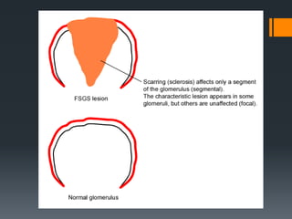
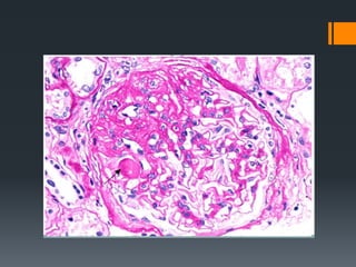
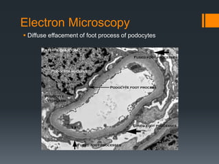
Focal Segmental
Glomerulosclerosis
Venkatesh Karthikeyan,
Velammal Medical College
Focal Segmental
Glomerulosclerosis
Focal – Less than 50% glomeruli is affected
Segmental – involving a portion of glomerular tuft
Sclerosis – abnormal hardening
Commonest cause of nephrotic sydrome
Causes
Idiopathic
Secondary
Loss of renal tissue (unilateral renal agenesis,
advanced stages of reflux nephropathy,
hypertensive nephropathy)
Associated with conditions like sickle cell
anemia, HIV, Heroin abuse, obesity
Mutations in genes like nephrin, podocin(ARD),
alpha – actinin 4(ADD)
Hallmark feature
 Degeneration and focal disruption of visceral epithelial cells
Light Microscopy
Sclerosis (extracellular accumulation of
collagenous matrix in mesangium or capillary
loops or both)
Hyalinosis (extracellular accumulation of
homogenous and eosinophilic material in
glomeruli)
Electron Microscopy
 Diffuse effacement of foot process of podocytes
Immunofluorescence microscopy
 IgM and C3 may be seen in the sclerotic areas and in
mesangium
Clinical Features
 Non-selective proteinuria
 Reduction in GFR
 Hypertension
 Poor response to corticosteroids
 Recurrence common in transplant patients
 Children have better prognosis
Collapsing Glomerulopathy
 Variant of FSG
 Usually associated with HIV and cystinosis
 Poor prognosis
 Collapse and sclerosis of the entire glomerular tuft
Other variants of FSG
 Glomerular tip lesion variant
 Cellular variant
 Perihilar variant
 Not otherwise specific (NOS) variant
Take home points
 Hallmark of FSG is degeneration and focal disruption of
visceral epithelial cells
 Hyalinosis and sclerosis seen
 Commonest cause for nephrotic syndrome
 Collapsing glomerulopathy is a variant of FSG seen in HIV
patients
 No Immunoglobulin or complement deposits seen on
immunofluorescence microscopy in Minimal change disease
unlike FSG
Thank you 
Dr Richard Bright
Father of Nephrology
Dr KS Chugh
Father of Nephrology in India

Focal Segmental Glomerulosclerosis - Pathology - FSGS

Editor's Notes

  • #2 Rudolf virchow, richard bright
  • #7  – representing leaked plasma proteins due to endothelial or capillary wall injury – hyalinization and sclerosis occludes capillary lumen and forms fibrous adhesion between sclerotic portions of glomoeruli and nearby parietal epithelium
  • #9 Thinning or shortening
  • #12 Lysosomal storage disorder characterised by abnormal accumulation of cystine