Introduction to Renal Pathology
•Download as PPT, PDF•
69 likes•17,830 views

This document provides guidance on sampling, allocating, and fixing renal biopsy tissue for light microscopy, immunofluorescence, and electron microscopy evaluation. It discusses obtaining adequate sample sizes from different renal regions and dividing tissue between fixatives. Common stains used for light microscopy like H&E, PAS, trichrome, and silver stains are also described. Normal renal histology of the glomerulus, interstitium, tubules, and vessels is outlined.

Recommended
More Related Content
What's hot
What's hot (20)
Viewers also liked
Similar to Introduction to Renal Pathology
Similar to Introduction to Renal Pathology (20)
More from edwinchowyw
More from edwinchowyw (20)
Recently uploaded
Recently uploaded (20)
Introduction to Renal Pathology
- 2. Tissue Sampling, Allocation, and Fixative
- 7. Stains
- 18. Approach-
- 26. Normal Histology
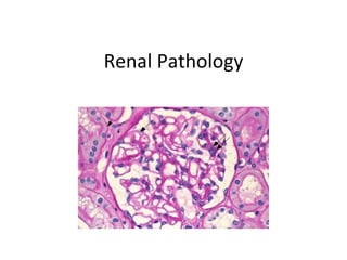
- 30. Image of a lobule of the glomerular tuft: red arrows indicate several mesangial areas in which there are 1 or 2 nuclei. Green arrows indicate nuclei of endothelial cells (Masson’s trichrome, X400).
- 32. The mesangial matrix also stains with the PAS, like the basal membranes, due to the affinity of PAS by type IV collagen (PAS, X300).
- 34. The GBM is seen perfectly smooth, without perpendicular irregularities nor projections (red arrows). The flat cytoplasm of the visceral epithelial cell can be seen; and in some points, it is possible also to see the cytoplasm of the endothelial cells. The nucleus of a podocyte appears pointed with the green arrow. The nuclei of the endothelial cells usually are found towards the mesangial portion of the capillary (blue arrow) (Methenamine-silver)
- 38. JGA: The yellow arrows indicate the macula densa, see the apical nuclei. Almost in contact with macula densa cells is the extraglomerular mesangium indicated with the black arrows. The green arrow marks the efferent arteriole and the blue arrow the afferent arteriole. The Peripolar cells are located exactly in the angle in which parietal epithelium contacts visceral epithelium (H&E, X.400).
- 44. Nerves accompanying the vessels
- 48. Brush border of the proximal tubules has affinity by the reagents used in the periodic acid of Schiff coloration (arrows). (PAS, X200).
- 49. The silver stain emphasizes the basement membranes of tubules and it allows us to delineate the contours very well. The prominent eosinophilic cytoplasm of the proximal tubules contrasts with the clearer and less abundant cytoplasm of the distal tubules (asterisks). (Methenamine-silver stain, X200).
- 54. Vessels
- 59. The arcuate arteries run by the interstitial space between the cortex and medulla, and they are accompanied by lymphatic vessels, nerves and veins (asterisk) (H&E, X100).
- 60. The cortical radial or interlobular arteries are branches of the arcuate and originate the afferent arterioles. Usually they have, depending of the thickness of their wall, several layers of muscular cells (H&E, X300).