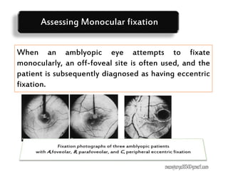
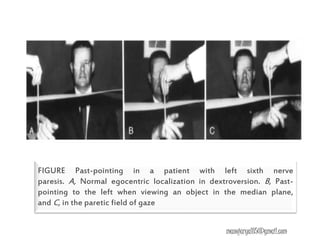
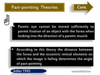
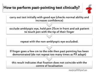
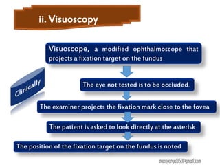
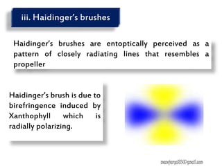
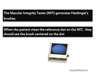
Monocular fixation -Past pointing test,
Visuoscopy, Haidinger’s brush, Fixation
disparity, After image transfer test
Manoj Aryal B. Optometry
manojaryal85@gmail.com
Past pointing Visuoscopy
Haidinger’s brush Fixation disparity
After image transfer test
Monocular fixation testing methods
Overview of diagnostic evaluation of strabismus
Diagnostic test sequences
Presentation layout
manojaryal85@gmail.com
Overview of
Diagnostic
evaluation of
strabismus
Management
plan
Case H/O
Diagnostic
summery
Diagnostic
test
sequences
manojaryal85@gmail.com
Refraction
MONOCULAR FIXATION
Visual acuity
Deviation variables Correspondence
Sensorimotor fusion
Visual efficiency
Diagnostic test
sequences
manojaryal85@gmail.com
A failure of an eye in monocular vision to take up
fixation with the fovea, but with some other point.
This hardly occurs except in clinical conditions as the
patient is generally not fixing with that eye anyway.
It is only shown when the better eye is covered
(Exception = microtropia with identity)
Eccentric Fixation
manojaryal85@gmail.com
Four Theories as to the cause of Eccentric
Fixation
Anomalous correspondence theory
(Chavasse, 1939, Cuppers, 1956)
Suppression Theory (Worth, 1906, Bangerter,1953)
Motor theory (Schor, 1978)
Pickwell (1981)
manojaryal85@gmail.com
1. Suppression Theory
Occurs when central acuity has dropped to a level
below that of the surrounding area, so that better
acuity results.
Now thought to be unlikely as foveal VA still seems
to be better than in the rest of the retina.
manojaryal85@gmail.com
2. Anomalous Correspondence theory
A change in the central area of localisation
resulting from a central scotoma in the
amblyopic eye
Major problem with this theory is that the
angle of anomaly is usually much greater than
angle of EF
EF secondary to the development of ARC
manojaryal85@gmail.com
Failure of the EOM to relax from the deviation (in
strabismus) = MUSCLE POTENTIATION.
This is a likely cause as habitual strabismic deviation
causes an adaptive after-effect which modifies the
subsequent monocular localisation
3. Motor theory
manojaryal85@gmail.com
4. Pickwell (1981)
A sequel to an enlargement of Panum’s fusional
area following decompensated heterophoria at an
early age – eventually leads to microtropia – a loss
of accurate correspondence
manojaryal85@gmail.com
Presence of EF will account for :
A portion of the patients reduced visual acuity
Can contaminate a number of other test
results, such as
the magnitude of deviation, and
the angle of anomaly in correspondence
testing.
May mask or fool the practitioner when
evaluating the patient for strabismus and
manojaryal85@gmail.com
When an amblyopic eye attempts to fixate
monocularly, an off-foveal site is often used, and the
patient is subsequently diagnosed as having eccentric
fixation.
Assessing Monocular fixation
manojaryal85@gmail.com
Location Noted by identifying what portion of the
retina is being used to fixate: nasal,
temporal, superior, or inferior retina.
There may be combination of both
horizontal and vertical components
Direction of the EF is usually in the direction
of the presenting strabismus
manojaryal85@gmail.com
Magnitude of EF is evaluated by:
 Using the calibrated target in the direct
ophthalmoscope
 By determining the distance between a perceived
entoptic phenomenon and a fixation test point
Weymouth and Flom (1961)
Discovered the formula for predicting visual acuity
from known EF.
manojaryal85@gmail.com
•Most EF is 3 prism diopter or less
•When magnitude of EF equals the magnitude of the
strabismus, no movement will be seen on the unilateral
cover test and the strabismus may be missed.
Predicting visual acuity from known EF
MAR=E(pd) +1
where,
Nasal EF =+E
Temporal EF=-E
manojaryal85@gmail.com
Tests for Monocular
Fixation
manojaryal85@gmail.com
ANOMALIES OF EGOCENTRIC LOCALIZATION
were first described by Von Graefel in patients with
recent paralyses of the extra ocular muscles and are
referred to as
"past-pointing" or "false orientation."
It occurs in recent paresis when affected eye is
looking monocularly in the direction of action of
paretic muscle
Past-pointing test
manojaryal85@gmail.com
Not found in muscle restriction and nonparetic
incomitancy
Misjudging the location of an object and pointing
too far in the same direction in which the object
was displaced
E.g.-in case of LLR palsy,px points a finger to see
object to left located further than its normal
position.
Past-pointing test Cont.
manojaryal85@gmail.com
FIGURE Past-pointing in a patient with left sixth nerve
paresis. A, Normal egocentric localization in dextroversion. B, Past-
pointing to the left when viewing an object in the median plane,
and C, in the paretic field of gaze
manojaryal85@gmail.com
Past-pointing Theories
Egocentric localization of visual objects in
subjective space is almost accurate as
long as there is proper correlation
between ocular innervation and its effect,
that is, as long as the amount of actual
eye movement corresponds to what is
intended
(Von Helmholtz,Bielschowsky, and Hofmann)
manojaryal85@gmail.com
 Paretic eye cannot be moved sufficiently to
permit fixation of an object with the fovea when
looking into the direction of a paretic muscle
 According to this theory the distance between
the fovea and the eccentric retinal elements on
which the image is falling determines the angle
of past-pointing
Past-pointing Theories
Adler 1945
Cont.
manojaryal85@gmail.com
Past-pointing Theories
Is based on the discovery of sensory receptors in the
extra ocular muscles.
There is proprioceptive feedback from the extra
ocular muscles to the central nervous system that
influences egocentric localization of objects
Cont.
manojaryal85@gmail.com
Thus, past-pointing is felt to be caused by a disparity
between the information received by the brain from
the extra ocular muscles and the amount of motor
impulses required to produce adequate movement
into the field of action of a paretic muscle
manojaryal85@gmail.com
How to perform past-pointing test clinically?
this result indicates that fixation does not coincide with the
centre of localisation
If finger goes a few cm to the side then past pointing has been
demonstrated (do not repeat too many times as PX adapt)
repeat with the non-amblyopic eye occluded.
occlude amblyopic eye, hold pen 25cm in front and ask patient
to touch pen with the tip of their finger
carry out test initially with good eye (checks normal ability and
increases confidence)
manojaryal85@gmail.com
ii. Visuoscopy
Visuoscope, a modified ophthalmoscope that
projects a fixation target on the fundus
The examiner projects the fixation mark close to the fovea
The eye not tested is to be occluded.
The patient is asked to look directly at the asterisk
The position of the fixation target on the fundus is noted
manojaryal85@gmail.com
Poor patient cooperation
Lack of distinct foveal reflex
manojaryal85@gmail.com
Haidinger’s brushes are entoptically perceived as a
pattern of closely radiating lines that resembles a
propeller
Haidinger’s brush is due to
birefringence induced by
Xanthophyll which is
radially polarizing.
iii. Haidinger’s brushes
manojaryal85@gmail.com
Haidinger’s brushes can be used to detect macular
edema.
The effect is less pronounced or absent in macular
edema. This can occur even before ophthalmoscopic
signs of macular edema.
Because Haidinger's brush corresponds to the macula, it
is sometimes used as a gross subjective test of macular
function.
Haidinger’s brushes Cont.
manojaryal85@gmail.com
Haidinger’s brush can determine whether amblyopic
patients fixate with their fovea or not (eccentric fixation)
since the fovea always corresponds to the center of the
hourglass and the center of rotation
Haidinger’s brushes Cont.
manojaryal85@gmail.com
Pointing task with HB. Patient is
instructed to align the point P with HB
superimposed on the fovea.
A-central fixation with brush, pointer
and fixation point aligned
B-eccentric fixation with primary
visual direction at the eccentric point
When the patient views the reference dot on the MIT, they
should see the brush centered on the dot
The Macular Integrity Tester (MIT) generates Haidinger’s
brushes.
manojaryal85@gmail.com
The practitioner can then measure the distance
between these two points and convert the millimeter
displacement into prism diopter, based on the testing
distance
In eccentric fixation, this is not the case because a
retinal locus other than the fovea is used for fixation
The direction and magnitude of the EF can be assessed
by asking the patient to report the location of the
propeller in relationship to the fixation point.
manojaryal85@gmail.com
Maxwell's spot. A round, dark-purplish spot
perceived when the eye is exposed to a homogenous
blue or purple field alternately with achromatic light,
can be used to evaluate the magnitude and direction
of EF, similarly to haidinger’s brush
Used in vision therapy to “tag” where the patient is
fixating
Can also be used to measure the density of macular
pigment. The darker Maxwell’s spot, the denser the
pigment
manojaryal85@gmail.com
Visual sensation persisting after the original
stimulus has been removed
Once created in each eye, their position in relation
to each other persists in an open or closed eye
conditions
Subjective test to determine and detect the
presence or absence of ARC
After Image Tests
manojaryal85@gmail.com
Introduction
Positive after image-which appear the same as the
original stimulus
Negative after image-in which the light areas of the
original stimulus appear dark
manojaryal85@gmail.com
Introduction Cont.
After Image Tests
After Image Tests
Principle
To produce after images by presenting a bright light
to each eye in turn.
The images can be obtained by using special slides in
the major amblyoscope or with a hand-held flash
apparatus.
manojaryal85@gmail.com
The apparatus consists of a linear light with central
black band within a circular black background
mounted on the flash apparatus.
The lines can be presented horizontally or vertically
The apparatus is flashed while the patient fixates the
black band
After Image Tests
Principle
manojaryal85@gmail.com
Cont.
After Image Tests
Procedures
First presented horizontally to the eye with better VA
and then vertically to the poorer eye for 15-20
seconds in a darkened room
Each eye fixates on the central black mark of a
glowing filament
manojaryal85@gmail.com
Looking the after image of two filaments on a
uniform surface(wall),px. sees a cross which shows
NRC, or the 2 filaments will be separated indicating
ARC.
Fellow eye is occluded.
Cont.
After Image Tests
Procedures
manojaryal85@gmail.com
Cont.
After Image Tests
Procedures
Alternatively the position of positive after image
can be assessed in dark
The patient looks at a black wall in normal
illumination and should see two negative linear
after images, each with a central gap corresponding
to the fixation and therefore representing the visual
direction of fovea.
manojaryal85@gmail.com
Px. indicates the relative position of two gaps in
the center of each after image
The gaps correspond to the visual direction of
each fovea if central fixation is present
Interpretation depends on the fixation behavior
Cont.
After Image Tests
Procedures
manojaryal85@gmail.com
Interpretation with central fixation
First assess with visuoscope
In px with NRC, two fovea have a common visual
direction, sees the gap superimposed
After Image Tests
manojaryal85@gmail.com
Px. with ARC and right exotropia ,vertical after
image is displaced to the right
manojaryal85@gmail.com
In ARC, two fovea no longer have a common visual
direction.Px with right esotropia sees vertical after
image displaced to the left
Interpretation with Eccentric fixation
After Image Tests
Asterisk lies between fovea and disc with
visuoscopy
With NRC(rare in px. with EF)or when the angle of
anomaly is not identical with the degree of
eccentricity, the after image in right eye will be
localized uncrossed to right
manojaryal85@gmail.com
In small angle strabismus, px may employ the same
extra-foveal retinal element for fixation that forms
a common visual direction with fovea of sound eye
under binocular conditions (ARC)
When degree of eccentricity of fixation is equal to
angle of anomaly, superimposition of gaps occur
indicating ARC.
Interpretation with Eccentric fixation Cont.
manojaryal85@gmail.com
Indications for use
After Image Tests
The test is difficult for the patient to perform and to
interpret, which limits it’s application
Testing conditions are entirely artificial and
abnormal retinal correspondence is only diagnosed if
it is well established
Other tests have largely superseded this test in
routine clinical examination.
manojaryal85@gmail.com
Fixation Disparity Test
The underconvergence or overconvergence with
respect to the plane of regard called fixation
disparity is measured in minutes of arc and
depends on the size of Panum’s fusional area.
Fixation disparity (FD) is the difference between
the convergence angle under binocular viewing
and the angle subtended by the target at the
centers of rotation.
OR
manojaryal85@gmail.com
It is a failure of the two visual axes to
simultaneously intersect the object of regard
during attempted binocular fixation.
It is also referred to as a micro strabismus because
the eyes are not binocularly aligned during
attempted fusion.
Retinal slip is another synonym
manojaryal85@gmail.com
The main difference between a strabismus and FD is
that the patient is not fusing with a strabismus but are
with FD.
Fixation disparity is the residual error of a partially
corrected phoria.
 Usually a large phoria underlies a small fixation
disparity
manojaryal85@gmail.com
Fixation disparity in which
the eyes underconverse in
relation to plane of regard
is called exo fixation
disparity, whereas fixation
disparity in which the eyes
overconverse Is called eso
fixation disparity
Fig. Exo FD
Fig. Eso FD manojaryal85@gmail.com
Components of fixation disparity curve
The horizontal axis intercept(associated
phoria)
The vertical axis intercept(FD amount)
Slope at the vertical axis intercept
The center of symmetry (inflection point)
Shape/type of curve type
manojaryal85@gmail.com
The horizontal axis intercept(Associated Phoria)
manojaryal85@gmail.com
The vertical axis intercept(FD amount)
manojaryal85@gmail.com
Slope at the vertical axis intercept
Steep fixation disparity curve
Indicates:
SVA mechanism that is too
slow or that has too little gain
given the stimulus time
constraint
slope - measured between 3 base-in & 3 base-out
Slope at the vertical axis intercept
Cont.
Flat fixation disparity curve
Indicates as a pair the disparity
vergence controller and the
SVA mechanism are operating
in comfortable ranges with
regard to both amplification
ability and speed
manojaryal85@gmail.com
The center of symmetry
(inflection point)
Signal the passage of the disparity vergence system from control
of the crossed disparity detector system to the uncrossed
disparity detector system(or vice versa)
It moves up & down(vertically) according to need for a
maintenance level of innervation( fixation disparity)
Moves side to
side(horizontally) according
to the direction and amount
of phoria and state of
adaptation.
Shape/type of curve type
Clinically identical(most common) curve
Consist of relatively flat
central portion with an
upsweep on the left and
down sweep on the
right
The central portion of
the curve owes it’s
flatness to the action of
the SVA mechanism
The flatness of the central portion is very much dependent on
the manner in which curve is generated
Faster the curve generated- the steeper the curve
When they are symptomatic the slope of FDC is typically steeper
than the average.
Usually asymptomatic.
Slope Value of approx. 1’ of arc per  discriminate b/w
symptomatic & asymptomatic Pts.
Pts. With steeper slopes respond well to Orthoptics when they
are symptomatic
The cortical images from the two eyes
increasingly flip and the fusion of these images
become increasingly less exact as the prism
power increases, finally diplopia occurs
manojaryal85@gmail.com
Fixation disparity
changed little over a
wide range of prism
power, then suddenly
made a large change
at one or both of the
extremes
manojaryal85@gmail.com
Most type II occur with Esophoria.
Respond better to prisms & to lens adds than to
Orthoptics.
An irregular FDC may be indicative of an
accommodative problem.
manojaryal85@gmail.com
Can also be managed
with Prism
Can be trained with Orthoptics but not as easily as Type I
Most often found in high
Exophoria
Fixation disparity
changed with forced
vergence only within a
narrow and outside the
interval levelled off at
nearly constant values
Indicating poorly developed binocular coordination
manojaryal85@gmail.com
Methods for measurement of
Fixation Disparity
Mallett FD test
It was designed not to measure FD but not determine
the amount of prism necessary to eliminate the FD and
is therefore an associated phoria test
Two FD units(mallet boxes) are available
A wall mounted unit for d/s testing
A hand held unit for near testing
manojaryal85@gmail.com
Both near and distance FD units incorporate a foveal
fusion lock as well as a peripheral fusion lock: the only
monocularly seen stimuli are in the para-foveal area
Both units required polaroid filter in front of the patient
eye
Illumination:
Strong enough to allow for the filters
manojaryal85@gmail.com
Is a self contained,
internally illuminated unit
The letters OXO are seen
by both eyes and
constitute a gross foveal
fusion lock
Two red strips are provided one above and one below
the X , are polarized so one strop is seen with each eye
Used to measure lateral associated phoria as well as
the vertical associated phoria
The letter X subtend an angle of 10min of arc at d/s of
6m(20/40)
Also presents OXO letters
Letter X subtends 18 min
of arc at 35 cm distance
Instead red, green stripes are provided
manojaryal85@gmail.com
The patient is told to concentrate on the X in the
middle of OXO and is asked if he/she can
simultaneously see two red for distance, two green
strips for near
manojaryal85@gmail.com
If top strip is directly above bottom strip i.e. if no
displacement between strips-patient has no lateral
phoria or has a compensated phoria.
If top strip (seen by LE) is to the right of bottom strip-
patient has Exo FD
manojaryal85@gmail.com
If top strip is seen to the left of bottom- Eso FD
If both strips are displaced- FD is shared between
two eyes
If only one strip is displaced:
One eye is strongly dominant( undisplaced strip belong
to dominant eye)
manojaryal85@gmail.com
The associated phoria or the
amount of plus sphere power
to be prescribed is the
smallest amount that will
bring about alignment.
Base out prism/plus sphere
can be introduced until the
strips are aligned.
Once FD Is eliminated, the
exophoria can be considered
fully compensated .
For convergence insufficiency
institute base out training, the
criterion being the elimination of
fixation disparity.
Introduce base in prism starting
with 1 prism diopter or minus
lens until two strips are aligned.
Exo –associated phoria Eso – associated phoria
manojaryal85@gmail.com
Includes two stimuli
One for vertical disparity measurement
One for horizontal disparity measurement
Each test stimuli includes a black aperture
The Disparometer
Two reduced Snellen’s charts
seen with both eyes and two
polarized Vernier lines seen
in the center of the black
aperture(each eye seeing
only one Vernier lines)
manojaryal85@gmail.com
First presents the patient with the target in
which Vernier lines have no disparity
If the patient reports that lines are not perfectly
aligned, the examiner presents stimuli having
varying amounts of disparity until the patient
reports alignment
By determining the amount of FD while forcing
vergence with base in and base out prisms
presented in steps 3 prism diopter, FD curve
can be plotted
manojaryal85@gmail.com
Other methods …
Compare reflex position in each eye in turn (other
eye occluded). The relative displacement of the
reflex by 1mm = approx. 11degrees or 20 PD
Eccentricity is not usually this great however making
EF difficult to detect by this method.
Corneal reflex test
manojaryal85@gmail.com
Bjerrum Screen
Method
In normal subjects the blind spot is the same angular
distance from fixation in both eyes.
Plot the blind spot carefully in both eyes and compare
positions
Degree of eccentricity can be measured by the
difference in angular distance of blind spot from
fixation in each eye
manojaryal85@gmail.com
Amsler Chart
Amblyopes often have
small foveal scotoma
which shows up as a
disturbance on Amsler
 Occurs centrally if
central localisation
 Eccentrically if EF
manojaryal85@gmail.com
Neutral density Filters
If a ND filter is added and no reduction in VA
occurs then EF is likely to be present
manojaryal85@gmail.com
Centricity of Fixation (central vs eccentric)
Directional bias (nasal, temporal etc.)
Quality of fixation (steady vs unsteady)
Magnitude
Pattern of fixation (drifts, saccades, nystagmus
Percent foveation (30second visuoscopy)
Zero retinomotor point
Assessment of Fixation
manojaryal85@gmail.com
Treatment of Monocular Fixation
As in amblyopia, have to encourage foveal fixation
Direct Occlusion alone may improve fixation but
often a slight eccentricity remains
Pleoptic Treatment – desensitises eccentrically
fixing area
After image transfer – use to locate foveal fixation
manojaryal85@gmail.com
Inverse occlusion
Infants with eccentric fixation who are too young
for treatment with Haidinger’s brushes
Continued as long as improvement is made
Rarely suitable in adult – eccentric fixation is too
firmly established
manojaryal85@gmail.com
Red filter
600 – 640 nm
Only stimulate cones
Patient will attempt to fix with area of the retina
which contains maximum no of cones
manojaryal85@gmail.com
Pleoptics Therapy
Pleos=full,optikos=sight
Aim is to actively stimulate macula in dense
amblyopia with eccentric fixation
Two methods,either Bangerter or Cupper
manojaryal85@gmail.com
Bangerter’s method
It was followed by intermittent stimulation of
macula with flashes of light
Performed by modified Gullstrand’s
Ophthalmoscope,called as Pleoptophore
Bangerter dazzled the extramacular retina including
the eccentric point by bright light protecting the
macula by a disc projected onto it.
manojaryal85@gmail.com
Cupper’s method
Cupper used Euthyscope which had discs of varying
sizes to create a central after image apart from
dazzling the eccentric point
He used the alternate flashing of room
illumination(Alternascope) to perpetuate the after
images(forming negative after image in light and
positive after image in dark)
patient perceives an afterimage resembling the
white doughnut
manojaryal85@gmail.com
Treat underlying amblyopia by occlusion of non-
squinting eye (if patient <6 years old).
In patients >6 years – correct refractive error,
otherwise do not treat the microtropia.
Surgery is not appropriate
manojaryal85@gmail.com
माता पिताको मुखबाट निस्के को आशिर्ाादलाई
भगर्ािले िनि टाल्ि सक्दैिि् ।
दिैँ
आयो
THA
NK
YOU

Monocular Fixation

  • 1.
    Monocular fixation -Pastpointing test, Visuoscopy, Haidinger’s brush, Fixation disparity, After image transfer test Manoj Aryal B. Optometry manojaryal85@gmail.com
  • 2.
    Past pointing Visuoscopy Haidinger’sbrush Fixation disparity After image transfer test Monocular fixation testing methods Overview of diagnostic evaluation of strabismus Diagnostic test sequences Presentation layout manojaryal85@gmail.com
  • 3.
    Overview of Diagnostic evaluation of strabismus Management plan CaseH/O Diagnostic summery Diagnostic test sequences manojaryal85@gmail.com
  • 4.
    Refraction MONOCULAR FIXATION Visual acuity Deviationvariables Correspondence Sensorimotor fusion Visual efficiency Diagnostic test sequences manojaryal85@gmail.com
  • 5.
    A failure ofan eye in monocular vision to take up fixation with the fovea, but with some other point. This hardly occurs except in clinical conditions as the patient is generally not fixing with that eye anyway. It is only shown when the better eye is covered (Exception = microtropia with identity) Eccentric Fixation manojaryal85@gmail.com
  • 6.
  • 7.
    Four Theories asto the cause of Eccentric Fixation Anomalous correspondence theory (Chavasse, 1939, Cuppers, 1956) Suppression Theory (Worth, 1906, Bangerter,1953) Motor theory (Schor, 1978) Pickwell (1981) manojaryal85@gmail.com
  • 8.
    1. Suppression Theory Occurswhen central acuity has dropped to a level below that of the surrounding area, so that better acuity results. Now thought to be unlikely as foveal VA still seems to be better than in the rest of the retina. manojaryal85@gmail.com
  • 9.
    2. Anomalous Correspondencetheory A change in the central area of localisation resulting from a central scotoma in the amblyopic eye Major problem with this theory is that the angle of anomaly is usually much greater than angle of EF EF secondary to the development of ARC manojaryal85@gmail.com
  • 10.
    Failure of theEOM to relax from the deviation (in strabismus) = MUSCLE POTENTIATION. This is a likely cause as habitual strabismic deviation causes an adaptive after-effect which modifies the subsequent monocular localisation 3. Motor theory manojaryal85@gmail.com
  • 11.
    4. Pickwell (1981) Asequel to an enlargement of Panum’s fusional area following decompensated heterophoria at an early age – eventually leads to microtropia – a loss of accurate correspondence manojaryal85@gmail.com
  • 12.
    Presence of EFwill account for : A portion of the patients reduced visual acuity Can contaminate a number of other test results, such as the magnitude of deviation, and the angle of anomaly in correspondence testing. May mask or fool the practitioner when evaluating the patient for strabismus and manojaryal85@gmail.com
  • 13.
    When an amblyopiceye attempts to fixate monocularly, an off-foveal site is often used, and the patient is subsequently diagnosed as having eccentric fixation. Assessing Monocular fixation manojaryal85@gmail.com
  • 14.
  • 15.
    Location Noted byidentifying what portion of the retina is being used to fixate: nasal, temporal, superior, or inferior retina. There may be combination of both horizontal and vertical components Direction of the EF is usually in the direction of the presenting strabismus manojaryal85@gmail.com
  • 16.
    Magnitude of EFis evaluated by:  Using the calibrated target in the direct ophthalmoscope  By determining the distance between a perceived entoptic phenomenon and a fixation test point Weymouth and Flom (1961) Discovered the formula for predicting visual acuity from known EF. manojaryal85@gmail.com
  • 17.
    •Most EF is3 prism diopter or less •When magnitude of EF equals the magnitude of the strabismus, no movement will be seen on the unilateral cover test and the strabismus may be missed. Predicting visual acuity from known EF MAR=E(pd) +1 where, Nasal EF =+E Temporal EF=-E manojaryal85@gmail.com
  • 18.
  • 19.
    ANOMALIES OF EGOCENTRICLOCALIZATION were first described by Von Graefel in patients with recent paralyses of the extra ocular muscles and are referred to as "past-pointing" or "false orientation." It occurs in recent paresis when affected eye is looking monocularly in the direction of action of paretic muscle Past-pointing test manojaryal85@gmail.com
  • 20.
    Not found inmuscle restriction and nonparetic incomitancy Misjudging the location of an object and pointing too far in the same direction in which the object was displaced E.g.-in case of LLR palsy,px points a finger to see object to left located further than its normal position. Past-pointing test Cont. manojaryal85@gmail.com
  • 21.
    FIGURE Past-pointing ina patient with left sixth nerve paresis. A, Normal egocentric localization in dextroversion. B, Past- pointing to the left when viewing an object in the median plane, and C, in the paretic field of gaze manojaryal85@gmail.com
  • 22.
    Past-pointing Theories Egocentric localizationof visual objects in subjective space is almost accurate as long as there is proper correlation between ocular innervation and its effect, that is, as long as the amount of actual eye movement corresponds to what is intended (Von Helmholtz,Bielschowsky, and Hofmann) manojaryal85@gmail.com
  • 23.
     Paretic eyecannot be moved sufficiently to permit fixation of an object with the fovea when looking into the direction of a paretic muscle  According to this theory the distance between the fovea and the eccentric retinal elements on which the image is falling determines the angle of past-pointing Past-pointing Theories Adler 1945 Cont. manojaryal85@gmail.com
  • 24.
    Past-pointing Theories Is basedon the discovery of sensory receptors in the extra ocular muscles. There is proprioceptive feedback from the extra ocular muscles to the central nervous system that influences egocentric localization of objects Cont. manojaryal85@gmail.com
  • 25.
    Thus, past-pointing isfelt to be caused by a disparity between the information received by the brain from the extra ocular muscles and the amount of motor impulses required to produce adequate movement into the field of action of a paretic muscle manojaryal85@gmail.com
  • 26.
    How to performpast-pointing test clinically? this result indicates that fixation does not coincide with the centre of localisation If finger goes a few cm to the side then past pointing has been demonstrated (do not repeat too many times as PX adapt) repeat with the non-amblyopic eye occluded. occlude amblyopic eye, hold pen 25cm in front and ask patient to touch pen with the tip of their finger carry out test initially with good eye (checks normal ability and increases confidence) manojaryal85@gmail.com
  • 27.
    ii. Visuoscopy Visuoscope, amodified ophthalmoscope that projects a fixation target on the fundus The examiner projects the fixation mark close to the fovea The eye not tested is to be occluded. The patient is asked to look directly at the asterisk The position of the fixation target on the fundus is noted manojaryal85@gmail.com
  • 28.
    Poor patient cooperation Lackof distinct foveal reflex manojaryal85@gmail.com
  • 29.
    Haidinger’s brushes areentoptically perceived as a pattern of closely radiating lines that resembles a propeller Haidinger’s brush is due to birefringence induced by Xanthophyll which is radially polarizing. iii. Haidinger’s brushes manojaryal85@gmail.com
  • 30.
    Haidinger’s brushes canbe used to detect macular edema. The effect is less pronounced or absent in macular edema. This can occur even before ophthalmoscopic signs of macular edema. Because Haidinger's brush corresponds to the macula, it is sometimes used as a gross subjective test of macular function. Haidinger’s brushes Cont. manojaryal85@gmail.com
  • 31.
    Haidinger’s brush candetermine whether amblyopic patients fixate with their fovea or not (eccentric fixation) since the fovea always corresponds to the center of the hourglass and the center of rotation Haidinger’s brushes Cont. manojaryal85@gmail.com Pointing task with HB. Patient is instructed to align the point P with HB superimposed on the fovea. A-central fixation with brush, pointer and fixation point aligned B-eccentric fixation with primary visual direction at the eccentric point
  • 32.
    When the patientviews the reference dot on the MIT, they should see the brush centered on the dot The Macular Integrity Tester (MIT) generates Haidinger’s brushes. manojaryal85@gmail.com
  • 33.
    The practitioner canthen measure the distance between these two points and convert the millimeter displacement into prism diopter, based on the testing distance In eccentric fixation, this is not the case because a retinal locus other than the fovea is used for fixation The direction and magnitude of the EF can be assessed by asking the patient to report the location of the propeller in relationship to the fixation point. manojaryal85@gmail.com
  • 34.
    Maxwell's spot. Around, dark-purplish spot perceived when the eye is exposed to a homogenous blue or purple field alternately with achromatic light, can be used to evaluate the magnitude and direction of EF, similarly to haidinger’s brush Used in vision therapy to “tag” where the patient is fixating Can also be used to measure the density of macular pigment. The darker Maxwell’s spot, the denser the pigment manojaryal85@gmail.com
  • 35.
  • 36.
    Visual sensation persistingafter the original stimulus has been removed Once created in each eye, their position in relation to each other persists in an open or closed eye conditions Subjective test to determine and detect the presence or absence of ARC After Image Tests manojaryal85@gmail.com Introduction
  • 37.
    Positive after image-whichappear the same as the original stimulus Negative after image-in which the light areas of the original stimulus appear dark manojaryal85@gmail.com Introduction Cont. After Image Tests
  • 38.
    After Image Tests Principle Toproduce after images by presenting a bright light to each eye in turn. The images can be obtained by using special slides in the major amblyoscope or with a hand-held flash apparatus. manojaryal85@gmail.com
  • 39.
    The apparatus consistsof a linear light with central black band within a circular black background mounted on the flash apparatus. The lines can be presented horizontally or vertically The apparatus is flashed while the patient fixates the black band After Image Tests Principle manojaryal85@gmail.com Cont.
  • 40.
    After Image Tests Procedures Firstpresented horizontally to the eye with better VA and then vertically to the poorer eye for 15-20 seconds in a darkened room Each eye fixates on the central black mark of a glowing filament manojaryal85@gmail.com
  • 41.
    Looking the afterimage of two filaments on a uniform surface(wall),px. sees a cross which shows NRC, or the 2 filaments will be separated indicating ARC. Fellow eye is occluded. Cont. After Image Tests Procedures manojaryal85@gmail.com
  • 42.
    Cont. After Image Tests Procedures Alternativelythe position of positive after image can be assessed in dark The patient looks at a black wall in normal illumination and should see two negative linear after images, each with a central gap corresponding to the fixation and therefore representing the visual direction of fovea. manojaryal85@gmail.com
  • 43.
    Px. indicates therelative position of two gaps in the center of each after image The gaps correspond to the visual direction of each fovea if central fixation is present Interpretation depends on the fixation behavior Cont. After Image Tests Procedures manojaryal85@gmail.com
  • 44.
    Interpretation with centralfixation First assess with visuoscope In px with NRC, two fovea have a common visual direction, sees the gap superimposed After Image Tests manojaryal85@gmail.com
  • 45.
    Px. with ARCand right exotropia ,vertical after image is displaced to the right manojaryal85@gmail.com In ARC, two fovea no longer have a common visual direction.Px with right esotropia sees vertical after image displaced to the left
  • 46.
    Interpretation with Eccentricfixation After Image Tests Asterisk lies between fovea and disc with visuoscopy With NRC(rare in px. with EF)or when the angle of anomaly is not identical with the degree of eccentricity, the after image in right eye will be localized uncrossed to right manojaryal85@gmail.com
  • 47.
    In small anglestrabismus, px may employ the same extra-foveal retinal element for fixation that forms a common visual direction with fovea of sound eye under binocular conditions (ARC) When degree of eccentricity of fixation is equal to angle of anomaly, superimposition of gaps occur indicating ARC. Interpretation with Eccentric fixation Cont. manojaryal85@gmail.com
  • 48.
    Indications for use AfterImage Tests The test is difficult for the patient to perform and to interpret, which limits it’s application Testing conditions are entirely artificial and abnormal retinal correspondence is only diagnosed if it is well established Other tests have largely superseded this test in routine clinical examination. manojaryal85@gmail.com
  • 49.
    Fixation Disparity Test Theunderconvergence or overconvergence with respect to the plane of regard called fixation disparity is measured in minutes of arc and depends on the size of Panum’s fusional area. Fixation disparity (FD) is the difference between the convergence angle under binocular viewing and the angle subtended by the target at the centers of rotation. OR manojaryal85@gmail.com
  • 50.
    It is afailure of the two visual axes to simultaneously intersect the object of regard during attempted binocular fixation. It is also referred to as a micro strabismus because the eyes are not binocularly aligned during attempted fusion. Retinal slip is another synonym manojaryal85@gmail.com
  • 51.
    The main differencebetween a strabismus and FD is that the patient is not fusing with a strabismus but are with FD. Fixation disparity is the residual error of a partially corrected phoria.  Usually a large phoria underlies a small fixation disparity manojaryal85@gmail.com
  • 52.
    Fixation disparity inwhich the eyes underconverse in relation to plane of regard is called exo fixation disparity, whereas fixation disparity in which the eyes overconverse Is called eso fixation disparity Fig. Exo FD Fig. Eso FD manojaryal85@gmail.com
  • 53.
    Components of fixationdisparity curve The horizontal axis intercept(associated phoria) The vertical axis intercept(FD amount) Slope at the vertical axis intercept The center of symmetry (inflection point) Shape/type of curve type manojaryal85@gmail.com
  • 54.
    The horizontal axisintercept(Associated Phoria) manojaryal85@gmail.com
  • 55.
    The vertical axisintercept(FD amount) manojaryal85@gmail.com
  • 56.
    Slope at thevertical axis intercept Steep fixation disparity curve Indicates: SVA mechanism that is too slow or that has too little gain given the stimulus time constraint slope - measured between 3 base-in & 3 base-out
  • 57.
    Slope at thevertical axis intercept Cont. Flat fixation disparity curve Indicates as a pair the disparity vergence controller and the SVA mechanism are operating in comfortable ranges with regard to both amplification ability and speed manojaryal85@gmail.com
  • 58.
    The center ofsymmetry (inflection point) Signal the passage of the disparity vergence system from control of the crossed disparity detector system to the uncrossed disparity detector system(or vice versa) It moves up & down(vertically) according to need for a maintenance level of innervation( fixation disparity) Moves side to side(horizontally) according to the direction and amount of phoria and state of adaptation.
  • 59.
    Shape/type of curvetype Clinically identical(most common) curve Consist of relatively flat central portion with an upsweep on the left and down sweep on the right The central portion of the curve owes it’s flatness to the action of the SVA mechanism
  • 60.
    The flatness ofthe central portion is very much dependent on the manner in which curve is generated Faster the curve generated- the steeper the curve When they are symptomatic the slope of FDC is typically steeper than the average. Usually asymptomatic. Slope Value of approx. 1’ of arc per  discriminate b/w symptomatic & asymptomatic Pts. Pts. With steeper slopes respond well to Orthoptics when they are symptomatic
  • 61.
    The cortical imagesfrom the two eyes increasingly flip and the fusion of these images become increasingly less exact as the prism power increases, finally diplopia occurs manojaryal85@gmail.com
  • 62.
    Fixation disparity changed littleover a wide range of prism power, then suddenly made a large change at one or both of the extremes manojaryal85@gmail.com
  • 63.
    Most type IIoccur with Esophoria. Respond better to prisms & to lens adds than to Orthoptics. An irregular FDC may be indicative of an accommodative problem. manojaryal85@gmail.com
  • 64.
    manojaryal85@gmail.com Can also bemanaged with Prism Can be trained with Orthoptics but not as easily as Type I Most often found in high Exophoria
  • 65.
    Fixation disparity changed withforced vergence only within a narrow and outside the interval levelled off at nearly constant values Indicating poorly developed binocular coordination manojaryal85@gmail.com
  • 66.
    Methods for measurementof Fixation Disparity Mallett FD test It was designed not to measure FD but not determine the amount of prism necessary to eliminate the FD and is therefore an associated phoria test Two FD units(mallet boxes) are available A wall mounted unit for d/s testing A hand held unit for near testing manojaryal85@gmail.com
  • 67.
    Both near anddistance FD units incorporate a foveal fusion lock as well as a peripheral fusion lock: the only monocularly seen stimuli are in the para-foveal area Both units required polaroid filter in front of the patient eye Illumination: Strong enough to allow for the filters manojaryal85@gmail.com
  • 68.
    Is a selfcontained, internally illuminated unit The letters OXO are seen by both eyes and constitute a gross foveal fusion lock
  • 69.
    Two red stripsare provided one above and one below the X , are polarized so one strop is seen with each eye Used to measure lateral associated phoria as well as the vertical associated phoria The letter X subtend an angle of 10min of arc at d/s of 6m(20/40)
  • 70.
    Also presents OXOletters Letter X subtends 18 min of arc at 35 cm distance Instead red, green stripes are provided manojaryal85@gmail.com
  • 71.
    The patient istold to concentrate on the X in the middle of OXO and is asked if he/she can simultaneously see two red for distance, two green strips for near manojaryal85@gmail.com
  • 72.
    If top stripis directly above bottom strip i.e. if no displacement between strips-patient has no lateral phoria or has a compensated phoria. If top strip (seen by LE) is to the right of bottom strip- patient has Exo FD manojaryal85@gmail.com
  • 73.
    If top stripis seen to the left of bottom- Eso FD If both strips are displaced- FD is shared between two eyes If only one strip is displaced: One eye is strongly dominant( undisplaced strip belong to dominant eye) manojaryal85@gmail.com
  • 74.
    The associated phoriaor the amount of plus sphere power to be prescribed is the smallest amount that will bring about alignment. Base out prism/plus sphere can be introduced until the strips are aligned. Once FD Is eliminated, the exophoria can be considered fully compensated . For convergence insufficiency institute base out training, the criterion being the elimination of fixation disparity. Introduce base in prism starting with 1 prism diopter or minus lens until two strips are aligned. Exo –associated phoria Eso – associated phoria manojaryal85@gmail.com
  • 75.
    Includes two stimuli Onefor vertical disparity measurement One for horizontal disparity measurement Each test stimuli includes a black aperture The Disparometer Two reduced Snellen’s charts seen with both eyes and two polarized Vernier lines seen in the center of the black aperture(each eye seeing only one Vernier lines) manojaryal85@gmail.com
  • 76.
    First presents thepatient with the target in which Vernier lines have no disparity If the patient reports that lines are not perfectly aligned, the examiner presents stimuli having varying amounts of disparity until the patient reports alignment By determining the amount of FD while forcing vergence with base in and base out prisms presented in steps 3 prism diopter, FD curve can be plotted manojaryal85@gmail.com
  • 77.
    Other methods … Comparereflex position in each eye in turn (other eye occluded). The relative displacement of the reflex by 1mm = approx. 11degrees or 20 PD Eccentricity is not usually this great however making EF difficult to detect by this method. Corneal reflex test manojaryal85@gmail.com
  • 78.
    Bjerrum Screen Method In normalsubjects the blind spot is the same angular distance from fixation in both eyes. Plot the blind spot carefully in both eyes and compare positions Degree of eccentricity can be measured by the difference in angular distance of blind spot from fixation in each eye manojaryal85@gmail.com
  • 79.
    Amsler Chart Amblyopes oftenhave small foveal scotoma which shows up as a disturbance on Amsler  Occurs centrally if central localisation  Eccentrically if EF manojaryal85@gmail.com
  • 80.
    Neutral density Filters Ifa ND filter is added and no reduction in VA occurs then EF is likely to be present manojaryal85@gmail.com
  • 81.
    Centricity of Fixation(central vs eccentric) Directional bias (nasal, temporal etc.) Quality of fixation (steady vs unsteady) Magnitude Pattern of fixation (drifts, saccades, nystagmus Percent foveation (30second visuoscopy) Zero retinomotor point Assessment of Fixation manojaryal85@gmail.com
  • 82.
    Treatment of MonocularFixation As in amblyopia, have to encourage foveal fixation Direct Occlusion alone may improve fixation but often a slight eccentricity remains Pleoptic Treatment – desensitises eccentrically fixing area After image transfer – use to locate foveal fixation manojaryal85@gmail.com
  • 83.
    Inverse occlusion Infants witheccentric fixation who are too young for treatment with Haidinger’s brushes Continued as long as improvement is made Rarely suitable in adult – eccentric fixation is too firmly established manojaryal85@gmail.com
  • 84.
    Red filter 600 –640 nm Only stimulate cones Patient will attempt to fix with area of the retina which contains maximum no of cones manojaryal85@gmail.com
  • 85.
    Pleoptics Therapy Pleos=full,optikos=sight Aim isto actively stimulate macula in dense amblyopia with eccentric fixation Two methods,either Bangerter or Cupper manojaryal85@gmail.com
  • 86.
    Bangerter’s method It wasfollowed by intermittent stimulation of macula with flashes of light Performed by modified Gullstrand’s Ophthalmoscope,called as Pleoptophore Bangerter dazzled the extramacular retina including the eccentric point by bright light protecting the macula by a disc projected onto it. manojaryal85@gmail.com
  • 87.
    Cupper’s method Cupper usedEuthyscope which had discs of varying sizes to create a central after image apart from dazzling the eccentric point He used the alternate flashing of room illumination(Alternascope) to perpetuate the after images(forming negative after image in light and positive after image in dark) patient perceives an afterimage resembling the white doughnut manojaryal85@gmail.com
  • 88.
    Treat underlying amblyopiaby occlusion of non- squinting eye (if patient <6 years old). In patients >6 years – correct refractive error, otherwise do not treat the microtropia. Surgery is not appropriate manojaryal85@gmail.com
  • 90.
    माता पिताको मुखबाटनिस्के को आशिर्ाादलाई भगर्ािले िनि टाल्ि सक्दैिि् । दिैँ आयो
  • 91.

Editor's Notes

  • #20 If the patient, while his sound eye is covered, is asked to point toward an object located in the field of action of the paretic muscle, his finger will point beyond the object and towards the field of action of the paretic muscle.
  • #50 To avoid problem of rivalry or confusion the two lines are positioned side by side or one above the another .such pairs of lines are known as nonius lines
  • #89 Monofixation syndrome is a form of subnormal binocular vision without bifixation characterized by small-angle strabismus, unilateral absolute facultative central suppression scotoma of less than 3º, and peripheral fusion. While monofixation syndrome can be a primary disorder of binocular vision, it is more commonly a secondary sensory status from various primary causes