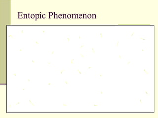
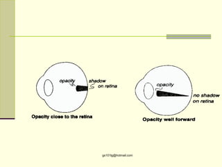
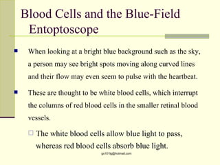
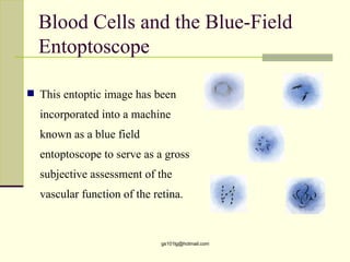
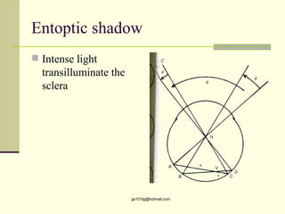
Entopic phenomena are visual sensations that originate from inside the eye. Some examples include seeing flashes of light when looking at a bright sky, which are caused by white blood cells moving through retinal blood vessels. Other entopic phenomena are produced by shadows cast on the retina from ocular structures and opacities, reflections and refractions of light within the eye, and pressure or movement of the eye. These phenomena are normal in most cases, but can sometimes indicate ocular issues like edema or retinal detachment. Understanding the various causes of entopic phenomena is important for eye health evaluations.
DIRECT DOWNLOAD LINK ❤❤https://healthkura.com/retinoscopy/❤❤
Dear viewers Check Out my other piece of works at ❤❤❤ https://healthkura.com ❤❤❤
Retinoscopy and Objective Refraction and Subjective Refraction in spherical ametropia and astigmatism
Retinoscopy (Principle & Techniques of Retinoscopy) and objective refraction, Subjective Refracition
Best presentation about retinoscopy and objective refraction techniques, and basis of subjective refraction. If you want to master the technique of retinoscopy, this presentation can be your guidance and partner in your journey to retinoscopy, objective refraction and subjective refraction.
Presentation Layout:
Retinoscope, types of retinoscope and uses of retinoscope
-Introduction to retinoscopy and objective refraction
-Retinoscopy
- In spherical ametropia
- In astigmatism
- Others: strabismus, amblyopia, pediatric pt.,
cycloplegic refraction
-Static and Dynamic Retinoscopy
-Problems seeing reflex during retinoscopy
-Errors in retinoscopy
Objective of retinoscopy and objective refraction
-To locate the far point of the eye conjugate to the retina
- Myopia or hyperopia
-Bring far point to the infinity by using appropriate lenses
- Determines amount of ametropia by retinoscopy and objective refraction
References:
-Clinical Procedures in Optometry by Eskridge, Amos and Bartlett ,
-Primary Care Optometry by Grosvenor T.,
-Borish’s Clinical Refraction by Benjamin W. J.,
-Theory And Practice Of Optics And Refraction by AK Khurana
-Retinoscopy-Student Manual by ICEE Refractive Error Training Package (2009)
-Clinical Optics and Refraction By Andrew Keirl, Caroline Christie
-Clinical Refraction Guide - A Kumar Bhootra
-Clinical Procedures in Primary Eye Care by David B. Elliott
-Internet
Follow me to get in touch with optometric and ophthalmic updates.
This slideshow talks about the History of Great Danes and gives information about the breed. During the presentation I will talk about the coat colors, characteristics, and health concerns about Great Danes and much more!
SPOTLIGHT ON THE PREMIUM CHANNEL – AcuFocusHealthegy
Presentation from OIS@ASCRS 2016
Nick Tarantino, OD, Chief Global Clinical & Regulatory Affairs Officer
Video Presentation:
https://www.youtube.com/watch?v=Nc4T9u62rBQ&list=PL1dmdBNnPTZJBhQxPOp0vdNg3s3wtN2yw&index=34
This is a slide show presentation I prepared for the Technical Support staff at Topcon Medical Systems to introduce and familiarize the art of refraction.
This presentation focuses in the most important tips in ocular physiology in a summarized manner .......you can watch the illustrated video in the following link
https://www.youtube.com/watch?v=eRbAOL37qus
Medicines Used for Glaucoma Management _Optom LectureGauriSShrestha
the most commonly prescribe treatment for glucoma is eye drops. These medicine decreases IOP to the level that prevents damage to the optic nerve by either decrease acqueous secretion from the ciliary body or facilitating acqueous drainage through the trabecular or uveoscleral outflow systems. This presentation outlines the principal eye medicine currently used in ophthalmic practice.
Ocular steroids-Dexamethasone, Betamethasone, Prednisolone and FlurometholoneGauriSShrestha
Corticosteroids are secreted by the adrenal cortex.
Adrenocorticotrophic hormone(ACTH) stimulates the adrenal cortex to produce corticosteroids.
Types of adrenal cortex-related steroids are glucocorticoids and mineralocorticoids. Corticosteroids have anti-inflammatory and immunosuppressive effects
NSAIDs are analgesic, antipyretic and anti-inflammatory drugs.
Non-narcotic, non-opioid, aspirin-like drugs
Primarily, acts on peripheral pain mechanism and do not depress the central nervous system
Some protective role against cancer and cardiovascular diseases (e.g., aspirin, antithrombotic effects)
Accounts for 5% of all drugs prescribe globally
30 million people consume NSAIDs every day
Available over the counter medicine in most countries
Ocular anti-infective agents: Antibiotics, Antivirals and antifungalsGauriSShrestha
Anti-infective agents are effective against a variety of infections (e.g., virus, rickettsiae, bacteria, fungi and protozoa) and cause competitive inhibition of a biochemical process of pathogens. Minimum inhibitory concentration (MICs) are often used for common anti-infective drugs. In the eye, route of drug administration is determined by the locus of infection. However, this ppt covers mostly topical and common antibacterial, antiviral and antifungal medicines.
Pediatric refraction is one of the challenging areas of optometry practice. I am so glad to share some of the important aspect of pediatric refraction.
New Directions in Targeted Therapeutic Approaches for Older Adults With Mantl...i3 Health
i3 Health is pleased to make the speaker slides from this activity available for use as a non-accredited self-study or teaching resource.
This slide deck presented by Dr. Kami Maddocks, Professor-Clinical in the Division of Hematology and
Associate Division Director for Ambulatory Operations
The Ohio State University Comprehensive Cancer Center, will provide insight into new directions in targeted therapeutic approaches for older adults with mantle cell lymphoma.
STATEMENT OF NEED
Mantle cell lymphoma (MCL) is a rare, aggressive B-cell non-Hodgkin lymphoma (NHL) accounting for 5% to 7% of all lymphomas. Its prognosis ranges from indolent disease that does not require treatment for years to very aggressive disease, which is associated with poor survival (Silkenstedt et al, 2021). Typically, MCL is diagnosed at advanced stage and in older patients who cannot tolerate intensive therapy (NCCN, 2022). Although recent advances have slightly increased remission rates, recurrence and relapse remain very common, leading to a median overall survival between 3 and 6 years (LLS, 2021). Though there are several effective options, progress is still needed towards establishing an accepted frontline approach for MCL (Castellino et al, 2022). Treatment selection and management of MCL are complicated by the heterogeneity of prognosis, advanced age and comorbidities of patients, and lack of an established standard approach for treatment, making it vital that clinicians be familiar with the latest research and advances in this area. In this activity chaired by Michael Wang, MD, Professor in the Department of Lymphoma & Myeloma at MD Anderson Cancer Center, expert faculty will discuss prognostic factors informing treatment, the promising results of recent trials in new therapeutic approaches, and the implications of treatment resistance in therapeutic selection for MCL.
Target Audience
Hematology/oncology fellows, attending faculty, and other health care professionals involved in the treatment of patients with mantle cell lymphoma (MCL).
Learning Objectives
1.) Identify clinical and biological prognostic factors that can guide treatment decision making for older adults with MCL
2.) Evaluate emerging data on targeted therapeutic approaches for treatment-naive and relapsed/refractory MCL and their applicability to older adults
3.) Assess mechanisms of resistance to targeted therapies for MCL and their implications for treatment selection
Factory Supply Best Quality Pmk Oil CAS 28578–16–7 PMK Powder in Stockrebeccabio
Factory Supply Best Quality Pmk Oil CAS 28578–16–7 PMK Powder in Stock
Telegram: bmksupplier
signal: +85264872720
threema: TUD4A6YC
You can contact me on Telegram or Threema
Communicate promptly and reply
Free of customs clearance, Double Clearance 100% pass delivery to USA, Canada, Spain, Germany, Netherland, Poland, Italy, Sweden, UK, Czech Republic, Australia, Mexico, Russia, Ukraine, Kazakhstan.Door to door service
Hot Selling Organic intermediates
Prix Galien International 2024 Forum ProgramLevi Shapiro
June 20, 2024, Prix Galien International and Jerusalem Ethics Forum in ROME. Detailed agenda including panels:
- ADVANCES IN CARDIOLOGY: A NEW PARADIGM IS COMING
- WOMEN’S HEALTH: FERTILITY PRESERVATION
- WHAT’S NEW IN THE TREATMENT OF INFECTIOUS,
ONCOLOGICAL AND INFLAMMATORY SKIN DISEASES?
- ARTIFICIAL INTELLIGENCE AND ETHICS
- GENE THERAPY
- BEYOND BORDERS: GLOBAL INITIATIVES FOR DEMOCRATIZING LIFE SCIENCE TECHNOLOGIES AND PROMOTING ACCESS TO HEALTHCARE
- ETHICAL CHALLENGES IN LIFE SCIENCES
- Prix Galien International Awards Ceremony
Title: Sense of Taste
Presenter: Dr. Faiza, Assistant Professor of Physiology
Qualifications:
MBBS (Best Graduate, AIMC Lahore)
FCPS Physiology
ICMT, CHPE, DHPE (STMU)
MPH (GC University, Faisalabad)
MBA (Virtual University of Pakistan)
Learning Objectives:
Describe the structure and function of taste buds.
Describe the relationship between the taste threshold and taste index of common substances.
Explain the chemical basis and signal transduction of taste perception for each type of primary taste sensation.
Recognize different abnormalities of taste perception and their causes.
Key Topics:
Significance of Taste Sensation:
Differentiation between pleasant and harmful food
Influence on behavior
Selection of food based on metabolic needs
Receptors of Taste:
Taste buds on the tongue
Influence of sense of smell, texture of food, and pain stimulation (e.g., by pepper)
Primary and Secondary Taste Sensations:
Primary taste sensations: Sweet, Sour, Salty, Bitter, Umami
Chemical basis and signal transduction mechanisms for each taste
Taste Threshold and Index:
Taste threshold values for Sweet (sucrose), Salty (NaCl), Sour (HCl), and Bitter (Quinine)
Taste index relationship: Inversely proportional to taste threshold
Taste Blindness:
Inability to taste certain substances, particularly thiourea compounds
Example: Phenylthiocarbamide
Structure and Function of Taste Buds:
Composition: Epithelial cells, Sustentacular/Supporting cells, Taste cells, Basal cells
Features: Taste pores, Taste hairs/microvilli, and Taste nerve fibers
Location of Taste Buds:
Found in papillae of the tongue (Fungiform, Circumvallate, Foliate)
Also present on the palate, tonsillar pillars, epiglottis, and proximal esophagus
Mechanism of Taste Stimulation:
Interaction of taste substances with receptors on microvilli
Signal transduction pathways for Umami, Sweet, Bitter, Sour, and Salty tastes
Taste Sensitivity and Adaptation:
Decrease in sensitivity with age
Rapid adaptation of taste sensation
Role of Saliva in Taste:
Dissolution of tastants to reach receptors
Washing away the stimulus
Taste Preferences and Aversions:
Mechanisms behind taste preference and aversion
Influence of receptors and neural pathways
Impact of Sensory Nerve Damage:
Degeneration of taste buds if the sensory nerve fiber is cut
Abnormalities of Taste Detection:
Conditions: Ageusia, Hypogeusia, Dysgeusia (parageusia)
Causes: Nerve damage, neurological disorders, infections, poor oral hygiene, adverse drug effects, deficiencies, aging, tobacco use, altered neurotransmitter levels
Neurotransmitters and Taste Threshold:
Effects of serotonin (5-HT) and norepinephrine (NE) on taste sensitivity
Supertasters:
25% of the population with heightened sensitivity to taste, especially bitterness
Increased number of fungiform papillae
These lecture slides, by Dr Sidra Arshad, offer a quick overview of physiological basis of a normal electrocardiogram.
Learning objectives:
1. Define an electrocardiogram (ECG) and electrocardiography
2. Describe how dipoles generated by the heart produce the waveforms of the ECG
3. Describe the components of a normal electrocardiogram of a typical bipolar leads (limb II)
4. Differentiate between intervals and segments
5. Enlist some common indications for obtaining an ECG
Study Resources:
1. Chapter 11, Guyton and Hall Textbook of Medical Physiology, 14th edition
2. Chapter 9, Human Physiology - From Cells to Systems, Lauralee Sherwood, 9th edition
3. Chapter 29, Ganong’s Review of Medical Physiology, 26th edition
4. Electrocardiogram, StatPearls - https://www.ncbi.nlm.nih.gov/books/NBK549803/
5. ECG in Medical Practice by ABM Abdullah, 4th edition
6. ECG Basics, http://www.nataliescasebook.com/tag/e-c-g-basics
Lung Cancer: Artificial Intelligence, Synergetics, Complex System Analysis, S...Oleg Kshivets
RESULTS: Overall life span (LS) was 2252.1±1742.5 days and cumulative 5-year survival (5YS) reached 73.2%, 10 years – 64.8%, 20 years – 42.5%. 513 LCP lived more than 5 years (LS=3124.6±1525.6 days), 148 LCP – more than 10 years (LS=5054.4±1504.1 days).199 LCP died because of LC (LS=562.7±374.5 days). 5YS of LCP after bi/lobectomies was significantly superior in comparison with LCP after pneumonectomies (78.1% vs.63.7%, P=0.00001 by log-rank test). AT significantly improved 5YS (66.3% vs. 34.8%) (P=0.00000 by log-rank test) only for LCP with N1-2. Cox modeling displayed that 5YS of LCP significantly depended on: phase transition (PT) early-invasive LC in terms of synergetics, PT N0—N12, cell ratio factors (ratio between cancer cells- CC and blood cells subpopulations), G1-3, histology, glucose, AT, blood cell circuit, prothrombin index, heparin tolerance, recalcification time (P=0.000-0.038). Neural networks, genetic algorithm selection and bootstrap simulation revealed relationships between 5YS and PT early-invasive LC (rank=1), PT N0—N12 (rank=2), thrombocytes/CC (3), erythrocytes/CC (4), eosinophils/CC (5), healthy cells/CC (6), lymphocytes/CC (7), segmented neutrophils/CC (8), stick neutrophils/CC (9), monocytes/CC (10); leucocytes/CC (11). Correct prediction of 5YS was 100% by neural networks computing (area under ROC curve=1.0; error=0.0).
CONCLUSIONS: 5YS of LCP after radical procedures significantly depended on: 1) PT early-invasive cancer; 2) PT N0--N12; 3) cell ratio factors; 4) blood cell circuit; 5) biochemical factors; 6) hemostasis system; 7) AT; 8) LC characteristics; 9) LC cell dynamics; 10) surgery type: lobectomy/pneumonectomy; 11) anthropometric data. Optimal diagnosis and treatment strategies for LC are: 1) screening and early detection of LC; 2) availability of experienced thoracic surgeons because of complexity of radical procedures; 3) aggressive en block surgery and adequate lymph node dissection for completeness; 4) precise prediction; 5) adjuvant chemoimmunoradiotherapy for LCP with unfavorable prognosis.
Tom Selleck Health: A Comprehensive Look at the Iconic Actor’s Wellness Journeygreendigital
Tom Selleck, an enduring figure in Hollywood. has captivated audiences for decades with his rugged charm, iconic moustache. and memorable roles in television and film. From his breakout role as Thomas Magnum in Magnum P.I. to his current portrayal of Frank Reagan in Blue Bloods. Selleck's career has spanned over 50 years. But beyond his professional achievements. fans have often been curious about Tom Selleck Health. especially as he has aged in the public eye.
Follow us on: Pinterest
Introduction
Many have been interested in Tom Selleck health. not only because of his enduring presence on screen but also because of the challenges. and lifestyle choices he has faced and made over the years. This article delves into the various aspects of Tom Selleck health. exploring his fitness regimen, diet, mental health. and the challenges he has encountered as he ages. We'll look at how he maintains his well-being. the health issues he has faced, and his approach to ageing .
Early Life and Career
Childhood and Athletic Beginnings
Tom Selleck was born on January 29, 1945, in Detroit, Michigan, and grew up in Sherman Oaks, California. From an early age, he was involved in sports, particularly basketball. which played a significant role in his physical development. His athletic pursuits continued into college. where he attended the University of Southern California (USC) on a basketball scholarship. This early involvement in sports laid a strong foundation for his physical health and disciplined lifestyle.
Transition to Acting
Selleck's transition from an athlete to an actor came with its physical demands. His first significant role in "Magnum P.I." required him to perform various stunts and maintain a fit appearance. This role, which he played from 1980 to 1988. necessitated a rigorous fitness routine to meet the show's demands. setting the stage for his long-term commitment to health and wellness.
Fitness Regimen
Workout Routine
Tom Selleck health and fitness regimen has evolved. adapting to his changing roles and age. During his "Magnum, P.I." days. Selleck's workouts were intense and focused on building and maintaining muscle mass. His routine included weightlifting, cardiovascular exercises. and specific training for the stunts he performed on the show.
Selleck adjusted his fitness routine as he aged to suit his body's needs. Today, his workouts focus on maintaining flexibility, strength, and cardiovascular health. He incorporates low-impact exercises such as swimming, walking, and light weightlifting. This balanced approach helps him stay fit without putting undue strain on his joints and muscles.
Importance of Flexibility and Mobility
In recent years, Selleck has emphasized the importance of flexibility and mobility in his fitness regimen. Understanding the natural decline in muscle mass and joint flexibility with age. he includes stretching and yoga in his routine. These practices help prevent injuries, improve posture, and maintain mobilit
Tom Selleck Health: A Comprehensive Look at the Iconic Actor’s Wellness Journey
Entopic phenomenon
1. Entopic Phenomenon in
Eye
Gauri S. Shrestha, M.Optom, FIACLE
Lecturer
B.P. Koirala Lions Centre for Ophthalmic Studies
2. What Does ‘Entopic Phenomenon’
Mean?
This is any sensation that comes from INSIDE the
eye
Ent-Optic: ‘inside the optics’
Visual sensation can also be raised from shadows
of opacities within the eye
Eg mechanical pressure on the globe
Entoptic phenomena are produced when
something other than light stimulates the retina
These sensation not directly due to the formation
of an optical image by the refracting system of the
eye
gs101lg@hotmail.com
3. What Is An Example?
Can be seen especially when looking at a bright
blue sky
What Does It Look Like?
Small, rapid pin-point sparks of light darting about
in the central vision.
We all have the potential to see this phenomenon,
but most of us ignore it.
gs101lg@hotmail.com
5. What Causes It?
Some people think these sparks are floaters.
They actually represent white blood cells moving
through the blood capillaries of the retina.
Red blood cells are not seen
Compact and close to the retina
This is a normal finding, and actually may indicate
normal retinal function
gs101lg@hotmail.com
6. Patients and Entopic Phenomenon
Some people become suddenly aware of this
phenomenon.
Sudden awareness can lead to the idea that there is a
problem with the eyes, when actually there is not
Sparkles that can be seen illuminating in the central
vision
Most visible when we look at something bright then close
our eyes or immediately look at something dark.
gs101lg@hotmail.com
7. Causes of Entoptic Phenomena
Refractive Effects Xanthophyll Effects
Tear film Maxwell's spot
Corneal corrugation Haidinger's brush
Diffraction Effects
Pressure Phosphenes
Corneal haloes
Corneal corona Digital Pressure
Ciliary corona Eye Movement
Asterism Moore’s Lightning
Shadows Streaks
Ocular opacities Electrical Phosphenes
Purkinje tree
Blue field
Battery stimulation
entoptoscope Blue arcs of the
gs101lg@hotmail.com
retina
8. Refractive effect
Small surface changes across the cornea can
redirect light outside the retinal image.
Tear film
When the eye blinks, a horizontal ridge of tears is
left momentarily where the lids came together
Some observers report a “shadow” effect seen as a
horizontal striation
Mucous strands can do the same thing, but they
last longer and move around with the blink
gs101lg@hotmail.com
9. Refractive effect
Corneal corrugations
Squeezing the lids tightly shut gives transient
ridges on the cornea that can give “shadow”
streaks, monocular diplopia, and even
decreased visual acuity, which in extreme
cases can last longer than an hour
gs101lg@hotmail.com
10. Diffraction effects: Corneal Halos
The stroma of the cornea is composed of collagen
fibrils between 19-34 nm in thickness.
The interfibrillar separation is much smaller than
the wavelength of light
light scattered by one fibril can’t constructively
interfere with another fibril.
No diffraction pattern is formed.
In addition, there is destructive interference
between scattered and non-scattered light,
further reduce the effects of the scatter
gs101lg@hotmail.com
12. Diffraction effects : Corneal Corona
With corneal edema, the regularity of the
collagen fibrils is disrupted and the normal
beneficial destructive interference no
longer occurs
With severe edema and water droplets
or water clefts in the epithelium, even
more scattering occurs.
With monochromatic light, an Airy-disc
like appearance is seen.
With white light, a white center will be
surrounded by chromatic rings or red-
yellow, purple, etc. This is called the
gs101lg@hotmail.com
corneal corona.
13. Ciliary corona
Spread of light around an isolated bright source of
light eg street lamp
Diffraction of particles with in the eye
No color fringe is seen
Only central disc is perceived
Diameter of disc depends upon the source
brightness
Normal phenomenon
gs101lg@hotmail.com
15. Diffraction effects: Corneal Colored
Halos
Corneal edema can be caused by increased
intraocular pressure that forces water into the
cornea producing water clefts, which act as
diffractive particles.
e.g., patients may report colored haloes around
small bright lights during episodes of acute
glaucoma.
Swimming in chlorinated pools and overwear of
contact lenses may give a similar effect.
gs101lg@hotmail.com
16. Diffraction effects : Lenticular Halo
The lens also has regularly-arranged fibers. With
the exception of the zonular area and the anterior
cellular area, its fibers are laid out in a radial
fashion.
The axial part of the lens is uniform, so no halo is
seen with small pupil diameters (< 3mm).
Under low light conditions or when dilated, the
effects of the peripheral zones become apparent
and a halo is seen.
gs101lg@hotmail.com
18. Emsley-Fincham test
A lenticular halo is normal!
Since the corneal and lenticular halos look similar
and the corneal halo is not normal, they must be
differentiated via the Emsley-Fincham test.
Move a stenopaeic slit across the pupil.
The corneal halo is reduced in brightness a bit no
matter where the slit.
The lenticular halo changes in shape as the slit
moves!
gs101lg@hotmail.com
20. Asterisms
Small bright objects against a dark background
usually have spikes surrounding their geometric
images.
An example of this is bright stars where the effect is
so prominent that artists frequently depict stars with
spikes.
This effect is assumed to be due to diffraction off
the suture lines of the crystalline lens.
gs101lg@hotmail.com
22. Shadows
The passage of light through the ocular media
may be affected by localized heterogeneities in
refractive index that scatter light, or by opacities
that absorb or scatter light.
gs101lg@hotmail.com
23. Shadows
1. Ocular opacities
2. Purkinje tree
3. Blue field entoptoscope
gs101lg@hotmail.com
24. Shadows
Unless an opacity is nearly the same size as the pupil or
close to the retina, it won’t cast a significant shadow.
Objects in a room lit by a single window won’t have
strong shadows, except for those near the walls.
We use a small light source to make the shadows denser
and more defined.
gs101lg@hotmail.com
26. Shadows and Parallax
By using parallax, the location of an opacity can
be localized.
If the opacity is behind the exit pupil, against
motion is seen.
If the opacity is in front of the exit pupil, with
motion is seen.
The farther away from the exit pupil, the more
motion seen.
gs101lg@hotmail.com
28. Ocular opacities
Visualization of striae, folds, vacuoles, cysts and corneal nerves,
lens vacuoles vitreous opacities, mucous, oil globules, lens
sutures, Muscae volitantes
Entoptic image of eye lashes and corneal opacity shows with
movement
Lenticular and vitreous opacity downward movement
• Corneal scars, lens opacities, intraocular foreign bodies, vitreal
floaters and blood cells would all be expected to cast shadows.
• The effect is strongest for opacities nearest to the retina because
objects near the retina project an umbra rather than just a
penumbra onto the retina.
gs101lg@hotmail.com
29. Opacities may not be noticed at all by the patient if
completely opaque.
•An example is asteroid hyalosis, which are calcium deposits in
the vitreous.
•To the doctor looking in, they look very bright and may make a
good view of the fundus difficult due to glare; but because they
are opaque, from the retinal side they are dark and the patient
may be unaware of them.
gs101lg@hotmail.com
32. Muscae Volitantes
Cellular debris, probably from the embryonic
hyaloid vascular system.
Cast shadows and refracts light into bright
circular spots and other shapes such as wavy
filaments or cobwebs.
Most commonly seen when viewing a bright
background like a bright sky or white wall.
They move on eye movement.
Become more noticeable with age as the vitreous
liquifies.
gs101lg@hotmail.com
33. Vitreous Opacities
Most vitreous opacities are harmless.
A sudden onset of floaters may be serious,
especially if accompanied by photopsia (flashes of
light).
The sudden appearance of a “film, haze or cloud”
of opacities may be caused by bleeding into the
vitreous or vitreous detachment.
Vitreous opacities may be removed by vitrectomy.
gs101lg@hotmail.com
34. Purkinje Tree
Because the branching retinal blood vessels are in
front of the photoreceptors, they can cast a shadow
that resembles a tree.
They are normally not seen, but a small bright
light can reveal them as a branched pattern
stopping short of the avascular zone around the
fovea
gs101lg@hotmail.com
35. Purkinje Tree
Since stable images on the retina quickly fade (the
Troxler effect), the Purkinje tree is best seen if the
light source is constantly moved over a large
angle.
Patients sometimes comment on the Purkinje tree
when they are examined with bright lights, such as
the slit lamp or BIO.
gs101lg@hotmail.com
39. Blood Cells and the Blue-Field
Entoptoscope
When looking at a bright blue background such as the sky,
a person may see bright spots moving along curved lines
and their flow may even seem to pulse with the heartbeat.
These are thought to be white blood cells, which interrupt
the columns of red blood cells in the smaller retinal blood
vessels.
The white blood cells allow blue light to pass,
whereas red blood cells absorb blue light.
gs101lg@hotmail.com
40. Blood Cells and the Blue-Field
Entoptoscope
This entoptic image has been
incorporated into a machine
known as a blue field
entoptoscope to serve as a gross
subjective assessment of the
vascular function of the retina.
gs101lg@hotmail.com
42. Maxwell’s Spot
If a blue filter is quickly placed in front of your eye as you
view a bright, uniform white background, a dark disk appears
in the macular area.
This is due to a Xanthophyll pigment (zeaxanthin) in the
macula.
This acts like a yellow filter, which excludes more of the blue
light than the surrounding retina does so that a relatively dark
spot appears in the part of the visual field that corresponds to
the macula. gs101lg@hotmail.com
43. Maxwell’s Spot
Maxwell’s spot is used in vision therapy to “tag” where the
patient is fixating.
Maxwell’s spot can also be used to measure the density of
macular pigment. The darker Maxwell’s spot, the denser the
pigment.
e.g., cigarette smoking reduces the amount of macular
pigment, which makes a person more susceptible to UV
damage and to ARMD.
gs101lg@hotmail.com
45. Haidinger’s Brushes
Propellar like figure seen in polarized light near the fixation
point
Uniformly illuminated white screen is viewed through a rotating
polarizer and blue filter= yellow brushes appear.
Haidinger’s brush is due to birefringence induced by
Xanthophyll, which is radially polarizing.
A competing theory is that radially oriented receptor cell
axons form a birefringent layer in the macula.
gs101lg@hotmail.com
46. Haidinger’s brushes
Some structure in eye behaves as radial analyzer
of blue filter-yellow macular pigment
(Xanthophyll) radial analyzer
Vertical vibration falls on analyzer and horizontal
vibration on plane of transmission perpendicular
to plane of incidence
Vertical element transmit more blue light- so blue
brush is seen
gs101lg@hotmail.com
47. Haidinger’s Brushes
Dichroism=the effect of absorption of light polarized in
one direction and transmission in the plane at right angles
The figure fades rapidly due to visual adaptation, so it
must be kept in view by rotating the Polaroid filter so that
the hourglass also appears to rotate and exposes new
retina.
gs101lg@hotmail.com
48. Haidinger’s Brushes
The effect is less pronounced or absent in macular
edema. This can occur even before ophthalmoscopic
signs of macular edema.
gs101lg@hotmail.com
49. Haidinger’s Brushes
Because Haidinger's brush corresponds to the macula, it is
sometimes used as a gross subjective test of macular
function and sometimes as a training technique in
amblyopia to improve fixation.
Haidinger’s brush can determine whether amblyopic
patients fixate with their foveas or not (eccentric fixation)
since the fovea always corresponds to the center of the
hourglass and the center of rotation.
gs101lg@hotmail.com
50. Pressure Phosphenes
1. Digital Pressure
2. Eye Movement Phosphenes
3. Moore’s Lightning Streaks
gs101lg@hotmail.com
51. Digital Pressure Phosphenes
Phosphenes of all kinds are weak stimuli and
therefore have to be viewed in the dark.
If pressure is applied in the dark to the side of
the eyeball through the closed lid, a circular
bright spot will be seen
The pressure directly activates retinal cells.
gs101lg@hotmail.com
54. Pressure Phosphenes
Pressure phosphenes are now being used to
monitor patient’s intraocular pressure at home
with a device called the Proview.
The patient applies pressure through the eyelid
until a pressure phosphene is seen. The pressure
needed to produce the phosphene is read off the
instrument.
gs101lg@hotmail.com
57. Pressure Phosphenes
Mechanical traction on the retina also can cause
phosphenes.
Patients will complain of photopsia -- flashes of
light.
This is why a complaint of flashes of light must
be treated with utmost concern. Retinal
detachments are ocular emergencies.
Vitreous liquifaction and detachment can also
cause photopsia.
gs101lg@hotmail.com
58. Eye Movement Phosphenes
If you close your eyes and move them all the way to the
left or right, then try to move them even further, you’ll
see a bright half-ring shaped light with a dark center on
the opposite side of the field.
This is due to the extreme contraction of the rectus
muscle deforming the globe a bit and mechanically
stimulating the photoreceptors under the muscle’s
insertion site.
gs101lg@hotmail.com
59. Moore’s Lightning Streaks
When the vitreous liquefies with age (syneresis), the
points of remaining adherence between vitreous and
retina may tug on the retina, especially during eye
movements.
This produces pressure phosphenes, which appear as
lightning streaks at points in the visual field that
correspond to the locations of adherence.
gs101lg@hotmail.com
60. Moore’s Lightning Streaks
These may be benign, but the clinician
should check carefully for the possibility of
retinal tears and detachments because they
are more likely to occur in patients who
experience these events.
gs101lg@hotmail.com
61. Electrical phosphenes
1. Battery stimulation
2. Blue arcs of the retina
gs101lg@hotmail.com
62. Electrical and X-ray phosphenes
Battery stimulation
If a low-voltage battery (<10 V) is placed in the mouth
between tongue and upper lip in the dark, a faint glow
will be seen over the visual field.
Do not try this with high voltage battery
X-rays stimulation of the retina (typically higher doses)
can also produce phosphenes.
gs101lg@hotmail.com
64. Blue Arcs of the Retina
If a red patch of light (such as a very small red LED light) is
viewed in a dark room monocularly, blue arcs will be seen
emerging from the light source and heading towards the blind
spot.
This is sometimes seen when looking at the red LEDs on a
digital clock in a darkened room.
The effect is subtle, but is a little easier to see if one fixates
slightly to the side of the red light.
The arcs follow the course of the ganglion cell axons in the
nerve fiber layer gs101lg@hotmail.com
69. Blue Arcs of the Retina
Some people explain the effect as due to an electrical
“short circuit” between the axons from ganglion cells
under the red stimulus and ganglion cells encountered
along the path of those axons.
Others claim it is due to the electrical signals in the
ganglion cells in the fiber bundles stimulated by the red
light abnormally causing the photoreceptors below them
to respond.
gs101lg@hotmail.com
71. Orientation and location of obstruction
The erect retinal shadow of
An axial opacity behind the
a pin, placed between the
exit pupil casting a shadow on
pinhole and the eye,
the retina in the centre of the
appears inverted
illuminated retinal area
Downward movement of pin Downward movement of
hole casts image in opposite pinhole casts image on the
side gs101lg@hotmail.com
same side
Bennett & Rabbetts (Clinical Visual Optics) Figure 22.8. (a) The Emsley-Fincham test for distinguishing a lenticular halo. The stenopaeic slit is passed across the pupil, isolating differently orientated groups of lens fibres responsible for the sections of the halo shown in (b).
Tunnacliffe Fig. 7.58 a & b (a) Shadow cast by an opacity close to the retina. Note there will probably be a detectable shadow on the retina. Such shadows are usually only seen when looking at a bright surface. (b) An opacity further forwards may not cast its shadow on the retina. (c) A lenticular opacity (cataract) may be made visible by placing a pinhole at the anterior focal point of the eye and looking at a bright surface.
Bennett & Rabbetts (Clinical Visual Optics) Figure 22.3. The use of parallax to locate the site of an opacity. The circles on the right represent the entoptic field and the apparent relative position of a central opacity as seen by the subject. (a) Opacity behind pupil, pinhole central. (b) Effect on (a) of downward movement of pinhole. (c) Opposite effect of pinhole shift when opacity is in front of pupil.
Ring shaped floater (probably vitreous detachment)