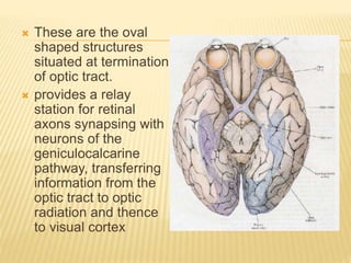
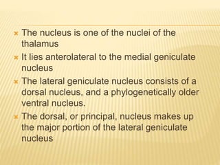
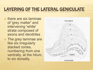
THE LATERAL GENICULATE NUCLEUS
MANOJ ARYAL
IOM,MMC
 These are the oval
shaped structures
situated at termination
of optic tract.
 provides a relay
station for retinal
axons synapsing with
neurons of the
geniculocalcarine
pathway, transferring
information from the
optic tract to optic
radiation and thence
to visual cortex
 there is a roughly 1:1 relationship between
retinal axons entering the lateral
geniculate nucleus and geniculocalcarine
neurons leaving it.
 Eighty per cent of the synaptic
connections of the lateral geniculate
nucleus are with retinofugal axons.
 The nucleus is one of the nuclei of the
thalamus
 It lies anterolateral to the medial geniculate
nucleus
 The lateral geniculate nucleus consists of a
dorsal nucleus, and a phylogenetically older
ventral nucleus.
 The dorsal, or principal, nucleus makes up
the major portion of the lateral geniculate
nucleus
 Much of the lateral
geniculate nucleus is
hidden, being
overlapped by the
pulvinar and visible
only in sections.
 In coronal section:
 it is like a peaked cap,
the peak projecting
laterally.
Schematic representation of a coronal
section
through the lateral geniculate body viewed
from its posterior
aspect.
 In horizontal section,
 related anteriorly to the optic tract which end
in it,
 laterally with the retrolenticular part of the
internal capsule,
 medially with the medial geniculate body,
 posteriorly with the hippocampal gyrus and
posterolaterally with the inferior cornu of the
lateral ventricle.
LAYERING OF THE LATERAL GENICULATE
 there are six laminae
of 'grey matter' and
intervening 'white'
strata composed of
axons and dendrites
 The grey lammae are
like six irregularly
stacked cones,
numbering from one
ventrally, at the hilum,
to six dorsally.
 The two inner layers consist of loosely
arranged large cells (the magnocellular
layers 1 and 2) and
 the four outer layers consist of polar staining
small and medium-sized cells (the
parvocellular layer 3-6)
 Crossed fibres of the
optic tract end
 in laminae 1, 4 and 6,
 uncrossed
 in 2, 3 and 5
so that fibres from
corresponding parts
of the two hemiretmae
(e.g. right temporal
and left nasal retina)
end in neighbouring
laminae.
Impulses from equivalent spots (a, b)
in the two retinae pass back m the optic
tract to the same region of the lateral
geniculate body .Crossed impulses (b)
terminate in laminae 1, 4 and 6 and
uncrossed Impulses (a) terminate in
laminae 2, 3 and
5.
Retinotopic
projection
 Fibres from each retina pass to both
magnocellular (1 and 2) and parvocellular (3-
6) laminae.
 This segregation is achieved within the
nucleus itself, since the crossed and
uncrossed fibres are still intermingled as they
enter the lateral geniculate nucleus
POSITION OF VISUAL FIBRES
 Macular fibres coming In optic tract occupy
two third of LGB
 Upper retinal fibres occupy the medial half of
the anterior one third of LGB.
 Lower retinal fibres occupy the lateral half of
anterior one third of the LGB.
FUNCTIONS
 RELAY FUNCTION
o Serves as relay station to relay visual information
from optic tract to visual cortex by the way of
geniculocalcarine tract.
 To gate the transmission of signal
o i.e. to control how much of signal be allowed to
pass to the cortex.
 LGB receives GATING control signals from
two major sources
 Cortigofugal fibres from the primary visual
cortex.
 Reticular area of mesencephalon.
Both of these are inhibitory and thus controls
the visual information that is allowed to pass.
BLOOD SUPPLY OF THE LATERAL
GENICULATE NUCLEUS
 Posterior cerebral and posterior choroidal
arteries.
 Anterior choroidal artery.

Lateral geniculate nucleus

  • 1.
    THE LATERAL GENICULATENUCLEUS MANOJ ARYAL IOM,MMC
  • 2.
     These arethe oval shaped structures situated at termination of optic tract.  provides a relay station for retinal axons synapsing with neurons of the geniculocalcarine pathway, transferring information from the optic tract to optic radiation and thence to visual cortex
  • 3.
     there isa roughly 1:1 relationship between retinal axons entering the lateral geniculate nucleus and geniculocalcarine neurons leaving it.  Eighty per cent of the synaptic connections of the lateral geniculate nucleus are with retinofugal axons.
  • 4.
     The nucleusis one of the nuclei of the thalamus  It lies anterolateral to the medial geniculate nucleus  The lateral geniculate nucleus consists of a dorsal nucleus, and a phylogenetically older ventral nucleus.  The dorsal, or principal, nucleus makes up the major portion of the lateral geniculate nucleus
  • 5.
     Much ofthe lateral geniculate nucleus is hidden, being overlapped by the pulvinar and visible only in sections.  In coronal section:  it is like a peaked cap, the peak projecting laterally. Schematic representation of a coronal section through the lateral geniculate body viewed from its posterior aspect.
  • 6.
     In horizontalsection,  related anteriorly to the optic tract which end in it,  laterally with the retrolenticular part of the internal capsule,  medially with the medial geniculate body,  posteriorly with the hippocampal gyrus and posterolaterally with the inferior cornu of the lateral ventricle.
  • 7.
    LAYERING OF THELATERAL GENICULATE  there are six laminae of 'grey matter' and intervening 'white' strata composed of axons and dendrites  The grey lammae are like six irregularly stacked cones, numbering from one ventrally, at the hilum, to six dorsally.
  • 8.
     The twoinner layers consist of loosely arranged large cells (the magnocellular layers 1 and 2) and  the four outer layers consist of polar staining small and medium-sized cells (the parvocellular layer 3-6)
  • 9.
     Crossed fibresof the optic tract end  in laminae 1, 4 and 6,  uncrossed  in 2, 3 and 5 so that fibres from corresponding parts of the two hemiretmae (e.g. right temporal and left nasal retina) end in neighbouring laminae. Impulses from equivalent spots (a, b) in the two retinae pass back m the optic tract to the same region of the lateral geniculate body .Crossed impulses (b) terminate in laminae 1, 4 and 6 and uncrossed Impulses (a) terminate in laminae 2, 3 and 5. Retinotopic projection
  • 10.
     Fibres fromeach retina pass to both magnocellular (1 and 2) and parvocellular (3- 6) laminae.  This segregation is achieved within the nucleus itself, since the crossed and uncrossed fibres are still intermingled as they enter the lateral geniculate nucleus
  • 11.
    POSITION OF VISUALFIBRES  Macular fibres coming In optic tract occupy two third of LGB  Upper retinal fibres occupy the medial half of the anterior one third of LGB.  Lower retinal fibres occupy the lateral half of anterior one third of the LGB.
  • 12.
    FUNCTIONS  RELAY FUNCTION oServes as relay station to relay visual information from optic tract to visual cortex by the way of geniculocalcarine tract.  To gate the transmission of signal o i.e. to control how much of signal be allowed to pass to the cortex.
  • 13.
     LGB receivesGATING control signals from two major sources  Cortigofugal fibres from the primary visual cortex.  Reticular area of mesencephalon. Both of these are inhibitory and thus controls the visual information that is allowed to pass.
  • 14.
    BLOOD SUPPLY OFTHE LATERAL GENICULATE NUCLEUS  Posterior cerebral and posterior choroidal arteries.  Anterior choroidal artery.