Downloaded 686 times


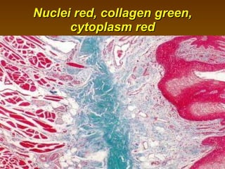


Masson's trichrome stain is a three-color staining protocol used to distinguish collagen from smooth muscle in histological samples. It works by using acid and dye solutions to differentially stain collagen fibers green and smooth muscle/cytoplasm red. The stain is commonly used to identify increased collagen deposition, such as in conditions like collagenous colitis that involve thickening of the subepithelial collagen layer in the intestine. When applied to samples, nuclei appear black/blue, muscle and cytoplasm appear red, and collagen appears green under microscopy.