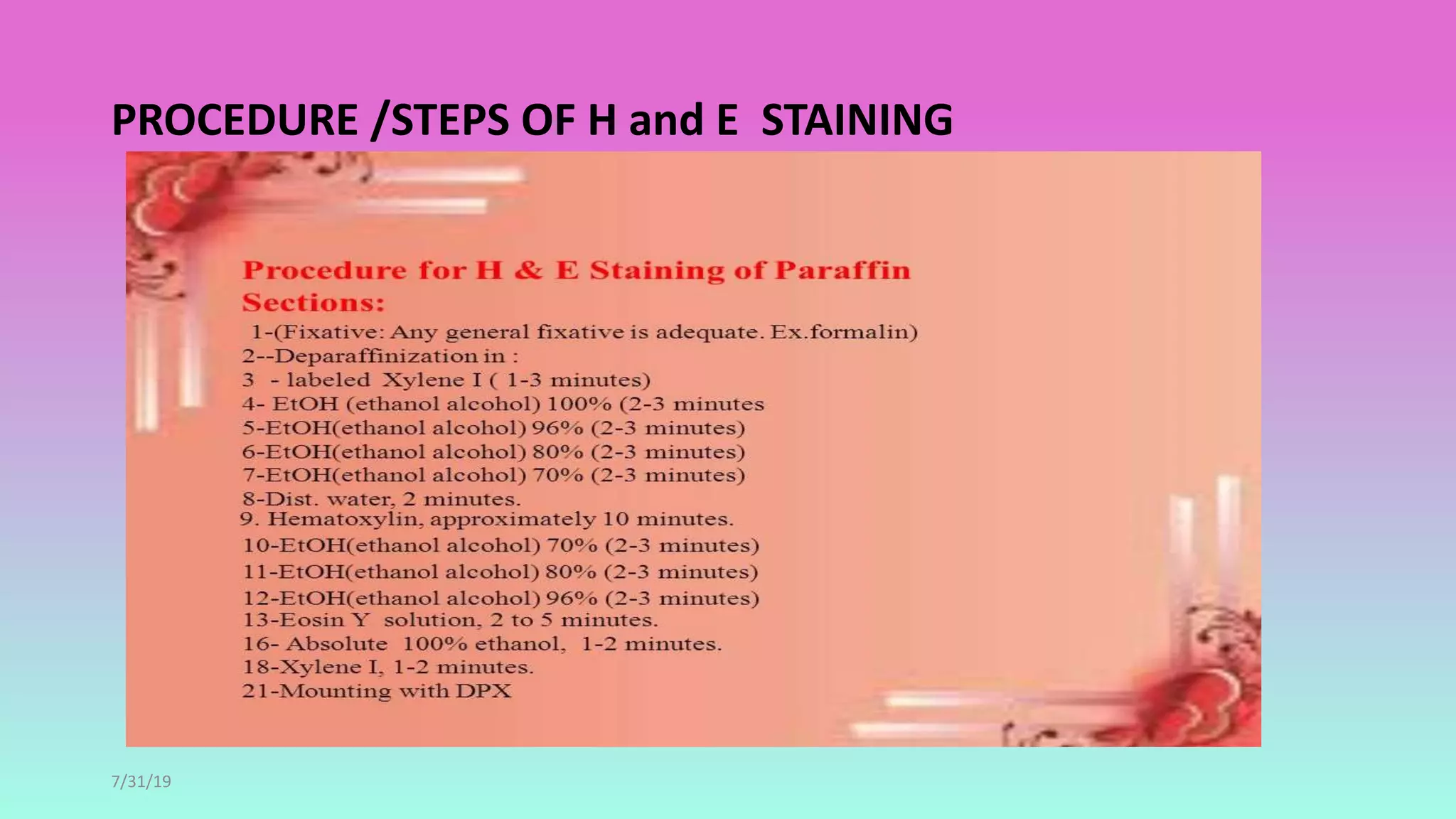
Hematoxylin and eosin (H&E) staining is the most common histological staining method. Hematoxylin stains cell nuclei blue by combining with oxidized hematin and a mordant like alum. Eosin stains cytoplasm and extracellular substances pink. For H&E staining, tissue sections are stained in hematoxylin, rinsed in acid alcohol to differentiate nuclei, rinsed in water to turn nuclei blue, and then stained in eosin to color non-nuclear structures pink, allowing easy visualization of cell morphology. H&E staining provides essential structural information and is useful for pathology examinations.
2. INTRODUCTION
• The histopathological sections that are prepared are colorless
and different components cannot be appreciated.
• Staining them with different color dyes having affinity od
special components of tissues helps in easy identification of
morphology.
• H & E staining is the most frequently used stain in histology.
3. HEMATOXYLIN
• It is extracted from bark of tree hematoylom campechianum.
• Hematoxylin itself is not a stain.
• It is a major oxidation product of hematin which is responsible for
natural color of dye.
4. • Hematin can be produced from hematoxylin from two ways:
• NATURAL OXIDATION
• By exposure to sunlight and air,but it is along process takes 3-4
months
• Solution retain the stain for longer time.
5. CHEMICAL OXIDATION
• Occur instantaneously with short half life
• Oxidation by sodium iodate most common ly used .eg –mayers
hematoxylin
• Oxidation by mercuric oxide eg harris hematoxylin.
6. • Hematin is a weak base dye having poor affinity dye for tissues and
inadequate as nuclear stain with mordant.
• MORDANT-Mordant is a substance which act as intermediate
between dye and tissue .it is a metal with valency of 2+.
• Examples are ammonium and potassium alum ferric salt.
7. PROPRTIES OF HEMATOXYLIN
• Hematoxylin has no staining property.
• Hematin with mordant such as ammonium or potassium alum forms
lake which function as cationic dye and stains anionic tissue
components.
• Hematin in aqueous solution can be acidic or alkaline dye
depending on PH.
8. Classification of hematoxylin solutions
• Alum hematoxylin-Harris and Mayers
• Iron hematoxylin –Weigerts and Verhoeffs
• Tungston hematoxylin
• Molybdenum hematoxylin
• Lead hematoxylin
9. • PROGRESSIVE STAINING
• When tissue is left in the stain just long enough to reach the
property end point. the slide have to be examined under
microscope at different intervals.
• REGRESSIVE STAINING
• The tissue is overstained and then restained until the end point is
reached.
10. • Harris hematoxylin is regressive stain the over stainig is removed by
acid alcohol .The removal of excess dye is called differentiation.
• The hematoxylin alum gives a reddish hue to the tissues because of
acidic ph.
• To convert the color to final blue alkaline ph is required.
• This process is called bluing.it is done either with tap water or
ammonium hydroxide.
12. METHOD
• Dissolve the hematoxylin in absolute alcohol and ammonium alum
in hot water.
• Mix the 2 solutions and heat to boiling.
• Remove from the flame and add mercuric oxide and cool rapidly.
• Glacial acetic acid if added gives brisk nuclear staining but life of the
solution is reduced.
• Hence if acetic acid is to be added it should be added in working
solutions.
13. EOSIN
• Eosin is the counter stain that stains the cytoplasm rose coloured.
• The most widely used eosin is eosin y( y-yellow).
• It is available in either water or alcohol soluble form.
• Most of the labs use the water soluble form of eosin y in an alcohol
water solution.
15. PREPARATION
• Dissolve the eosin in water and then add to this 95% alcohol.
• To the final mixture add few drops of acetic acid which increase the
staining intensity of eosin.
• When ready to use the stain should be cloudy if clear add a few
drops of acetic acid.
• The solution should be standardized by stainig the control slide.