Downloaded 410 times


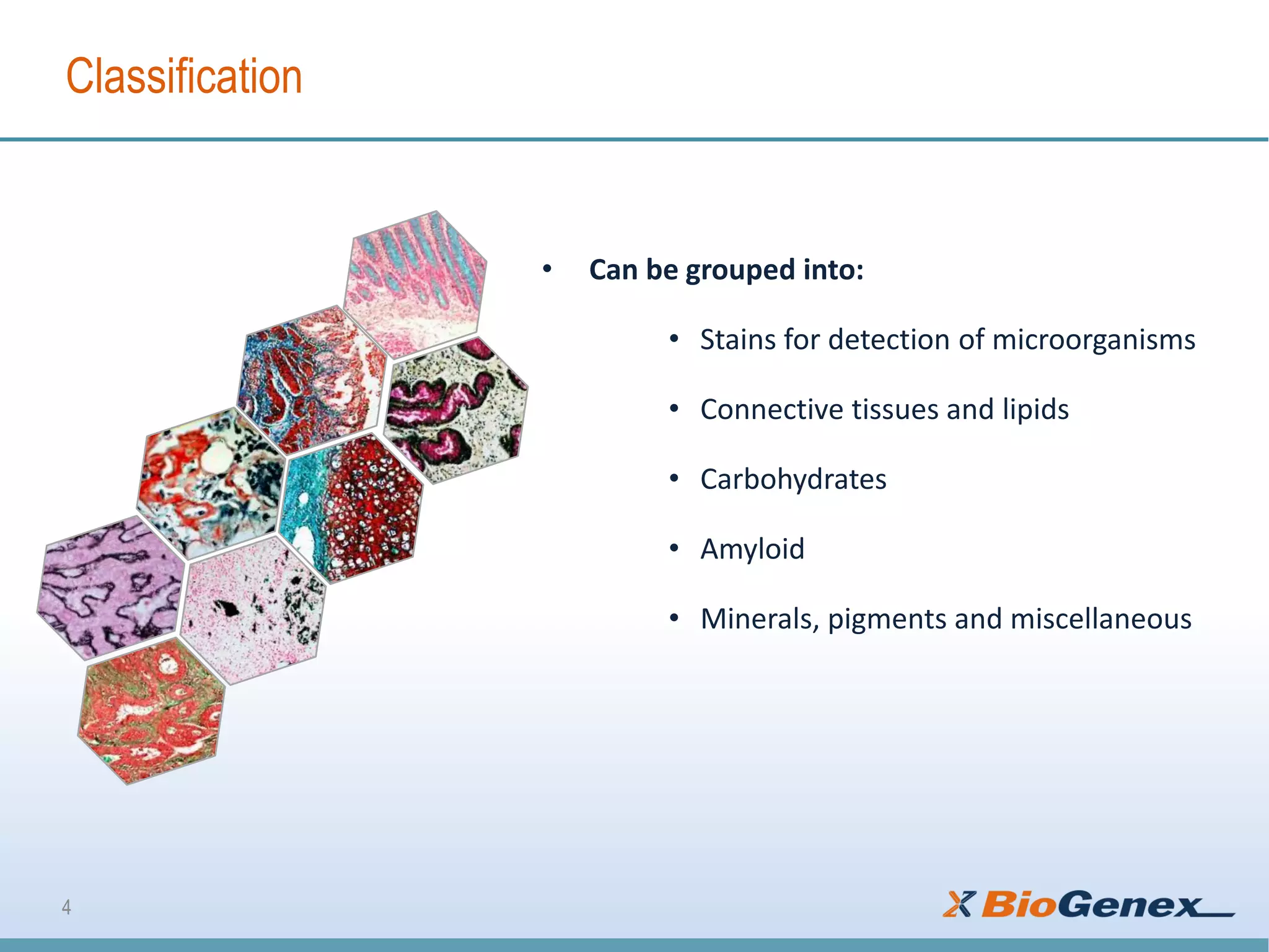


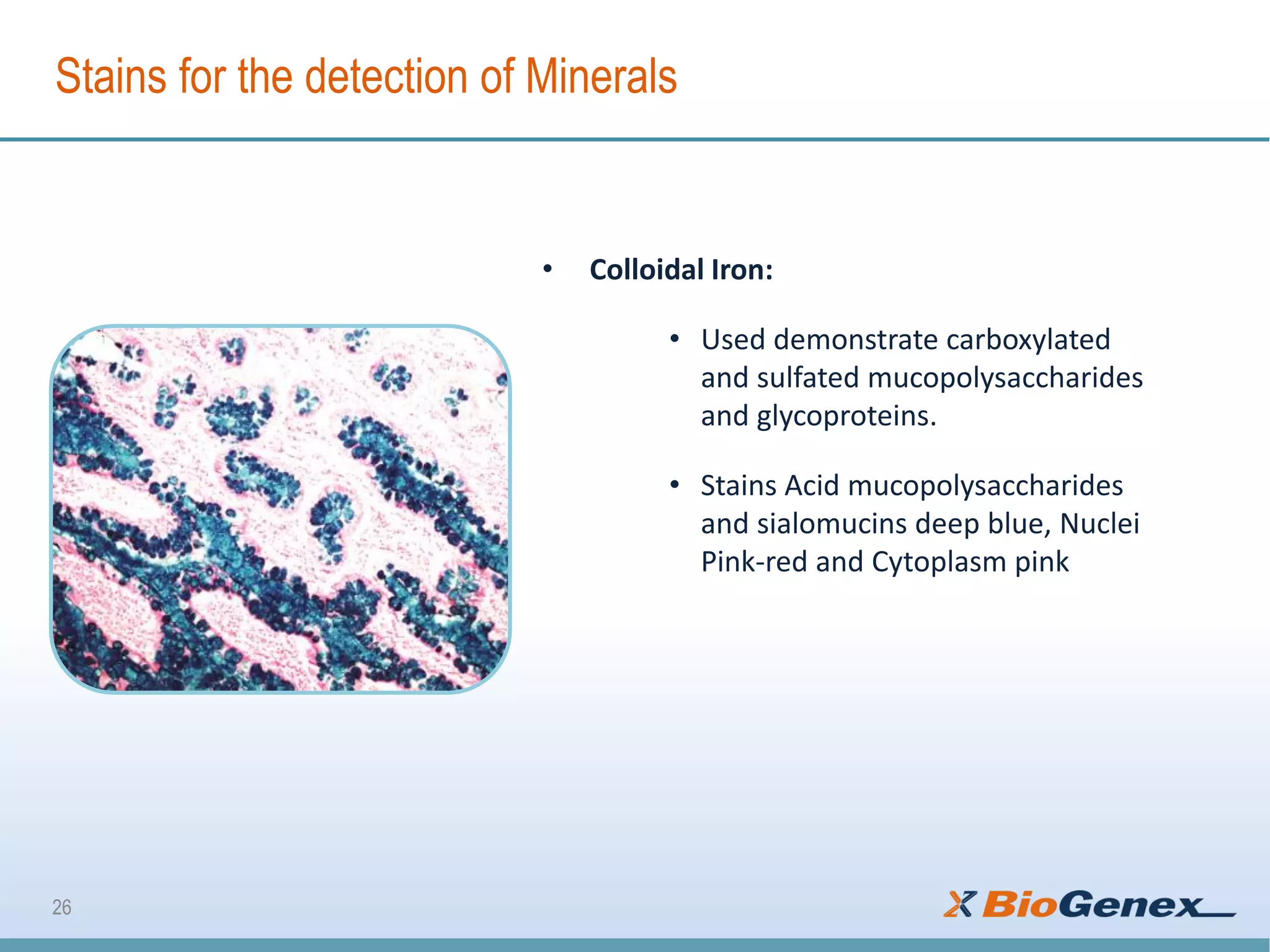


The document outlines various special staining techniques used in histology, highlighting their purposes in detecting microorganisms, connective tissues, carbohydrates, and minerals. It details specific stains such as Gram and Giemsa for bacteria, Gomori's trichrome for connective tissue differentiation, and Alcian blue for mucins. Additionally, it mentions the use of stains for visualizing substances like iron and calcium in tissue specimens.