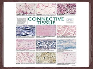
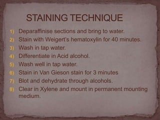
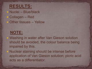
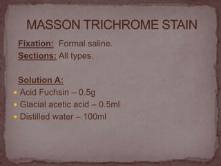
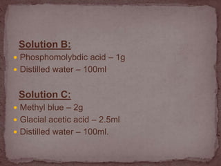
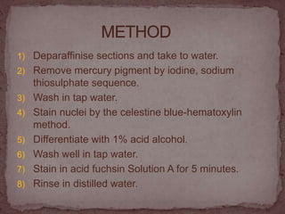
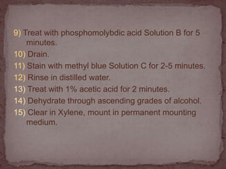
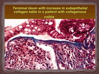
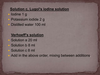
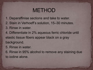
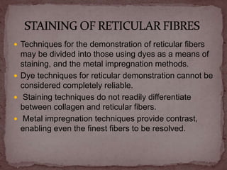
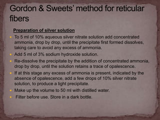
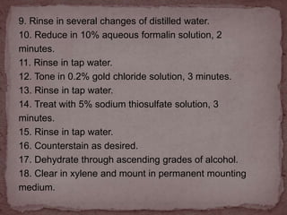
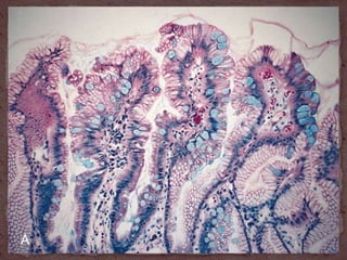
-Dr Sowmya Srinivas
 CONNECTIVE TISSUE STAINS
 CARBOHYDRATE STAINS
 LIPID STAINS
 NUCLEIC ACIDS STAINS
 PROTEINS
 MICRO ORGANISMS
 Connective tissue (CT) is one of the four types of
biological tissue that support, connect, or separate
different types of tissues and organs in the body.
 It develops from the mesoderm.
 The other three types are epithelial, muscle, and nervous
tissue.
 Connective tissue is found in between other tissues
everywhere in the body, including the nervous system.
 All connective tissue apart from blood and lymph consists
of three main components: fibers (elastic and
collagenous fibers), ground substance and cells
The connective tissues are divided into the following
groups:
 Connective tissue proper – includes loose or areolar,
dense, adipose, reticular
 Cartilage – hyaline elastic and fibrocartilage
 Bone – spongy or cancellous and dense or cortical
 Blood
 Blood-forming – hematopoietic.
Connective tissue includes-
 Collagen
 Elastin
 Reticulin
 Basement membrane
 Muscle
Collagen:
 Purpose: Bind bones and other tissues to each
other.
 Components: Alpha polypeptide chains.
 Location: tendon, ligament, skin, cornea,
cartilage, bone, blood vessels, gut, and
intervertebral disc.
Collagen may be differentially stained by any of
the following:
 Van Gieson's stain
 Masson's trichrome stain
 Mallory's trichrome stain
 Aniline blue stain
 Eosin
 Reticulin stain
 Van Gieson's Stain is a mixture of picric acid and
acid fuchsin.
 It is the simplest method of differential staining of
collagen and other connective tissue.
 It was introduced to histology by American
neuropsychiatrist and pathologist Ira Van Gieson.
Solution:
 Saturated aqueous solution of picric acid
(approximately 1 %)- 100 ml
 1 % acid fuchsin - 10 ml
The above quantities may be prepared and kept
as a stock solution, but it is better to use a
freshly prepared solution containing 5 ml of
saturated aqueous solution of picric acid and
0.75 ml of 1 % acid fuchsin which gives more
precise and sharp staining.
1) Deparaffinise sections and bring to water.
2) Stain with Weigert’s hematoxylin for 40 minutes.
3) Wash in tap water.
4) Differentiate in Acid alcohol.
5) Wash well in tap water.
6) Stain in Van Gieson stain for 3 minutes
7) Blot and dehydrate through alcohols.
8) Clear in Xylene and mount in permanent mounting
medium.
RESULTS:
 Nuclei – Blue/black
 Collagen – Red
 Other tissues – Yellow
NOTE:
 Washing in water after Van Gieson solution
should be avoided, the colour balance being
impaired by this.
 Nuclear staining should be intense before
application of Van Gieson solution; picric acid
acts as a differentiator.
Fixation: Formal saline.
Sections: All types.
Solution A:
 Acid Fuchsin – 0.5g
 Glacial acetic acid – 0.5ml
 Distilled water – 100ml
Solution B:
 Phosphomolybdic acid – 1g
 Distilled water – 100ml
Solution C:
 Methyl blue – 2g
 Glacial acetic acid – 2.5ml
 Distilled water – 100ml.
1) Deparaffinise sections and take to water.
2) Remove mercury pigment by iodine, sodium
thiosulphate sequence.
3) Wash in tap water.
4) Stain nuclei by the celestine blue-hematoxylin
method.
5) Differentiate with 1% acid alcohol.
6) Wash well in tap water.
7) Stain in acid fuchsin Solution A for 5 minutes.
8) Rinse in distilled water.
9) Treat with phosphomolybdic acid Solution B for 5
minutes.
10) Drain.
11) Stain with methyl blue Solution C for 2-5 minutes.
12) Rinse in distilled water.
13) Treat with 1% acetic acid for 2 minutes.
14) Dehydrate through ascending grades of alcohol.
15) Clear in Xylene, mount in permanent mounting
medium.
 Nuclei – Blue/black
 Cytoplasm, muscle and RBC’s – Red
 Collagen – Blue.
 Verhoeff's method
 Orcein technique
 Weigert’s Resorcin-fuchsin
 Aldehyde fuchsin method.
 Verhoeff’s method is the classical method for elastic
fibers and works well after all routine fixatives.
Coarse fibers are intensely stained, but the staining
of the fine fibers may be less than satisfactory. The
differentiation step is critical to the success of this
method
Preparation of stain:
Solution a
 Hematoxylin 5 g
 Absolute alcohol 100 ml
Solution b
 Ferric chloride 10 g
 Distilled water 100 ml
Solution c, Lugol’s iodine solution
 Iodine 1 g
 Potassium iodide 2 g
 Distilled water 100 ml
Verhoeff’s solution
 Solution a 20 ml
 Solution b 8 ml
 Solution c 8 ml
 Add in the above order, mixing between additions.
1. Deparaffinise sections and take to water.
2. Stain in Verhoeff’s solution, 15–30 minutes.
3. Rinse in water.
4. Differentiate in 2% aqueous ferric chloride until
elastic tissue fibers appear black on a gray
background.
5. Rinse in water.
6. Rinse in 95% alcohol to remove any staining due
to iodine alone.
7. Counterstain as desired (van Gieson is
conventional, although eosin may be used).
8. Blot to remove excess stain.
9. Dehydrate rapidly through ascending grades of
alcohol.
10. Clear in xylene and mount in permanent mounting
medium.
Results:
Elastic tissue fibers - black
Other tissues according to counterstain.
 Techniques for the demonstration of reticular fibers
may be divided into those using dyes as a means of
staining, and the metal impregnation methods.
 Dye techniques for reticular demonstration cannot be
considered completely reliable.
 Staining techniques do not readily differentiate
between collagen and reticular fibers.
 Metal impregnation techniques provide contrast,
enabling even the finest fibers to be resolved.
Preparation of silver solution
 To 5 ml of 10% aqueous silver nitrate solution add concentrated
ammonia, drop by drop, until the precipitate first formed dissolves,
taking care to avoid any excess of ammonia.
 Add 5 ml of 3% sodium hydroxide solution.
 Re-dissolve the precipitate by the addition of concentrated ammonia,
drop by drop, until the solution retains a trace of opalescence.
 If at this stage any excess of ammonia is present, indicated by the
absence of opalescence, add a few drops of 10% silver nitrate
solution, to produce a light precipitate.
 Make up the volume to 50 ml with distilled water.
 Filter before use. Store in a dark bottle.
Method:
1. Deparaffinize sections and take to water.
2. Treat with 1% potassium permanganate solution,
5 minutes.
3. Rinse in tap water.
4. Bleach in 1% oxalic acid solution.
5. Rinse in tap water.
6. Treat with 2.5% iron alum solution for at least 15
minutes.
7. Wash well in several changes of distilled water.
8. Place in a Coplin jar of silver solution, 2 minutes.
9. Rinse in several changes of distilled water.
10. Reduce in 10% aqueous formalin solution, 2
minutes.
11. Rinse in tap water.
12. Tone in 0.2% gold chloride solution, 3 minutes.
13. Rinse in tap water.
14. Treat with 5% sodium thiosulfate solution, 3
minutes.
15. Rinse in tap water.
16. Counterstain as desired.
17. Dehydrate through ascending grades of alcohol.
18. Clear in xylene and mount in permanent mounting
medium.
 Reticular fibers - black
 Nuclei - black or unstained
 Other elements according to counterstain
Preparation of silver solution:
 To 10 ml of 10% potassium hydroxide solution add 40 ml
of 10% silver nitrate solution.
 Allow the precipitate to settle and decant the supernatant.
 Wash the precipitate several times with distilled water.
 Add ammonia drop by drop until the precipitate has just
dissolved.
 Add further 10% silver nitrate solution until a little
precipitate remains.
 Dilute to 100 ml and filter.
 Store in a dark bottle.
1. Deparaffinize sections and take to water.
2. Treat with 1% potassium permanganate solution,
2 minutes.
3. Rinse in tap water.
4. Bleach in 2% potassium metabisulfate solution.
5. Rinse in tap water.
6. Treat with 2% iron alum, 2 minutes.
7. Wash in several changes of distilled water.
8. Place in Coplin jar of silver solution, 1 minute.
9. Wash in several changes of distilled water.
10. Reduce in 4% aqueous formalin solution, 3
minutes.
11. Rinse in tap water.
12. Tone in 0.2% gold chloride solution, 10 minutes.
13. Rinse in tap water.
14. Treat with 2% potassium metabisulfate solution,
1 minute.
15. Rinse in tap water.
16. Treat with 2% sodium thiosulfate solution,
1 minute.
17. Rinse in tap water.
18. Counterstain as desired (van Gieson or eosin is
suitable).
19. Dehydrate through ascending grades of alcohol.
20. Clear in xylene and mount in permanent mounting
medium.
Results:
Reticular fibers - black
Nuclei - gray
Other tissues - according to counterstain
Periodic acid solution:
 Periodic acid 1 g
 Distilled water 100 ml
Preparation of Schiff reagent:
 Dissolve 1 g of basic fuchsin and 1.9 g of sodium
metabisulfite in 100 ml of 0.15 M hydrochloric acid.
 Shake the solution at intervals or on a mechanical shaker
for 2 hours.
 The solution should be clear and yellow to light brown in
colour.
 Add 500 mg of activated charcoal and shake for 1 to
2 minutes.
 Filter the solution through a No. 1 Whatman filter into
a bottle.
 The filtered solution should be clear and colorless.
 If the solution is yellow, repeat the charcoal
decolorization using a fresh lot of activated charcoal.
 Store at 4°C. Solution is stable for several months.
1. Dewax in xylene and rehydrate through graded
ethanol to distilled water.
2. Oxidize with periodic acid for 5 minutes.
3. Rinse in several changes of distilled water.
4. Cover the sections with Schiff reagent for 15
minutes.
5. Rinse in running tap water for 5–10 minutes.
6. Stain the nuclei with hematoxylin. Differentiate and
blue the sections.
7. Dehydrate in graded ethanol and clear with
xylene.
8. Coverslip.
Results:
Glycogen, neutral/ sialomucins - magenta
Various glycoproteins - magenta
Nuclei - blue
Alcian blue solution:
 Alcian blue 8GX 1 g
 3% acetic acid solution 100 ml
Nuclear fast red:
 Aluminium sulphate Al2(SO4)3•18H2O 5 g
 Distilled water 100 ml
 Nuclear fast red 0.1 g
Dissolve the aluminium sulphate in the water with
heat. Add the nuclear fast red to water while still hot
and filter.
1. Dewax in xylene and rehydrate through graded
ethanol to distilled water.
2. Stain in the alcian blue solution for 30 minutes.
3. Rinse in running tap water for 5 minutes.
4. Counterstain in nuclear fast red for 10 minutes.
5. Wash in running tap water for 1 minute.
6. Dehydrate in graded ethanol.
7. Clear in xylene and mount in a miscible medium.
 Acid mucins (sulfomucins and sialomucins) - blue
 Proteoglycans and hyaluronic acid - blue
 Nuclei - red
Special stains by sowmya

Special stains by sowmya

  • 1.
  • 2.
     CONNECTIVE TISSUESTAINS  CARBOHYDRATE STAINS  LIPID STAINS  NUCLEIC ACIDS STAINS  PROTEINS  MICRO ORGANISMS
  • 3.
     Connective tissue(CT) is one of the four types of biological tissue that support, connect, or separate different types of tissues and organs in the body.  It develops from the mesoderm.  The other three types are epithelial, muscle, and nervous tissue.  Connective tissue is found in between other tissues everywhere in the body, including the nervous system.  All connective tissue apart from blood and lymph consists of three main components: fibers (elastic and collagenous fibers), ground substance and cells
  • 4.
    The connective tissuesare divided into the following groups:  Connective tissue proper – includes loose or areolar, dense, adipose, reticular  Cartilage – hyaline elastic and fibrocartilage  Bone – spongy or cancellous and dense or cortical  Blood  Blood-forming – hematopoietic.
  • 10.
    Connective tissue includes- Collagen  Elastin  Reticulin  Basement membrane  Muscle
  • 11.
    Collagen:  Purpose: Bindbones and other tissues to each other.  Components: Alpha polypeptide chains.  Location: tendon, ligament, skin, cornea, cartilage, bone, blood vessels, gut, and intervertebral disc.
  • 12.
    Collagen may bedifferentially stained by any of the following:  Van Gieson's stain  Masson's trichrome stain  Mallory's trichrome stain  Aniline blue stain  Eosin  Reticulin stain
  • 13.
     Van Gieson'sStain is a mixture of picric acid and acid fuchsin.  It is the simplest method of differential staining of collagen and other connective tissue.  It was introduced to histology by American neuropsychiatrist and pathologist Ira Van Gieson.
  • 14.
    Solution:  Saturated aqueoussolution of picric acid (approximately 1 %)- 100 ml  1 % acid fuchsin - 10 ml The above quantities may be prepared and kept as a stock solution, but it is better to use a freshly prepared solution containing 5 ml of saturated aqueous solution of picric acid and 0.75 ml of 1 % acid fuchsin which gives more precise and sharp staining.
  • 15.
    1) Deparaffinise sectionsand bring to water. 2) Stain with Weigert’s hematoxylin for 40 minutes. 3) Wash in tap water. 4) Differentiate in Acid alcohol. 5) Wash well in tap water. 6) Stain in Van Gieson stain for 3 minutes 7) Blot and dehydrate through alcohols. 8) Clear in Xylene and mount in permanent mounting medium.
  • 16.
    RESULTS:  Nuclei –Blue/black  Collagen – Red  Other tissues – Yellow NOTE:  Washing in water after Van Gieson solution should be avoided, the colour balance being impaired by this.  Nuclear staining should be intense before application of Van Gieson solution; picric acid acts as a differentiator.
  • 18.
    Fixation: Formal saline. Sections:All types. Solution A:  Acid Fuchsin – 0.5g  Glacial acetic acid – 0.5ml  Distilled water – 100ml
  • 19.
    Solution B:  Phosphomolybdicacid – 1g  Distilled water – 100ml Solution C:  Methyl blue – 2g  Glacial acetic acid – 2.5ml  Distilled water – 100ml.
  • 20.
    1) Deparaffinise sectionsand take to water. 2) Remove mercury pigment by iodine, sodium thiosulphate sequence. 3) Wash in tap water. 4) Stain nuclei by the celestine blue-hematoxylin method. 5) Differentiate with 1% acid alcohol. 6) Wash well in tap water. 7) Stain in acid fuchsin Solution A for 5 minutes. 8) Rinse in distilled water.
  • 21.
    9) Treat withphosphomolybdic acid Solution B for 5 minutes. 10) Drain. 11) Stain with methyl blue Solution C for 2-5 minutes. 12) Rinse in distilled water. 13) Treat with 1% acetic acid for 2 minutes. 14) Dehydrate through ascending grades of alcohol. 15) Clear in Xylene, mount in permanent mounting medium.
  • 22.
     Nuclei –Blue/black  Cytoplasm, muscle and RBC’s – Red  Collagen – Blue.
  • 25.
     Verhoeff's method Orcein technique  Weigert’s Resorcin-fuchsin  Aldehyde fuchsin method.  Verhoeff’s method is the classical method for elastic fibers and works well after all routine fixatives. Coarse fibers are intensely stained, but the staining of the fine fibers may be less than satisfactory. The differentiation step is critical to the success of this method
  • 26.
    Preparation of stain: Solutiona  Hematoxylin 5 g  Absolute alcohol 100 ml Solution b  Ferric chloride 10 g  Distilled water 100 ml
  • 27.
    Solution c, Lugol’siodine solution  Iodine 1 g  Potassium iodide 2 g  Distilled water 100 ml Verhoeff’s solution  Solution a 20 ml  Solution b 8 ml  Solution c 8 ml  Add in the above order, mixing between additions.
  • 28.
    1. Deparaffinise sectionsand take to water. 2. Stain in Verhoeff’s solution, 15–30 minutes. 3. Rinse in water. 4. Differentiate in 2% aqueous ferric chloride until elastic tissue fibers appear black on a gray background. 5. Rinse in water. 6. Rinse in 95% alcohol to remove any staining due to iodine alone.
  • 29.
    7. Counterstain asdesired (van Gieson is conventional, although eosin may be used). 8. Blot to remove excess stain. 9. Dehydrate rapidly through ascending grades of alcohol. 10. Clear in xylene and mount in permanent mounting medium. Results: Elastic tissue fibers - black Other tissues according to counterstain.
  • 32.
     Techniques forthe demonstration of reticular fibers may be divided into those using dyes as a means of staining, and the metal impregnation methods.  Dye techniques for reticular demonstration cannot be considered completely reliable.  Staining techniques do not readily differentiate between collagen and reticular fibers.  Metal impregnation techniques provide contrast, enabling even the finest fibers to be resolved.
  • 33.
    Preparation of silversolution  To 5 ml of 10% aqueous silver nitrate solution add concentrated ammonia, drop by drop, until the precipitate first formed dissolves, taking care to avoid any excess of ammonia.  Add 5 ml of 3% sodium hydroxide solution.  Re-dissolve the precipitate by the addition of concentrated ammonia, drop by drop, until the solution retains a trace of opalescence.  If at this stage any excess of ammonia is present, indicated by the absence of opalescence, add a few drops of 10% silver nitrate solution, to produce a light precipitate.  Make up the volume to 50 ml with distilled water.  Filter before use. Store in a dark bottle.
  • 34.
    Method: 1. Deparaffinize sectionsand take to water. 2. Treat with 1% potassium permanganate solution, 5 minutes. 3. Rinse in tap water. 4. Bleach in 1% oxalic acid solution. 5. Rinse in tap water. 6. Treat with 2.5% iron alum solution for at least 15 minutes. 7. Wash well in several changes of distilled water. 8. Place in a Coplin jar of silver solution, 2 minutes.
  • 35.
    9. Rinse inseveral changes of distilled water. 10. Reduce in 10% aqueous formalin solution, 2 minutes. 11. Rinse in tap water. 12. Tone in 0.2% gold chloride solution, 3 minutes. 13. Rinse in tap water. 14. Treat with 5% sodium thiosulfate solution, 3 minutes. 15. Rinse in tap water. 16. Counterstain as desired. 17. Dehydrate through ascending grades of alcohol. 18. Clear in xylene and mount in permanent mounting medium.
  • 36.
     Reticular fibers- black  Nuclei - black or unstained  Other elements according to counterstain
  • 39.
    Preparation of silversolution:  To 10 ml of 10% potassium hydroxide solution add 40 ml of 10% silver nitrate solution.  Allow the precipitate to settle and decant the supernatant.  Wash the precipitate several times with distilled water.  Add ammonia drop by drop until the precipitate has just dissolved.  Add further 10% silver nitrate solution until a little precipitate remains.  Dilute to 100 ml and filter.  Store in a dark bottle.
  • 40.
    1. Deparaffinize sectionsand take to water. 2. Treat with 1% potassium permanganate solution, 2 minutes. 3. Rinse in tap water. 4. Bleach in 2% potassium metabisulfate solution. 5. Rinse in tap water. 6. Treat with 2% iron alum, 2 minutes. 7. Wash in several changes of distilled water.
  • 41.
    8. Place inCoplin jar of silver solution, 1 minute. 9. Wash in several changes of distilled water. 10. Reduce in 4% aqueous formalin solution, 3 minutes. 11. Rinse in tap water. 12. Tone in 0.2% gold chloride solution, 10 minutes. 13. Rinse in tap water. 14. Treat with 2% potassium metabisulfate solution, 1 minute. 15. Rinse in tap water. 16. Treat with 2% sodium thiosulfate solution, 1 minute.
  • 42.
    17. Rinse intap water. 18. Counterstain as desired (van Gieson or eosin is suitable). 19. Dehydrate through ascending grades of alcohol. 20. Clear in xylene and mount in permanent mounting medium. Results: Reticular fibers - black Nuclei - gray Other tissues - according to counterstain
  • 45.
    Periodic acid solution: Periodic acid 1 g  Distilled water 100 ml Preparation of Schiff reagent:  Dissolve 1 g of basic fuchsin and 1.9 g of sodium metabisulfite in 100 ml of 0.15 M hydrochloric acid.  Shake the solution at intervals or on a mechanical shaker for 2 hours.  The solution should be clear and yellow to light brown in colour.
  • 46.
     Add 500mg of activated charcoal and shake for 1 to 2 minutes.  Filter the solution through a No. 1 Whatman filter into a bottle.  The filtered solution should be clear and colorless.  If the solution is yellow, repeat the charcoal decolorization using a fresh lot of activated charcoal.  Store at 4°C. Solution is stable for several months.
  • 47.
    1. Dewax inxylene and rehydrate through graded ethanol to distilled water. 2. Oxidize with periodic acid for 5 minutes. 3. Rinse in several changes of distilled water. 4. Cover the sections with Schiff reagent for 15 minutes. 5. Rinse in running tap water for 5–10 minutes. 6. Stain the nuclei with hematoxylin. Differentiate and blue the sections.
  • 48.
    7. Dehydrate ingraded ethanol and clear with xylene. 8. Coverslip. Results: Glycogen, neutral/ sialomucins - magenta Various glycoproteins - magenta Nuclei - blue
  • 51.
    Alcian blue solution: Alcian blue 8GX 1 g  3% acetic acid solution 100 ml Nuclear fast red:  Aluminium sulphate Al2(SO4)3•18H2O 5 g  Distilled water 100 ml  Nuclear fast red 0.1 g Dissolve the aluminium sulphate in the water with heat. Add the nuclear fast red to water while still hot and filter.
  • 52.
    1. Dewax inxylene and rehydrate through graded ethanol to distilled water. 2. Stain in the alcian blue solution for 30 minutes. 3. Rinse in running tap water for 5 minutes. 4. Counterstain in nuclear fast red for 10 minutes. 5. Wash in running tap water for 1 minute. 6. Dehydrate in graded ethanol. 7. Clear in xylene and mount in a miscible medium.
  • 53.
     Acid mucins(sulfomucins and sialomucins) - blue  Proteoglycans and hyaluronic acid - blue  Nuclei - red